Copyright
©2014 Baishideng Publishing Group Inc.
World J Orthop. Nov 18, 2014; 5(5): 694-698
Published online Nov 18, 2014. doi: 10.5312/wjo.v5.i5.694
Published online Nov 18, 2014. doi: 10.5312/wjo.v5.i5.694
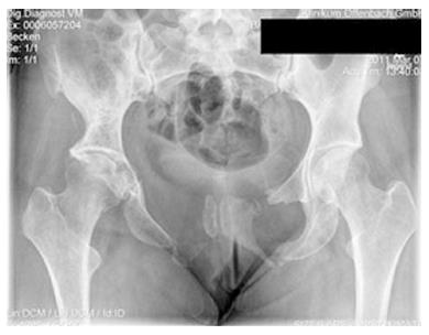
Figure 1 Case example: X-ray (A) and computed tomography scan (B), of a 26 years old female, with vanishing bone disease of the pelvis.
She presented with mild groin pain without any further symptoms.
Figure 2 X-ray of the pelvis of the previous patient.
Three years later she remained asymptomatic with only mild discomfort to the groin and no further symptoms.
- Citation: Nikolaou VS, Chytas D, Korres D, Efstathopoulos N. Vanishing bone disease (Gorham-Stout syndrome): A review of a rare entity. World J Orthop 2014; 5(5): 694-698
- URL: https://www.wjgnet.com/2218-5836/full/v5/i5/694.htm
- DOI: https://dx.doi.org/10.5312/wjo.v5.i5.694


