
















©2014 Baishideng Publishing Group Inc.
World J Orthop. Sep 18, 2014; 5(4): 486-495
Published online Sep 18, 2014. doi: 10.5312/wjo.v5.i4.486
Published online Sep 18, 2014. doi: 10.5312/wjo.v5.i4.486
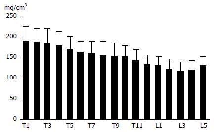
Figure 1 The trabecular bone mineral density of the thoracic and lumbar vertebrae[22].
The bone mineral density tends to decrease from the first thoracic to third lumbar vertebra.
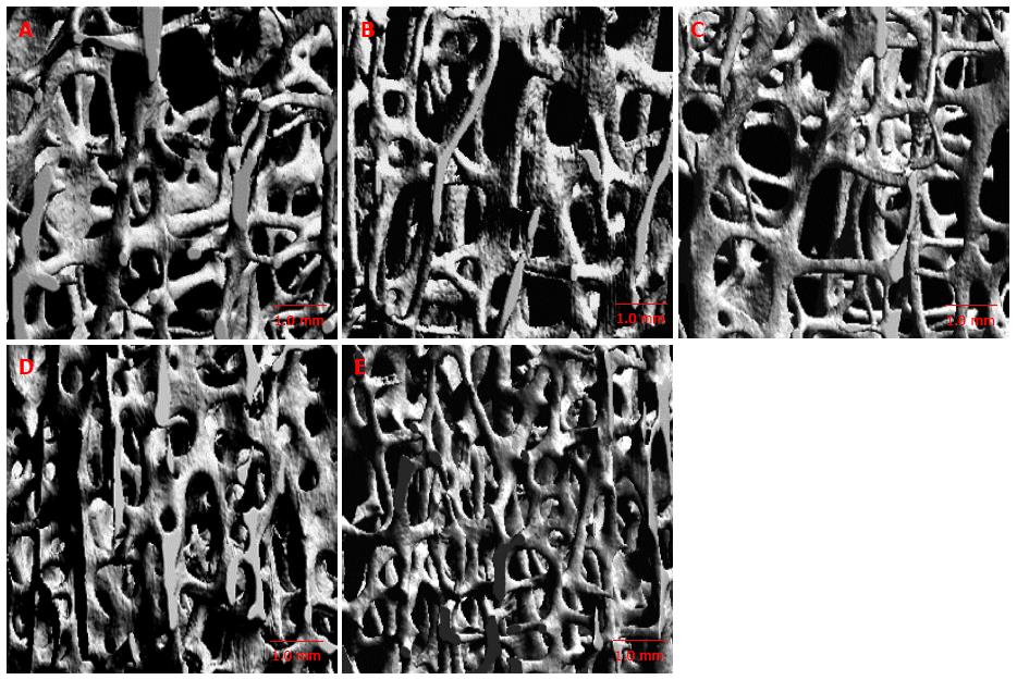
Figure 2 Micro-computed tomography image in different regions of vertebral body[25].
A: Anterosuperior; B: Anteroinferior; C: Central; D: Posterosuperior; E: Posteroinferior regions. The trabecular bone is lower in the anterosuperior and central regions.
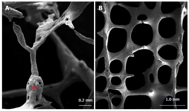
Figure 3 Scanning electron microscopic image of vertebral trabecular bone[25].
The microcallus (MC) is seen on the vertical trabecula (A). Tertical trabeculae are relatively thicker than the horizontal ones (B).
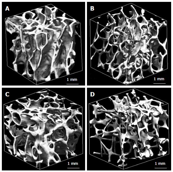
Figure 4 Trabecular microstructure of femoral neck from a man aged 62 years (A), a man aged 92 years (B), a woman aged 62 years (C), and a woman aged 92 years (D)[49].
The trabecular bone is higher in a man aged 62 years and is lower in a woman aged 92 years.
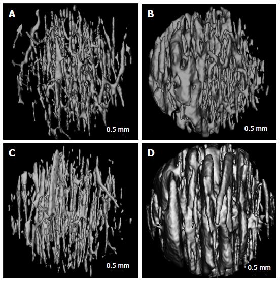
Figure 5 Three D reconstructed images of the canal networks in the inferior femoral neck cortex from a man aged 62 years (A), a man aged 92 years (B), a woman aged 62 years (C), and a woman aged 92 years (D)[49].
There are more enlarged canals in the 92 years than those of the 62 years.
- Citation: Chen H, Kubo KY. Bone three-dimensional microstructural features of the common osteoporotic fracture sites. World J Orthop 2014; 5(4): 486-495
- URL: https://www.wjgnet.com/2218-5836/full/v5/i4/486.htm
- DOI: https://dx.doi.org/10.5312/wjo.v5.i4.486