©2014 Baishideng Publishing Group Co.
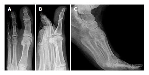
Figure 1 Radiographic images of a hallux rigidus grade 2.
A: Dorso-plantar view; B: Oblique view; C: Stress radiographs in dorsiflexion revealing bony impingement.
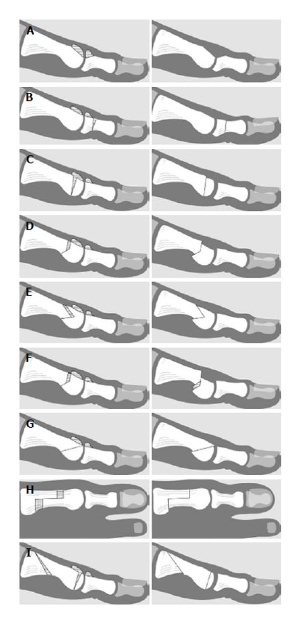
Figure 2 Diagrammatic presentations.
A: A Cheilectomy; B: A proximal phalanx osteotomy (Moberg); C: A dorsal closing wedge osteotomy (Watermann); D: A Watermann Green procedure; E: A Youngswick procedure; F: A Reverdin Green osteotomy; G: A distal oblique sliding osteotomy; H: The Sagittal Z osteotomy; I: A Drago procedure.
- Citation: Polzer H, Polzer S, Brumann M, Mutschler W, Regauer M. Hallux rigidus: Joint preserving alternatives to arthrodesis - a review of the literature. World J Orthop 2014; 5(1): 6-13
- URL: https://www.wjgnet.com/2218-5836/full/v5/i1/6.htm
- DOI: https://dx.doi.org/10.5312/wjo.v5.i1.6


