
















Copyright: ©Author(s) 2026.
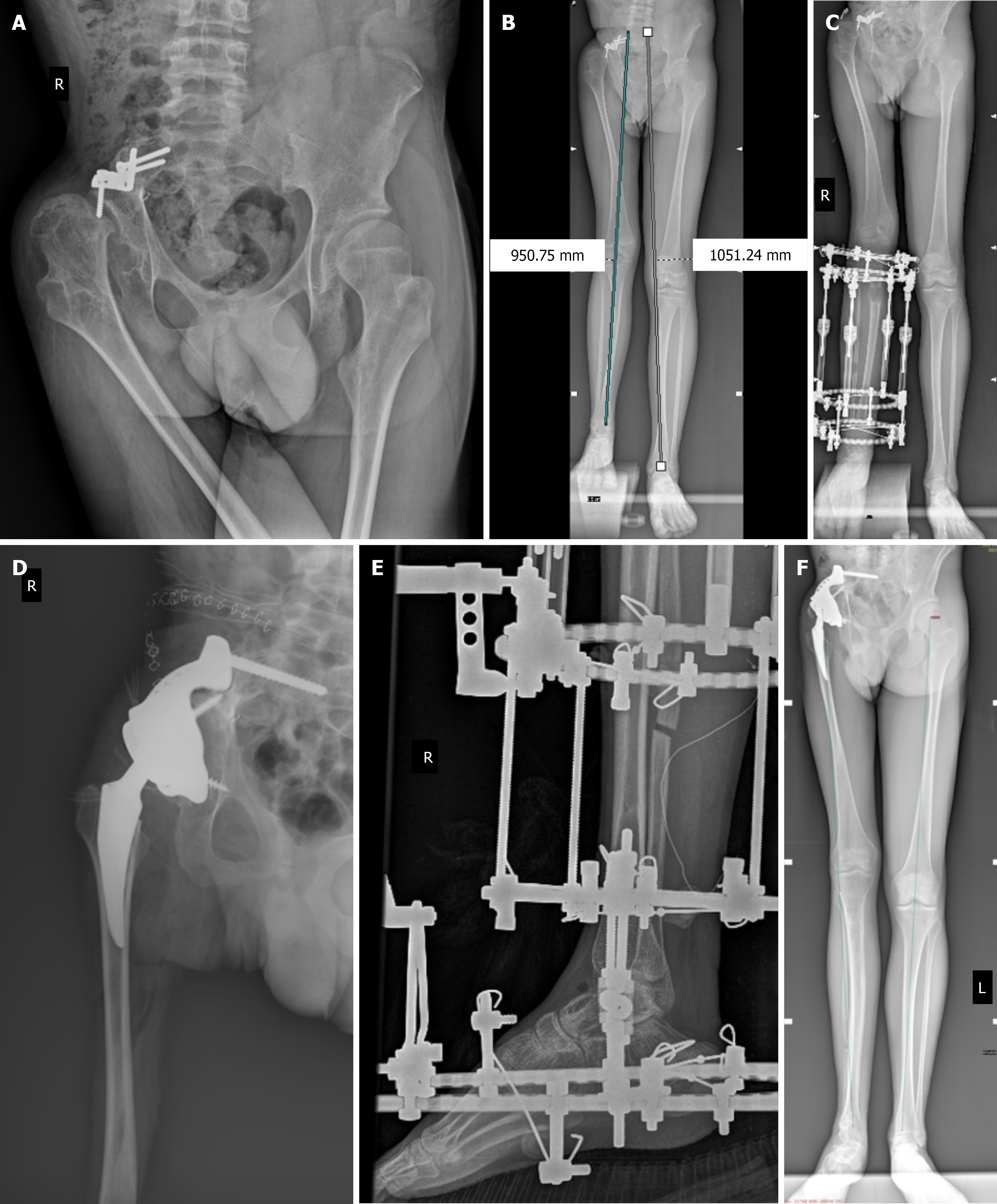
Figure 1 Imaging examinations.
A: Radiograph obtained at initial presentation in 2017 demonstrating pelvic deformity following internal hemipelvectomy, acetabular dysplasia, and proximal femoral deformity with lateral subluxation of the femoral head. Fixation hardware, including a plate and screws from the previous pelvic resection, is visible; B: Radiograph demonstrating limb length discrepancy between the lower extremities and lateral deviation of the mechanical axis in both lower limbs; C: Radiograph demonstrating tibial and fibular lengthening using an Ilizarov external fixator; the first stage of treatment; D: Postoperative radiograph demonstrating total hip arthroplasty with a custom-made acetabular component and femoral stem; the second stage of treatment; E: Radiograph demonstrating an Ilizarov external fixator used for correction of equinus deformity, combined with Hoke tenotomy; the third stage of treatment; F: Final radiographic outcome demonstrating improved limb alignment and maintained implant position after completion of the three-stage reconstructive treatment.
- Citation: Mirgos MP, Wrześniak Z, Pulik Ł, Górski R, Łęgosz P. Restoration of ambulation after internal hemipelvectomy using a three-stage reconstructive strategy: A case report and review of literature. World J Orthop 2026; 17(7): 121971
- URL: https://www.wjgnet.com/2218-5836/full/v17/i7/121971.htm
- DOI: https://dx.doi.org/10.5312/wjo.121971