
















Copyright: ©Author(s) 2026.
World J Orthop. May 18, 2026; 17(5): 119240
Published online May 18, 2026. doi: 10.5312/wjo.v17.i5.119240
Published online May 18, 2026. doi: 10.5312/wjo.v17.i5.119240
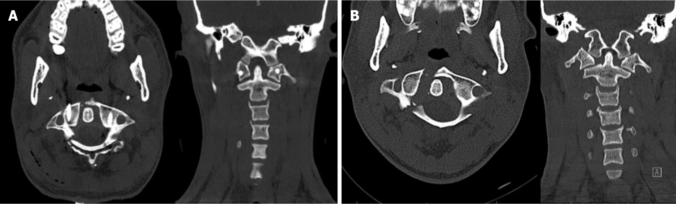
Figure 1 Preoperative and direct postoperative computed tomography scan.
A: Preoperative computed tomography (CT) scan showing the displaced fractures involving both the anterior and posterior arches of the atlas; B: Direct postoperative CT shows reduction of the fracture with appropriate positioning of the lateral mass screws and rods.
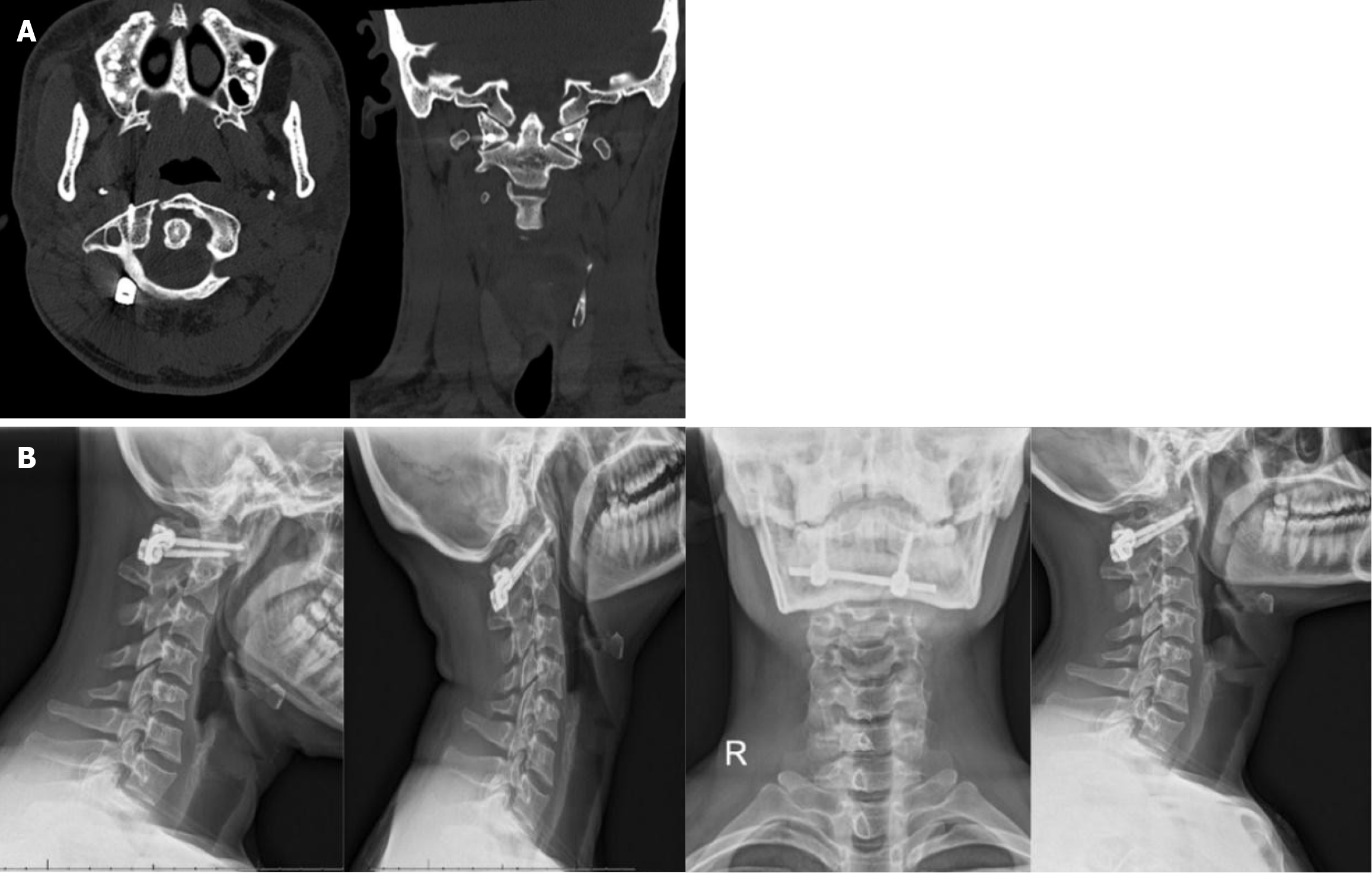
Figure 2 Re-examination 1 year and 9 months postoperative.
A: 1 year and 9 months Postoperative computed tomography scan shows bony union of the posterior arch with satisfactory consolidation; B: 1 year and 9 months dynamic flexion-extension cervical radiographs show atlantoaxial stability.
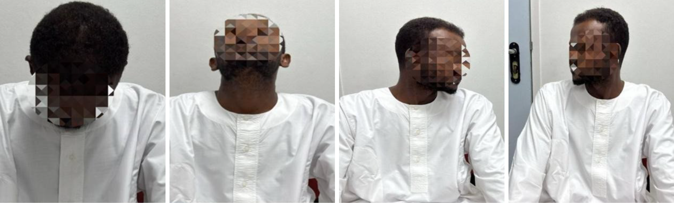
Figure 3 One year and nine months postoperative clinical examination show full cervical range of motion to the end point.
- Citation: Alhamal HA, Alhaddad S, Alsaleh B, Güneş L, Alzawad A, Azzam L, Alhabib M, Alhalal I, Alhamal H. Motion preserving fixation of an unstable Jefferson fracture using C1 lateral mass screws and rods: A case report. World J Orthop 2026; 17(5): 119240
- URL: https://www.wjgnet.com/2218-5836/full/v17/i5/119240.htm
- DOI: https://dx.doi.org/10.5312/wjo.v17.i5.119240