
















Copyright: ©Author(s) 2026.
World J Orthop. May 18, 2026; 17(5): 115855
Published online May 18, 2026. doi: 10.5312/wjo.v17.i5.115855
Published online May 18, 2026. doi: 10.5312/wjo.v17.i5.115855
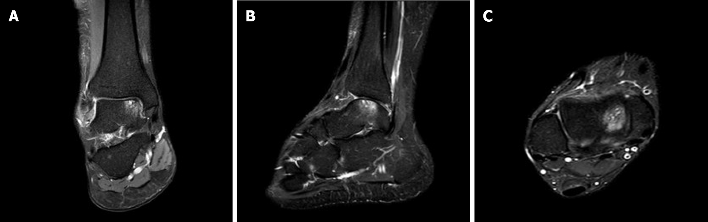
Figure 1 Preoperative magnetic resonance imaging demonstrating osteochondral defect of the medial talar dome (Raikin zone 4).
A: Coronal T2-weighted image showing the defect with surrounding bone marrow edema; B: Sagittal T2-weighted image demonstrating the depth and extent of the lesion; C: Axial T2-weighted image showing the medial location of the defect directly lateral to the medial malleolus.
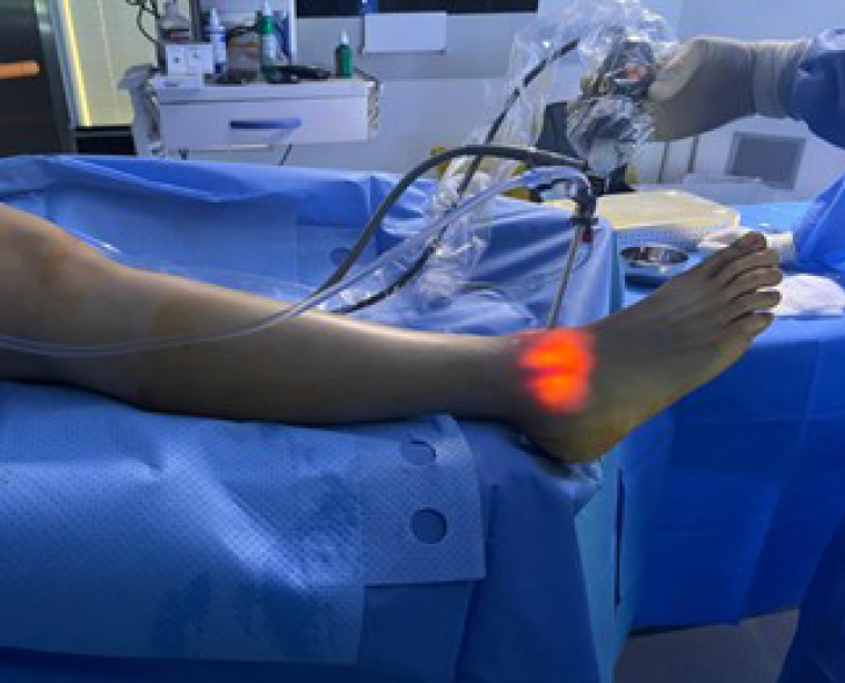
Figure 2
Diagnostic arthroscopy of the right ankle.
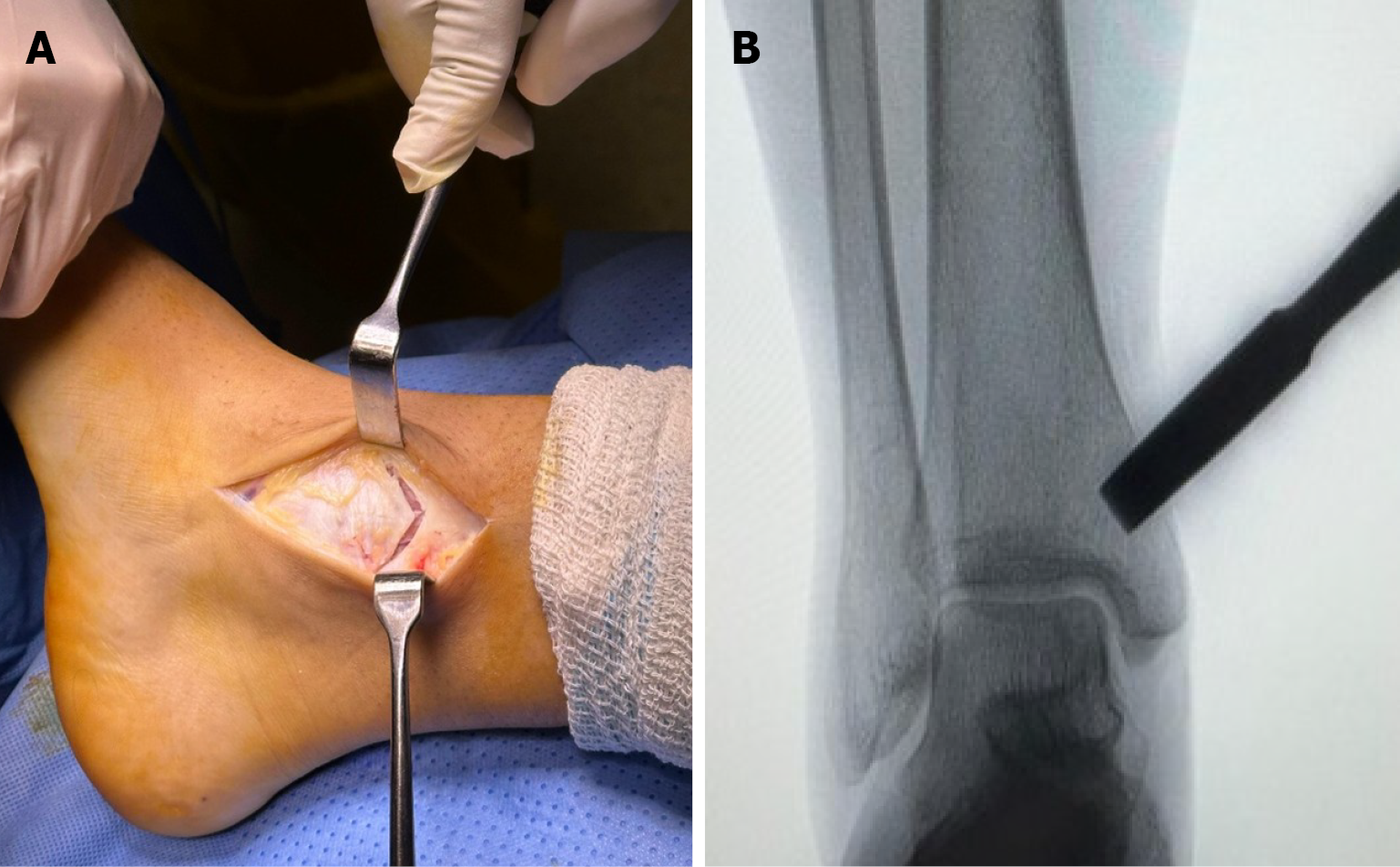
Figure 3 Medial malleolar osteotomy technique.
A: Completion of the osteotomy using a sharp osteotome after preliminary drilling and sawing; B: C-arm fluoroscopic confirmation of optimal osteotomy position and alignment before reflection of the malleolar fragment.
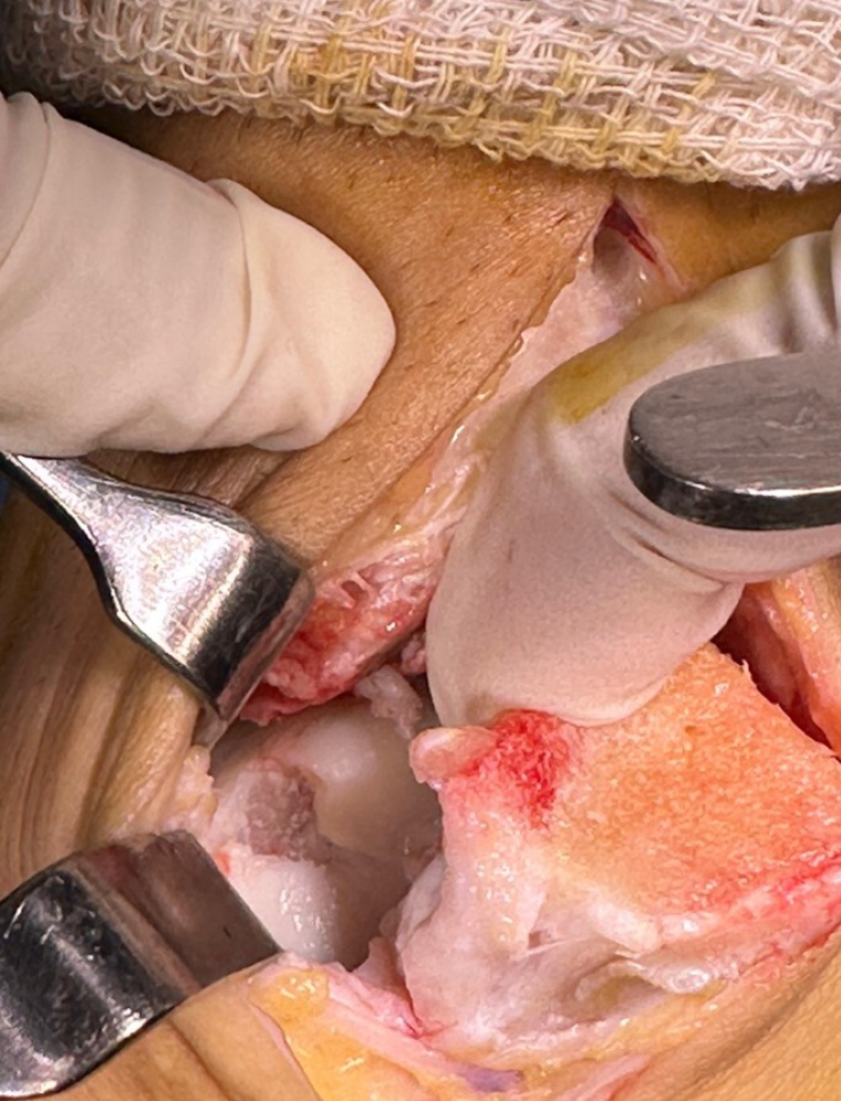
Figure 4
Surgical exposure after medial malleolar osteotomy showing excellent visualization of the osteochondral defect in the medial talar dome (Raikin zone 4) with the malleolar fragment retracted inferiorly.
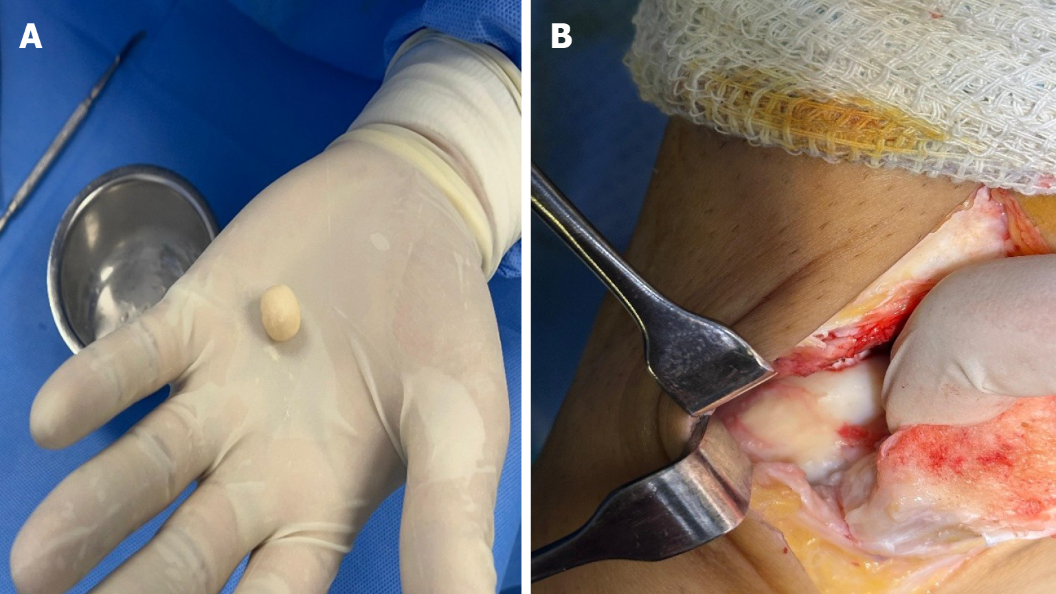
Figure 5 Allograft preparation and implantation.
A: Surgeon holding the prepared allograft, appearing as a small, moldable ball ready for implantation; B: The allograft filling the prepared osteochondral defect, showing complete coverage and appropriate contouring.
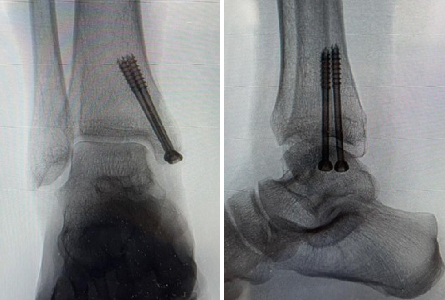
Figure 6 Fluoroscopic images showing osteotomy fixation.
Lateral ankle view demonstrating optimal positioning of two 4.5-mm cannulated titanium screws with anatomical reduction of the medial malleolus.
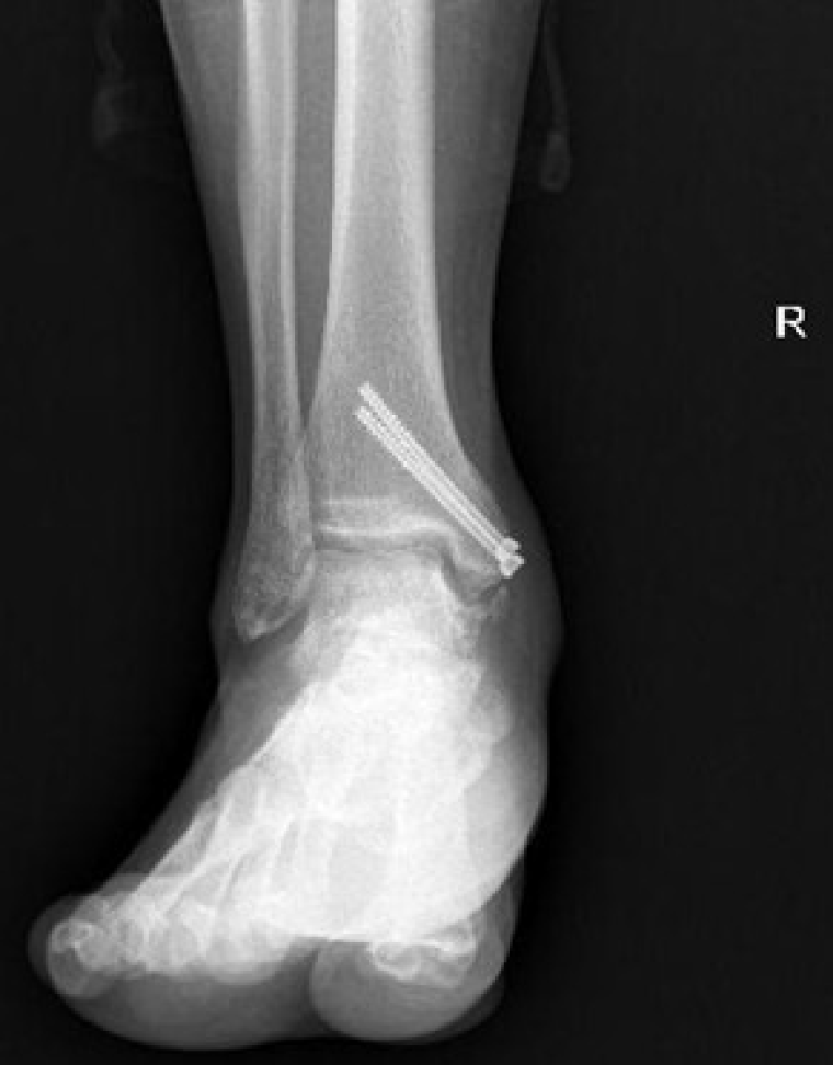
Figure 7 Anteroposterior plain radiograph of the right ankle at 12-month follow-up for the patient with residual intermittent pain.
The image demonstrates a stable osteosynthesis and a completely healed medial malleolar osteotomy site, with clear evidence of significant osteocartilaginous overgrowth of the allograft repair tissue projecting from the medial talar dome.
- Citation: Elalfy MM, Embaby OM, Aladl AN, Abdelrazek M, Youssef K, Abulaban O, Abushal MH. Cartilage allograft transplantation for challenging medial talar dome osteochondral defects: A retrospective case series utilizing medial malleolar osteotomy. World J Orthop 2026; 17(5): 115855
- URL: https://www.wjgnet.com/2218-5836/full/v17/i5/115855.htm
- DOI: https://dx.doi.org/10.5312/wjo.v17.i5.115855