
















Copyright: ©Author(s) 2026.
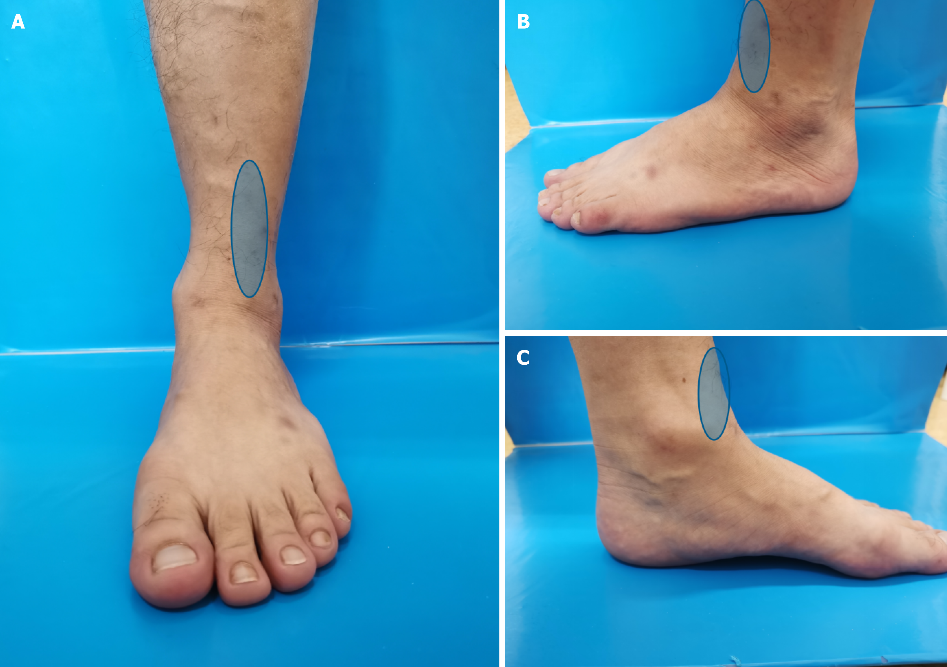
Figure 1 Clinical photographs showing the sites of persistent pain or tenderness at the anterior syndesmosis (blue areas).
A-C: The symptomatic area viewed from three directions.
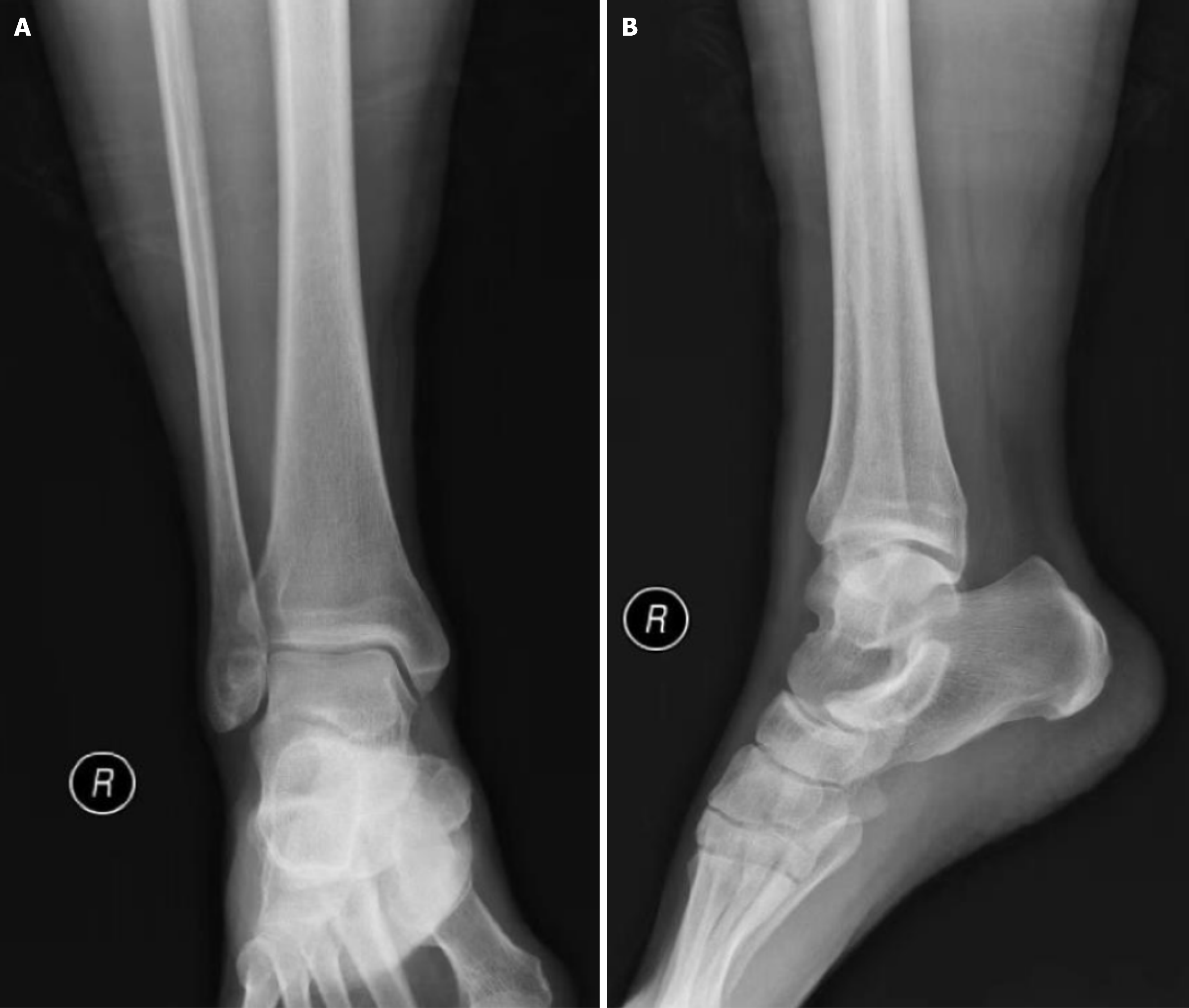
Figure 2 Non-weight-bearing ankle radiographs showing no definite syndesmotic diastasis.
A: Anteroposterior view; B: Lateral view.
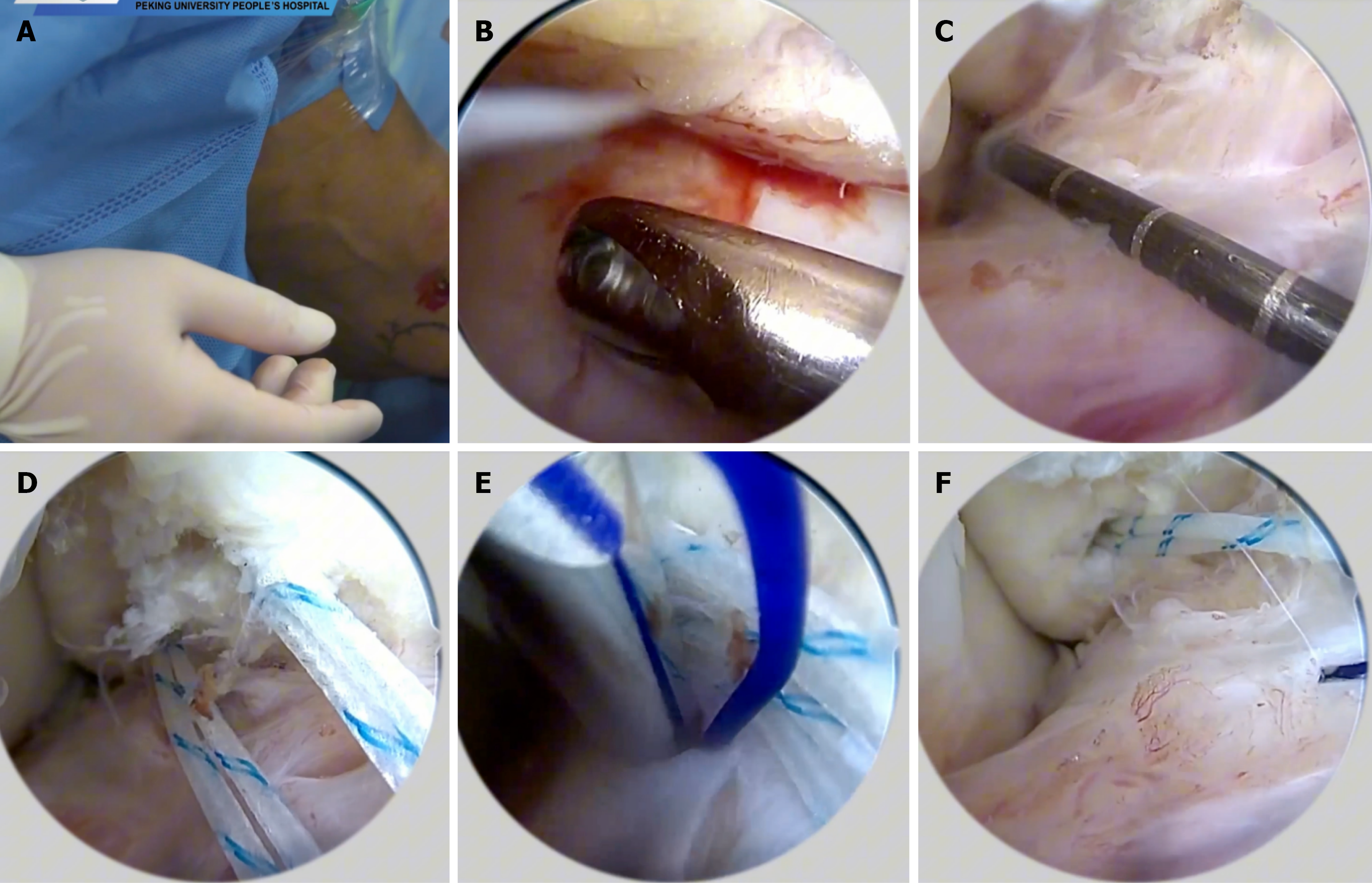
Figure 3 Intraoperative steps of arthroscopic Broström repair.
A-F: Sequential steps of the arthroscopic repair.
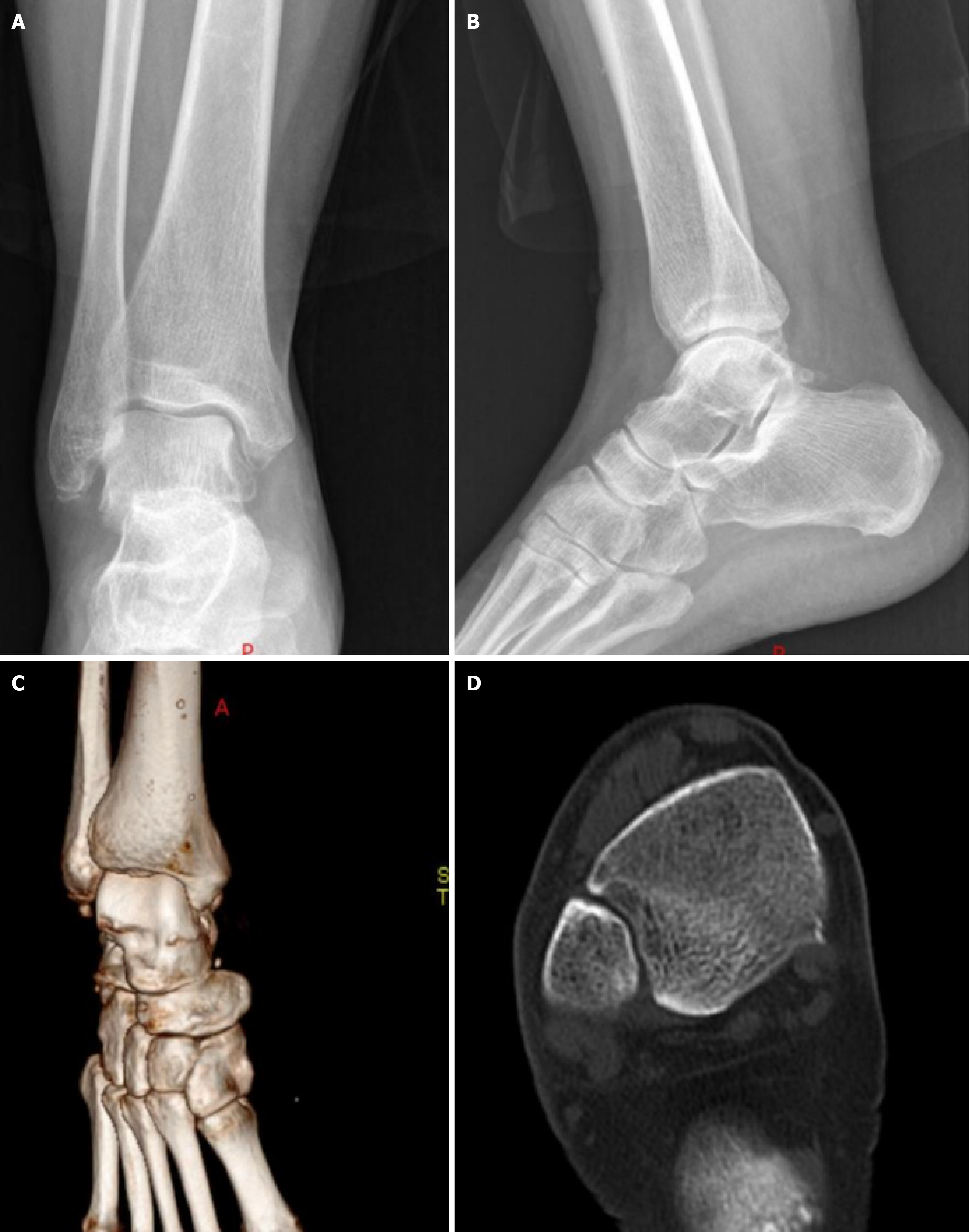
Figure 4 Baseline radiographs and computed tomography images obtained immediately after injury in November 2019 from the illustrative patient.
A: Anteroposterior radiograph; B: Lateral radiograph; C: Three-dimensional computed tomography reconstruction; D: Axial computed tomography image.
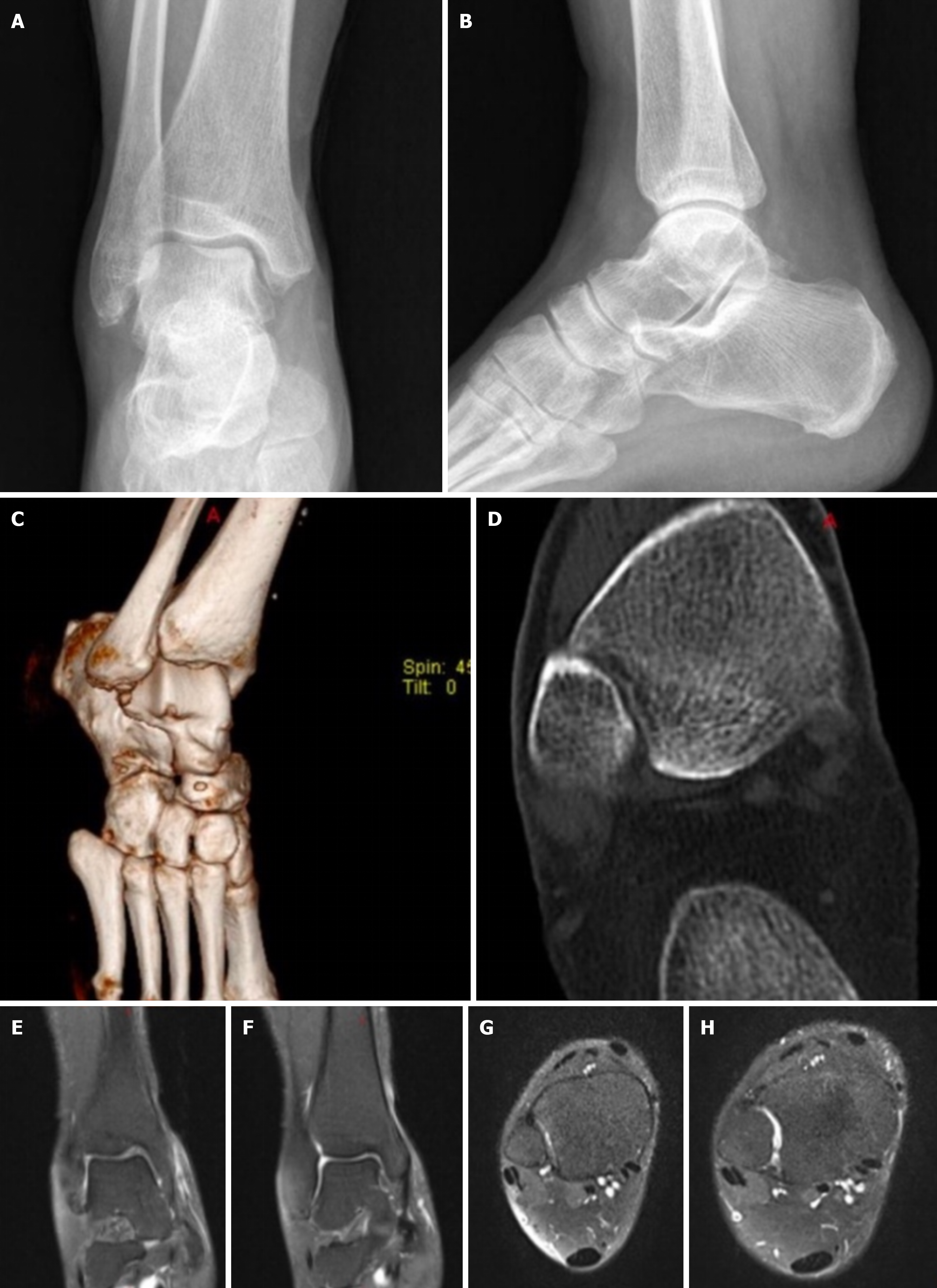
Figure 5 Follow-up radiographs, computed tomography images, and magnetic resonance images obtained in May 2020, approximately 6 months after injury, from the same illustrative patient shown in Figure 4.
A: Anteroposterior radiograph; B: Lateral radiograph; C: Three-dimensional computed tomography reconstruction; D: Axial computed tomography image; E and F: Coronal magnetic resonance images; G and H: Axial magnetic resonance images.
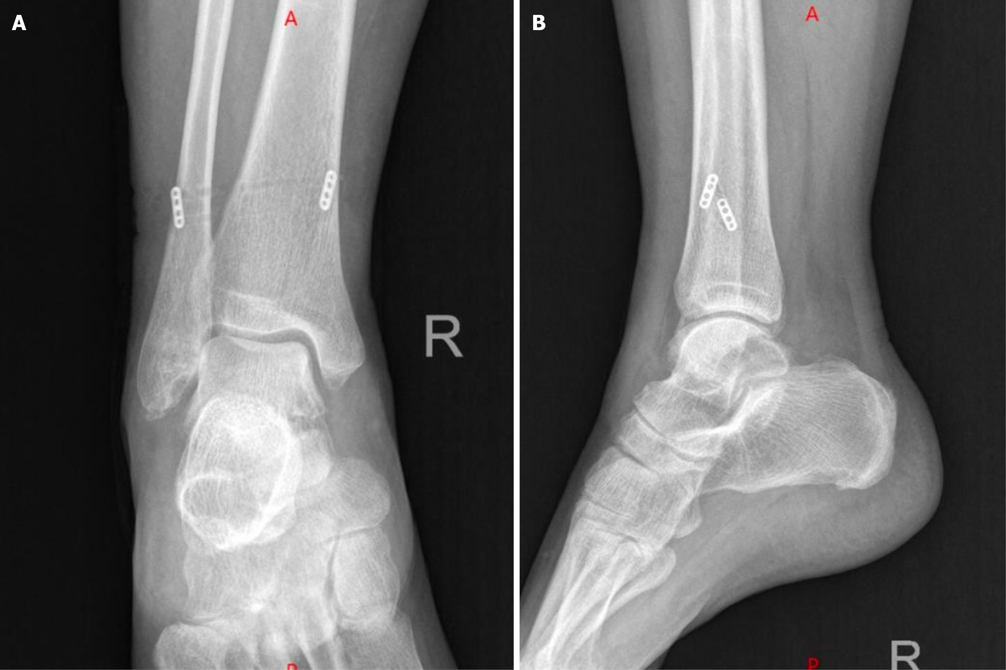
Figure 6 Postoperative radiographs after suture-button stabilization and Broström-Gould repair.
A: Anteroposterior view; B: Lateral view.
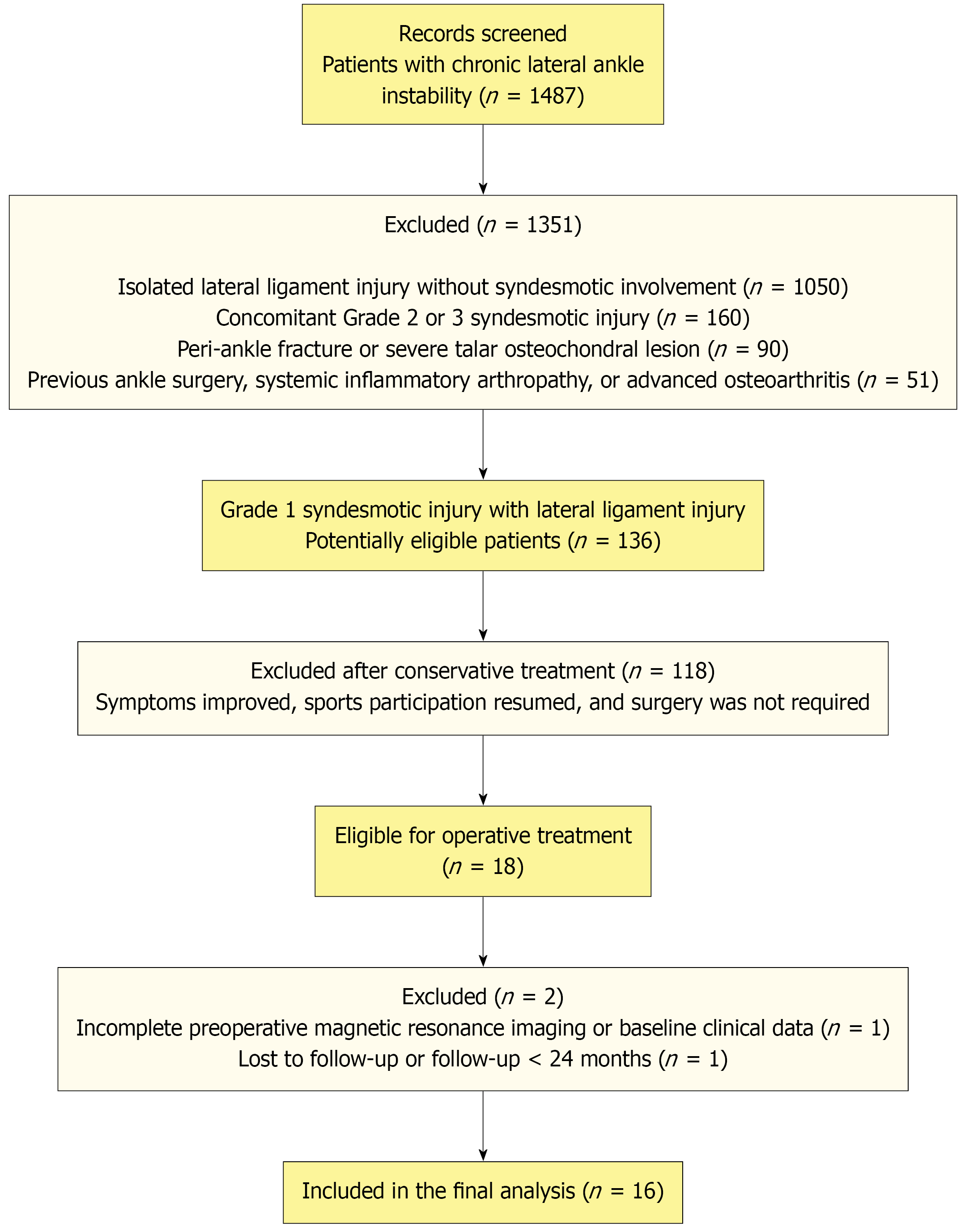
Figure 7
Flow diagram of patient screening, exclusion, and final inclusion.
- Citation: Li GC, Du CB, Lu H, Ju JB, Xu HL. Occult syndesmotic injury in chronic lateral ankle instability treated with Broström repair and suture-button stabilization. World J Orthop 2026; 17(10): 125993
- URL: https://www.wjgnet.com/2218-5836/full/v17/i10/125993.htm
- DOI: https://dx.doi.org/10.5312/wjo.125993