
















Copyright: ©Author(s) 2026.
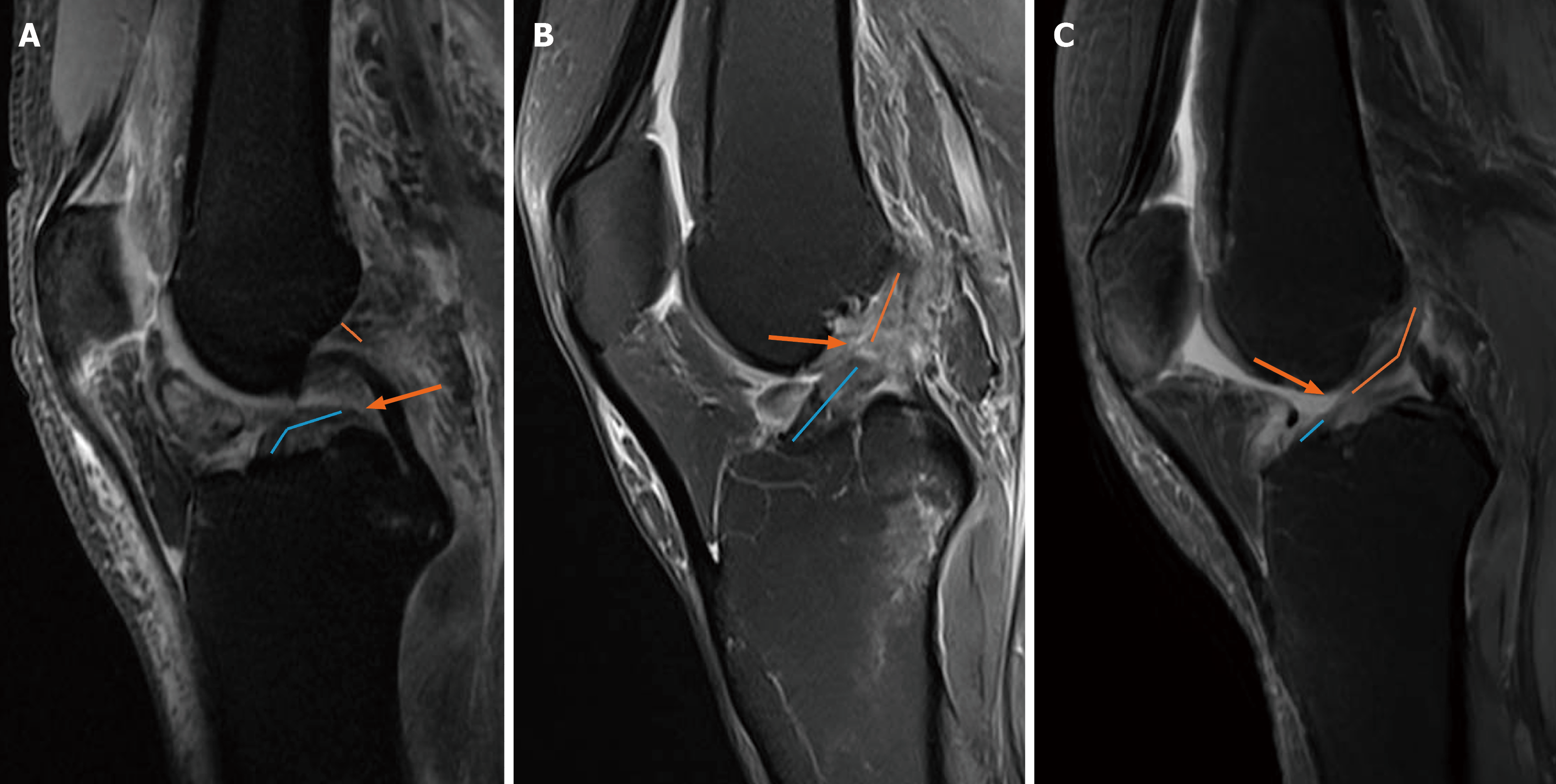
Figure 1 Preoperative sagittal T2-weighted fat-suppressed magnetic resonance imaging images of three older adults (≥ 50 years) with anterior cruciate ligament tears at different locations.
A: Tear near the femoral insertion of the anterior cruciate ligament (ACL); B: Midsubstance tear of the ACL; C: Tear near the tibial insertion of the ACL.
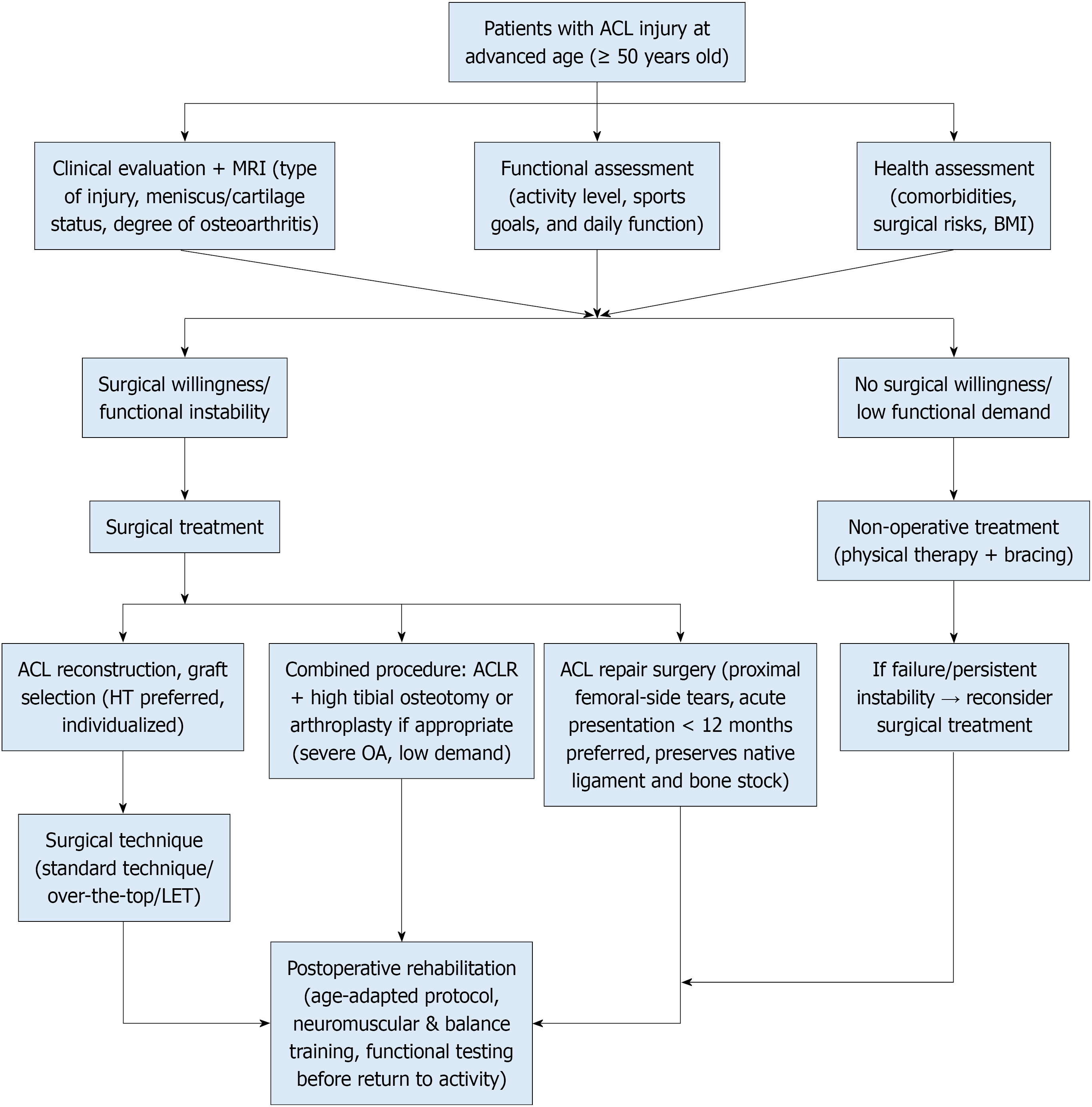
Figure 2 Clinical decision-making algorithm for anterior cruciate ligament injuries in older adult patients.
ACL: Anterior cruciate ligament; MRI: Magnetic resonance imaging; BMI: Body mass index; HT: Hamstring tendon; LET: Lateral extra-articular tenodesis; ACLR: Anterior cruciate ligament reconstruction; OA: Osteoarthritis.
- Citation: Jia YF, Wang HN, Xu LQ, Sun R. Treatment of anterior cruciate ligament injuries in older adults: A clinical review. World J Orthop 2026; 17(10): 125554
- URL: https://www.wjgnet.com/2218-5836/full/v17/i10/125554.htm
- DOI: https://dx.doi.org/10.5312/wjo.125554