
















Copyright: ©Author(s) 2026.
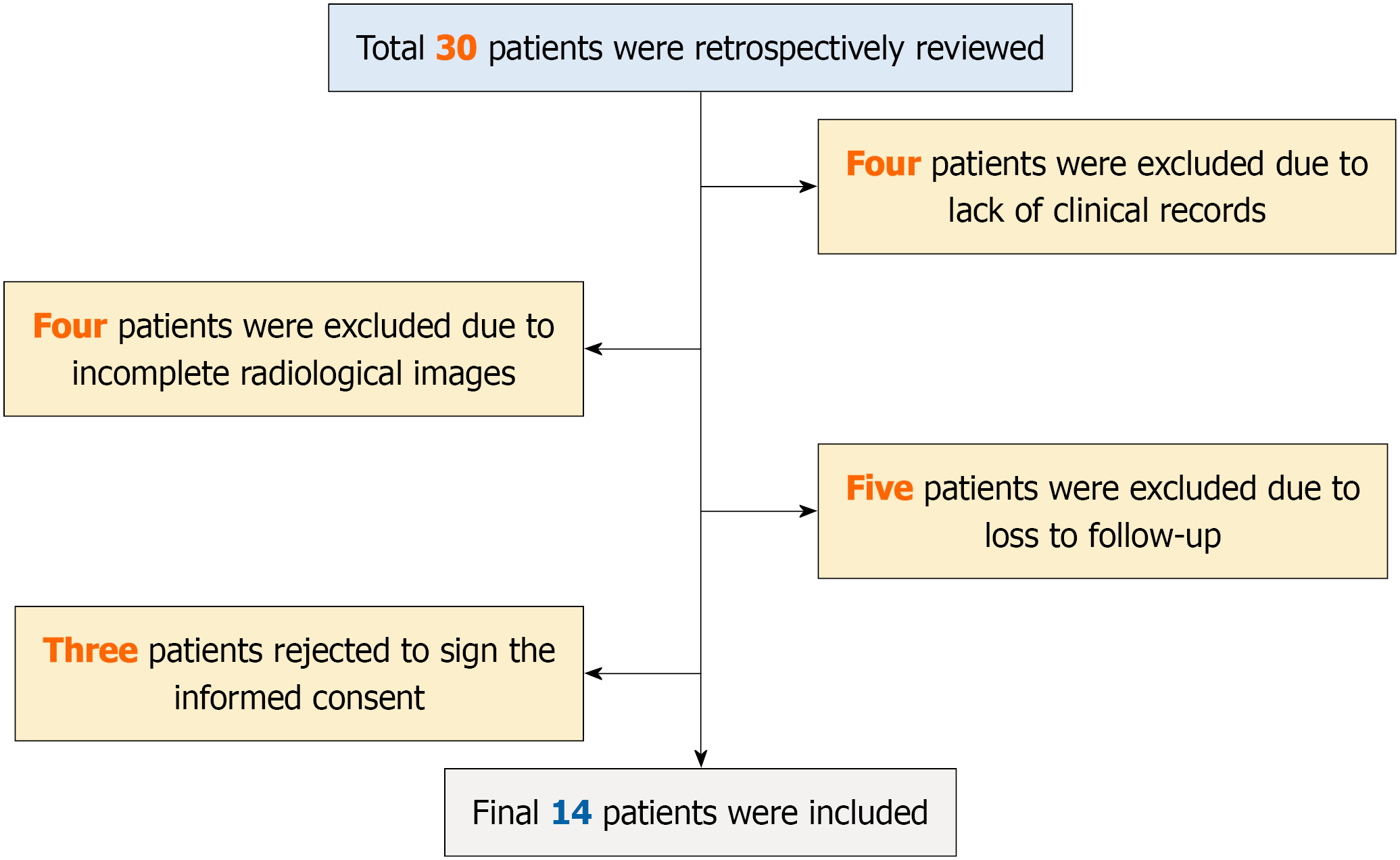
Figure 1
The participant flow chart of this study.
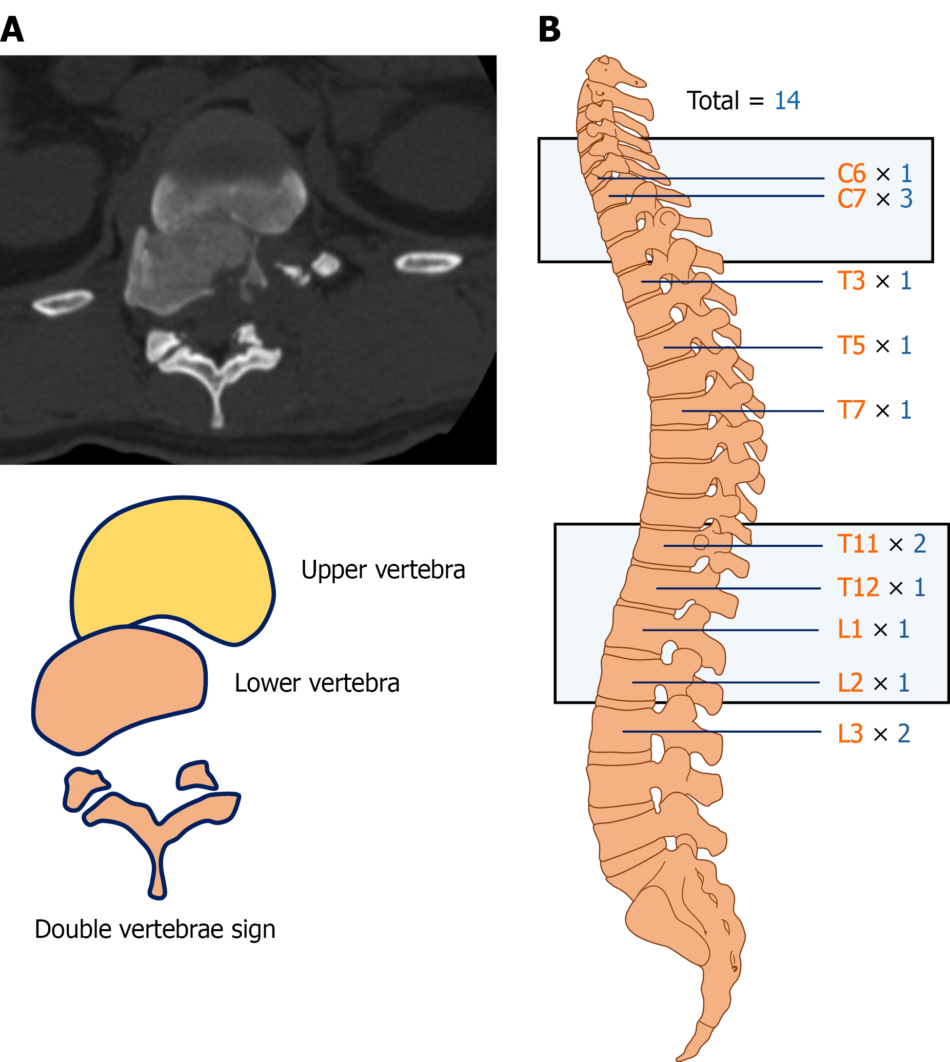
Figure 2 Overview of spondyloptosis in this series.
A: The double-vertebra sign on imaging indicates spondyloptosis; B: Definite vertebra location and distribution among patients in this cohort.
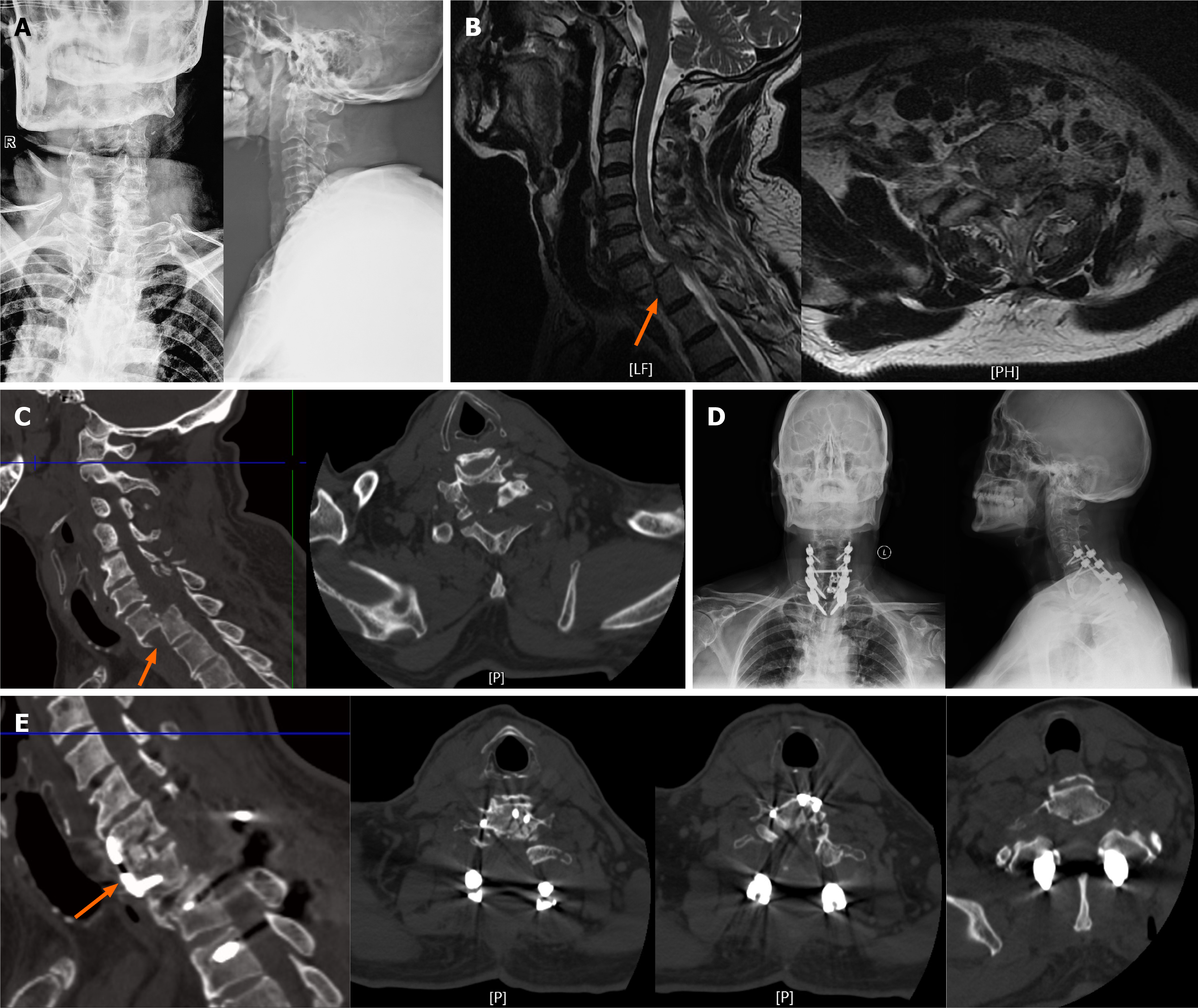
Figure 3 Representative case of cervical spondyloptosis.
A 63-year-old man sustained an injury after falling from a height. A and B: Preoperative radiographs and computed tomography (CT) showed C7 spondyloptosis (orange arrow); C: Magnetic resonance imaging demonstrated spinal cord compression. The patient subsequently underwent combined anterior–posterior fixation and discectomy with bone grafting (orange arrow); D and E: Postoperative radiographs and CT images demonstrated vertebral reduction and favorable internal fixation (orange arrow).
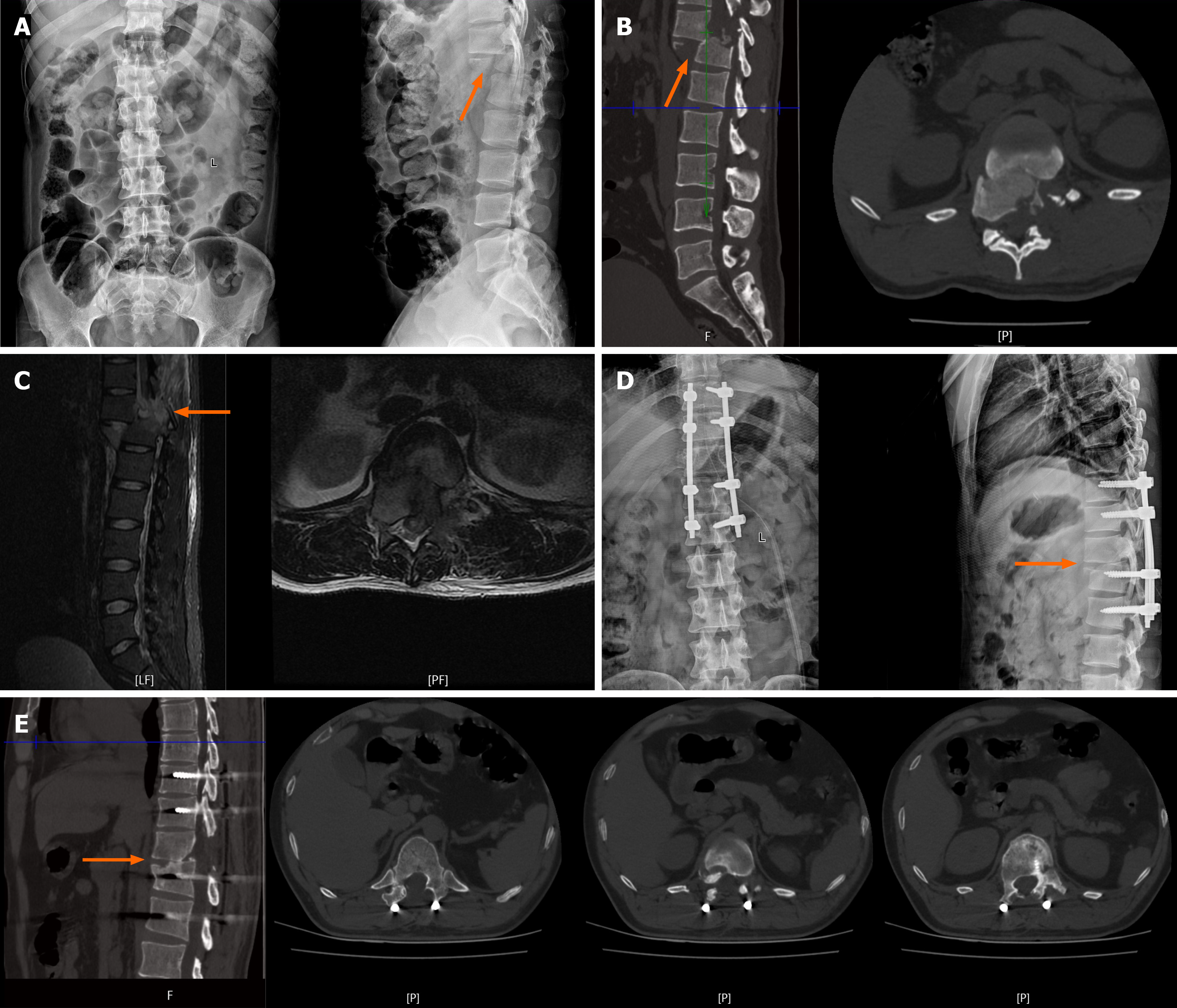
Figure 4 Representative case of thoracic spondyloptosis.
A 55-year-old man was injured in a motor vehicle accident. A and B: Preoperative radiographs and computed tomography (CT) images showed T11 spondyloptosis (orange arrow); C: Magnetic resonance imaging showed spinal cord compression and soft-tissue swelling. The patient subsequently underwent posterior pedicle screw fixation (orange arrow); D and E: Postoperative radiographs and CT images demonstrated vertebral reduction and favorable internal fixation (orange arrow).
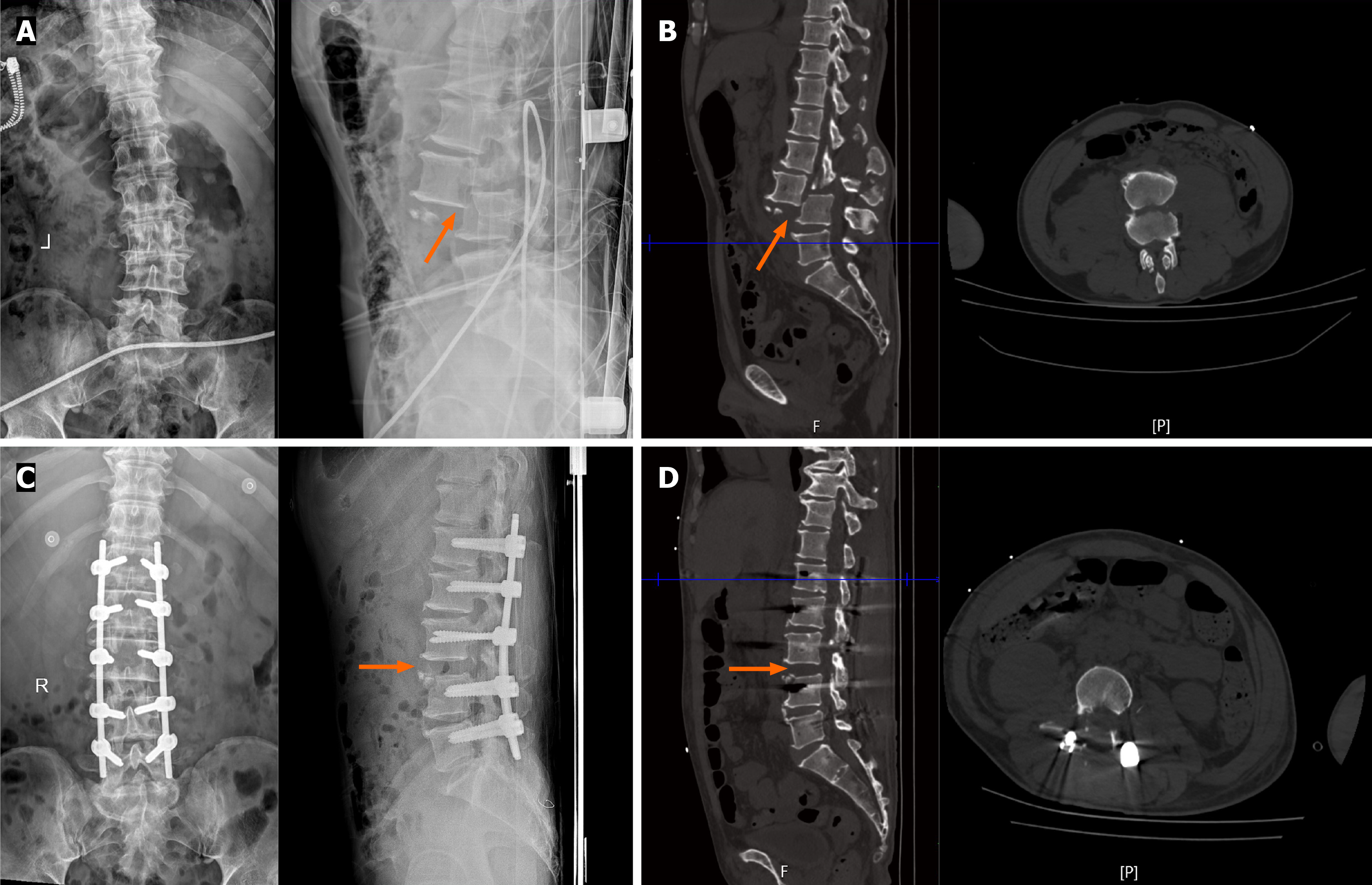
Figure 5 Representative case in lumbar spondyloptosis.
A 50-year-old man was injured in a motor vehicle accident. A: Preoperative radiographs (orange arrow); B: Computed tomography (CT) image showed L3 spondyloptosis with double-vertebra sign. The patient subsequently underwent posterior pedicle screw fixation (orange arrow); C and D: Postoperative radiographs and CT images demonstrated vertebral reduction and favorable internal fixation (orange arrow).
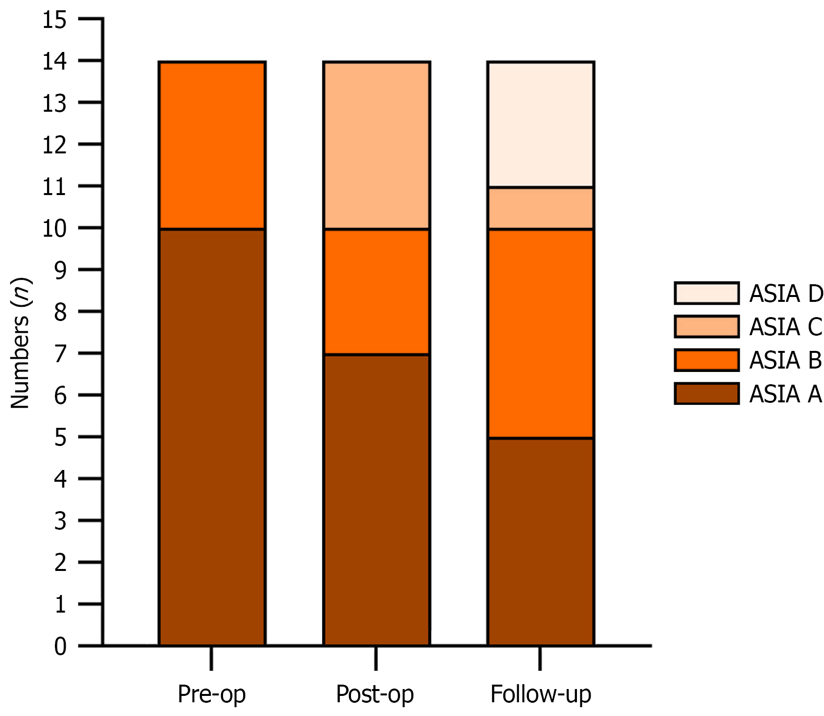
Figure 6 Presentation of preoperative, postoperative, and follow-up American Spinal Injury Association scale.
ASIA: American Spinal Injury Association.
- Citation: Yu XS, Mao ZH, Qiu C, Zhang LZ, Chang ZQ. Perioperative management and neurological outcomes of traumatic spondyloptosis: A retrospective case series of 14 patients. World J Orthop 2026; 17(10): 125014
- URL: https://www.wjgnet.com/2218-5836/full/v17/i10/125014.htm
- DOI: https://dx.doi.org/10.5312/wjo.125014