
















Copyright: ©Author(s) 2026.
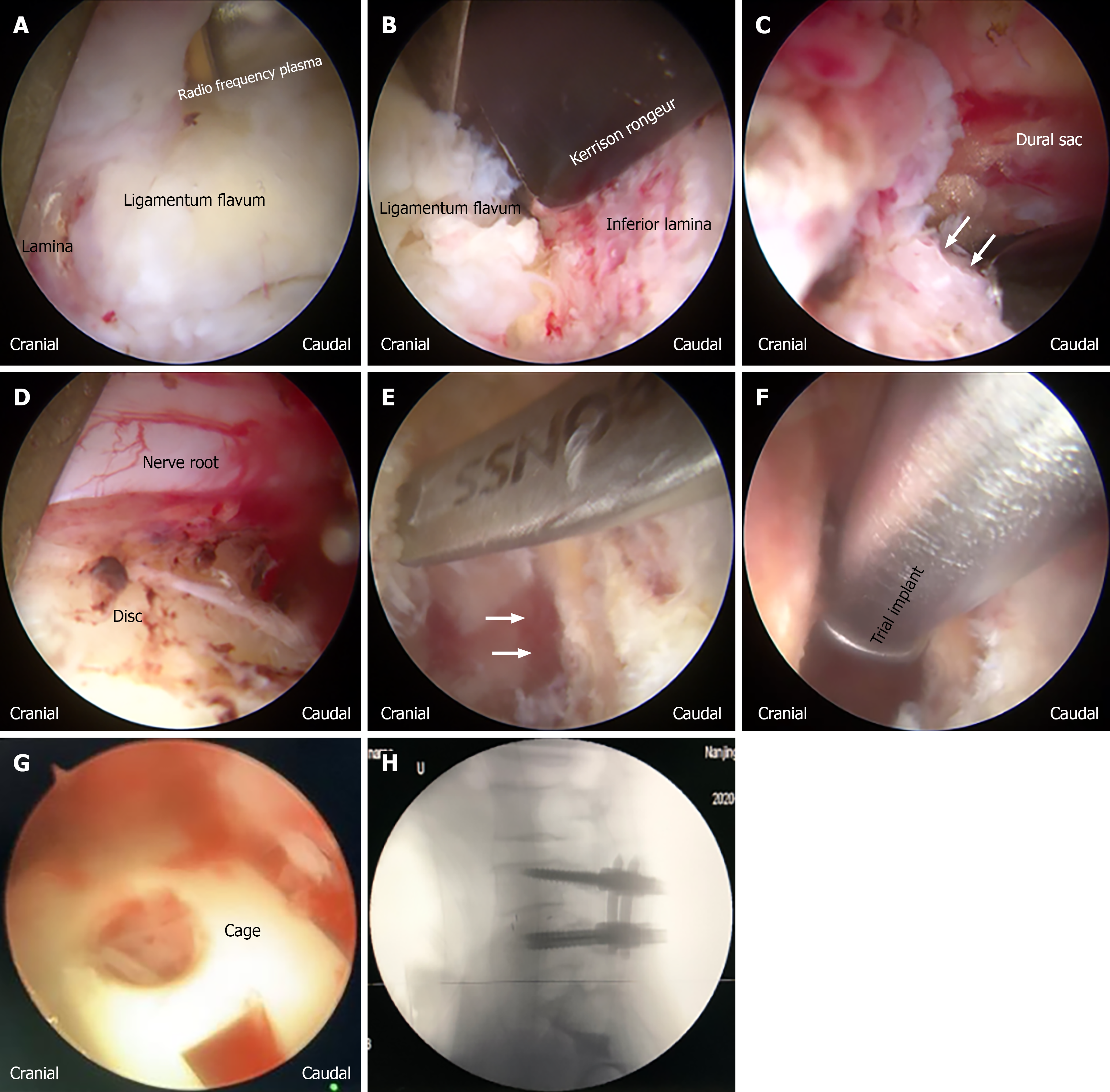
Figure 1 Key steps of biportal endoscopic transforaminal lumbar interbody fusion procedure, shown in chronological order.
A: Exposure of the ligamentum flavum after soft tissue clearance using radiofrequency plasma; B: Resection of the inferior edge of the lamina with a Kerrison rongeur; C: Resection of the medial aspect of the facet joint (white arrows indicate the resected facet margin), with exposure of the dural sac; D: Protection of the traversing nerve root, with the underlying disc exposed; E: Endplate preparation after discectomy (white arrows indicate the prepared endplate surface); F: Insertion of a trial implant to determine the appropriate cage size; G: Placement of the interbody cage under endoscopic guidance; H: Final pedicle screw and rod fixation confirmed by intraoperative fluoroscopy.
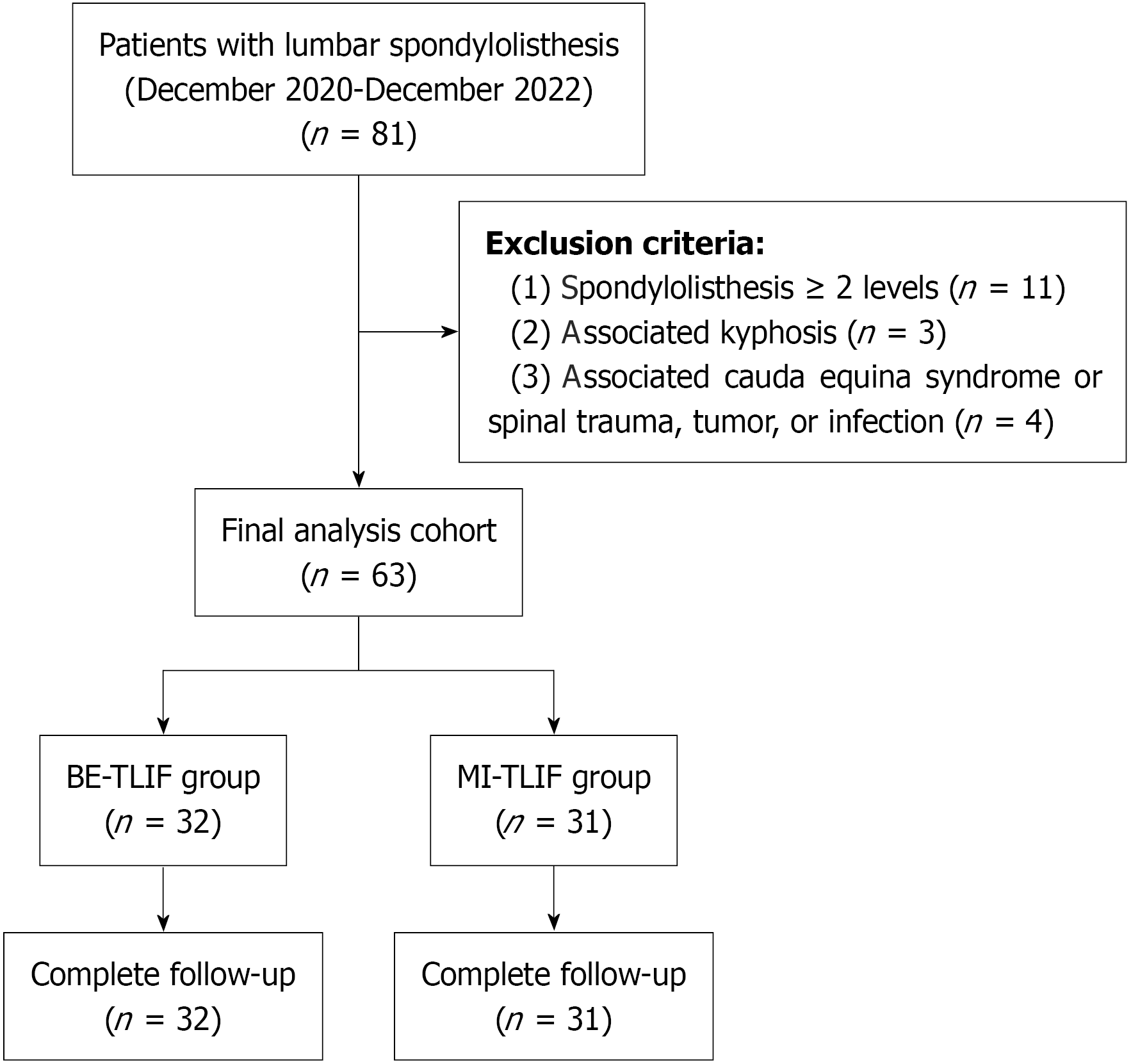
Figure 2 Participant flow diagram.
A total of 81 patients with lumbar spondylolisthesis who underwent surgery between December 2020 and December 2022 were screened. Eighteen patients were excluded according to the prespecified exclusion criteria: 11 with spondylolisthesis involving ≥ 2 levels, 3 with associated kyphosis, and 4 with associated cauda equina syndrome or spinal trauma, tumor, or infection. The remaining 63 patients were enrolled and allocated to the biportal endoscopic transforaminal lumbar interbody fusion group (n = 32) or the minimally invasive transforaminal lumbar interbody fusion group (n = 31). All patients completed the follow‑up and were included in the final analysis. BE-TLIF: Biportal endoscopic transforaminal lumbar interbody fusion; MI-TLIF: Minimally invasive transforaminal lumbar interbody fusion.
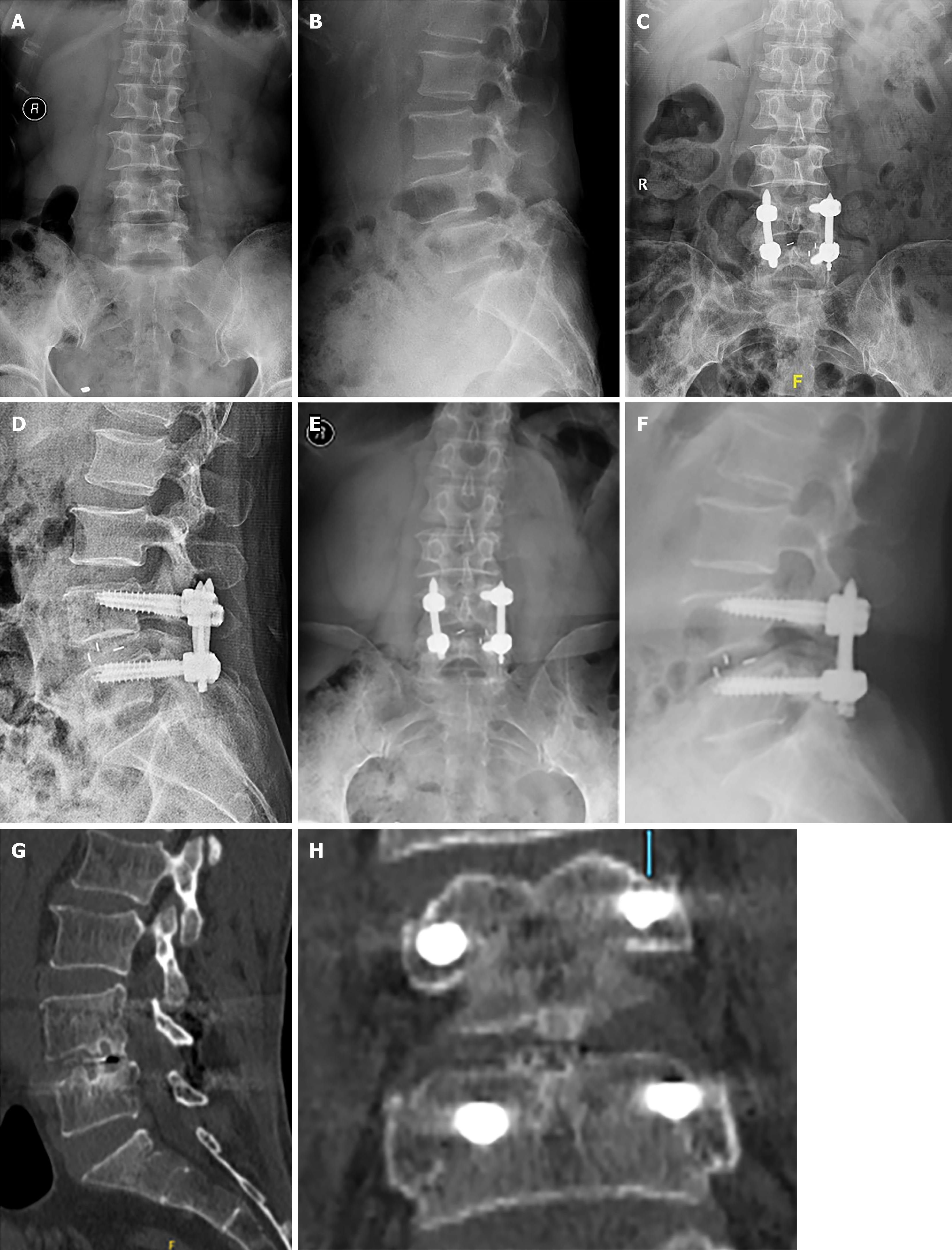
Figure 3 A 54-year-old female patient with L4 lumbar spondylolisthesis (Meyerding grade I) from the biportal endoscopic transforaminal lumbar interbody fusion group.
A and B: Preoperative anteroposterior and lateral radiographs; C and D: Postoperative 3-month anteroposterior and lateral radiographs; E and F: Anteroposterior and lateral X-ray at the final follow-up after operation showed good reduction; G and H: Computed tomography scans at the final follow-up after operation showed that the reconstruction of the trabeculae between the upper and lower endplates, and the position of the Cage was good.
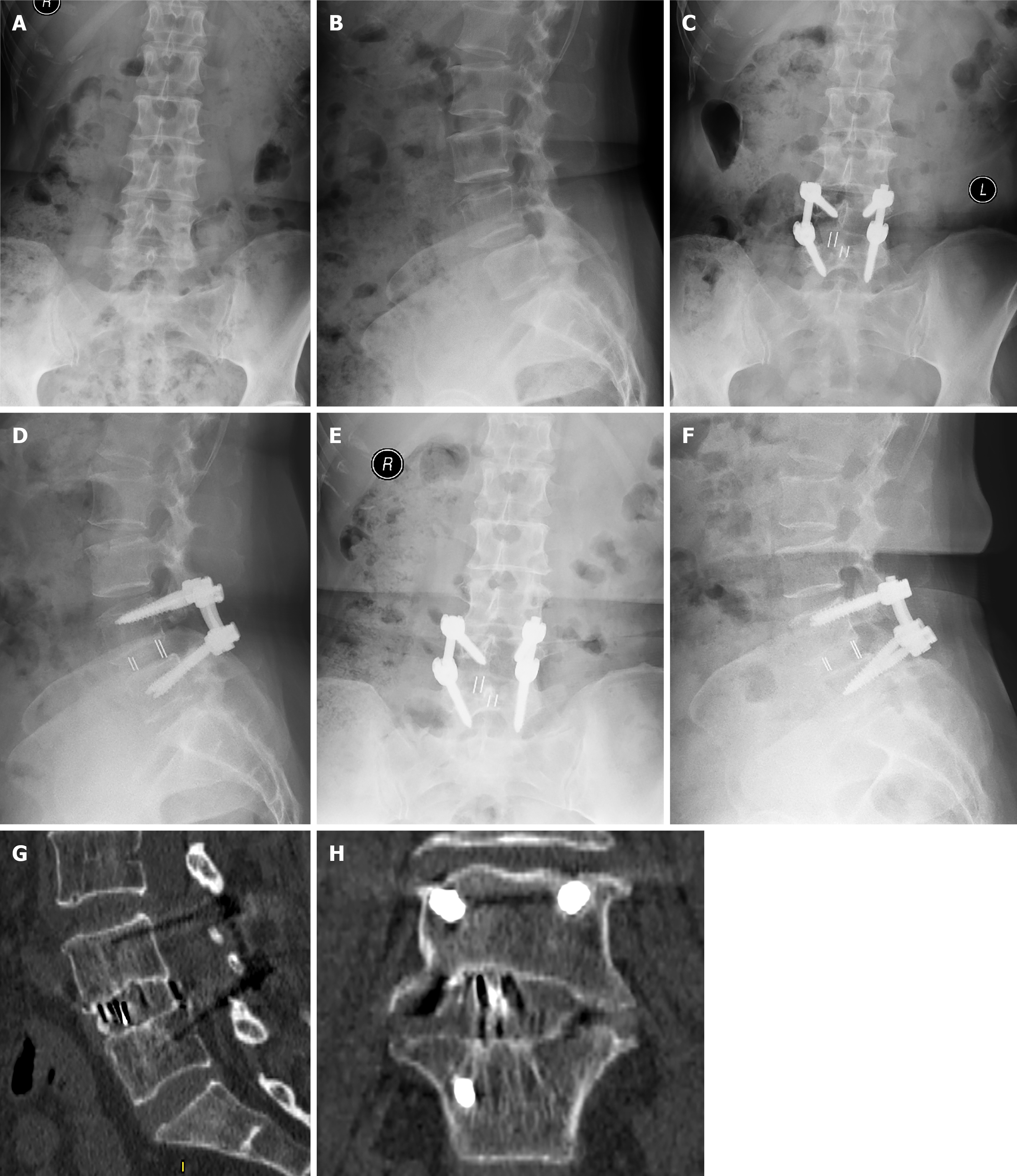
Figure 4 A 56-year-old female patient with L4 lumbar spondylolisthesis (Meyerding grade I) in minimally invasive transforaminal lumbar interbody fusion group.
A and B: Preoperative antero-posterior and lateral radiographs; C and D: Postoperative 3-month antero-posterior and lateral radiographs; E and F: Antero-posterior and lateral X-ray at the final follow-up after operation showed good reduction; G and H: Computed tomography scans at the final follow-up after operation show good trabecular reconstruction between the upper and lower endplates and a good Cage position.
- Citation: He P, Sun J, Liu XW, Gao Q, Xu B, Cong Y, Zhao WL. Comparison of biportal endoscopic vs minimally invasive transforaminal lumbar interbody fusion for single-level low-grade lumbar spondylolisthesis: A retrospective cohort study. World J Orthop 2026; 17(10): 124542
- URL: https://www.wjgnet.com/2218-5836/full/v17/i10/124542.htm
- DOI: https://dx.doi.org/10.5312/wjo.124542