©The Author(s) 2025.
World J Orthop. Jun 18, 2025; 16(6): 108454
Published online Jun 18, 2025. doi: 10.5312/wjo.v16.i6.108454
Published online Jun 18, 2025. doi: 10.5312/wjo.v16.i6.108454
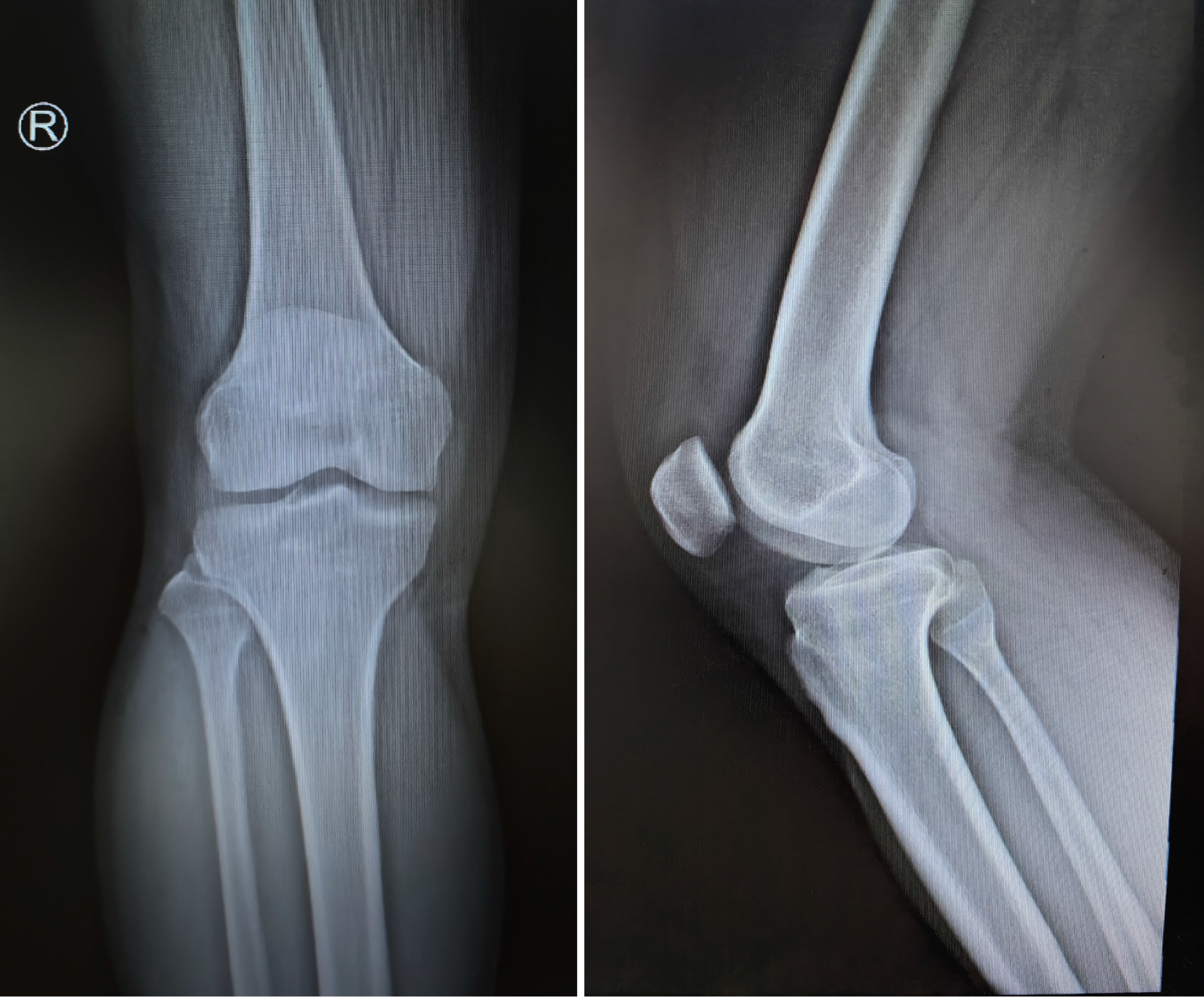
Figure 1
No obvious abnormalities were detected on X-ray imaging.
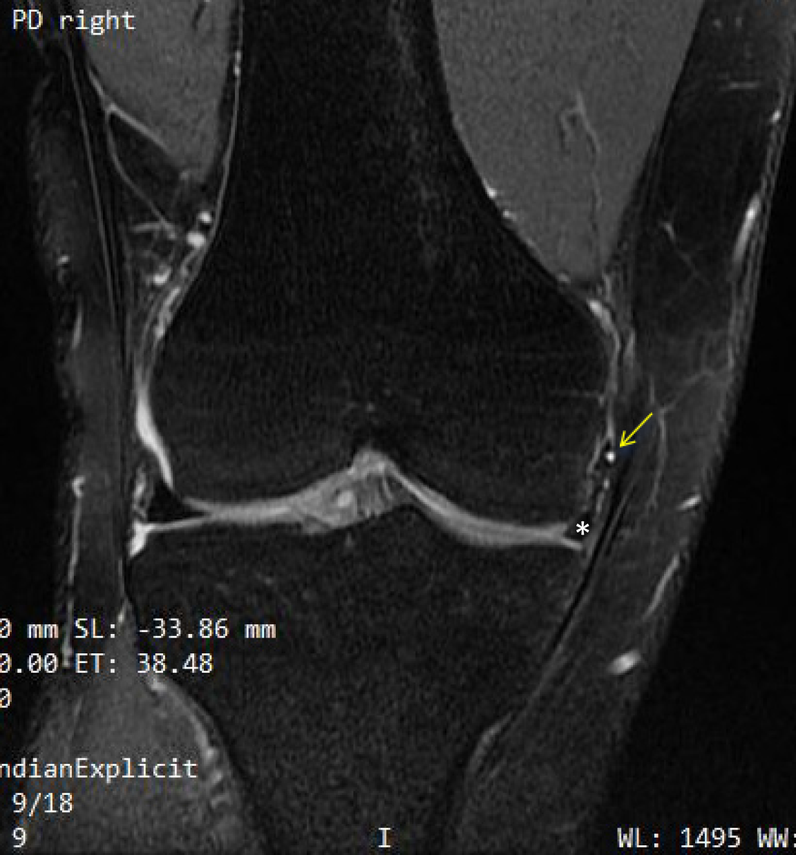
Figure 2
The magnetic resonance T2-weighted image shows a torn and flipped medial meniscus (indicated by the white asterisks) and a meniscus cyst is located on the free-edge of the flipped medial meniscus (indicated by the yellow arrow).
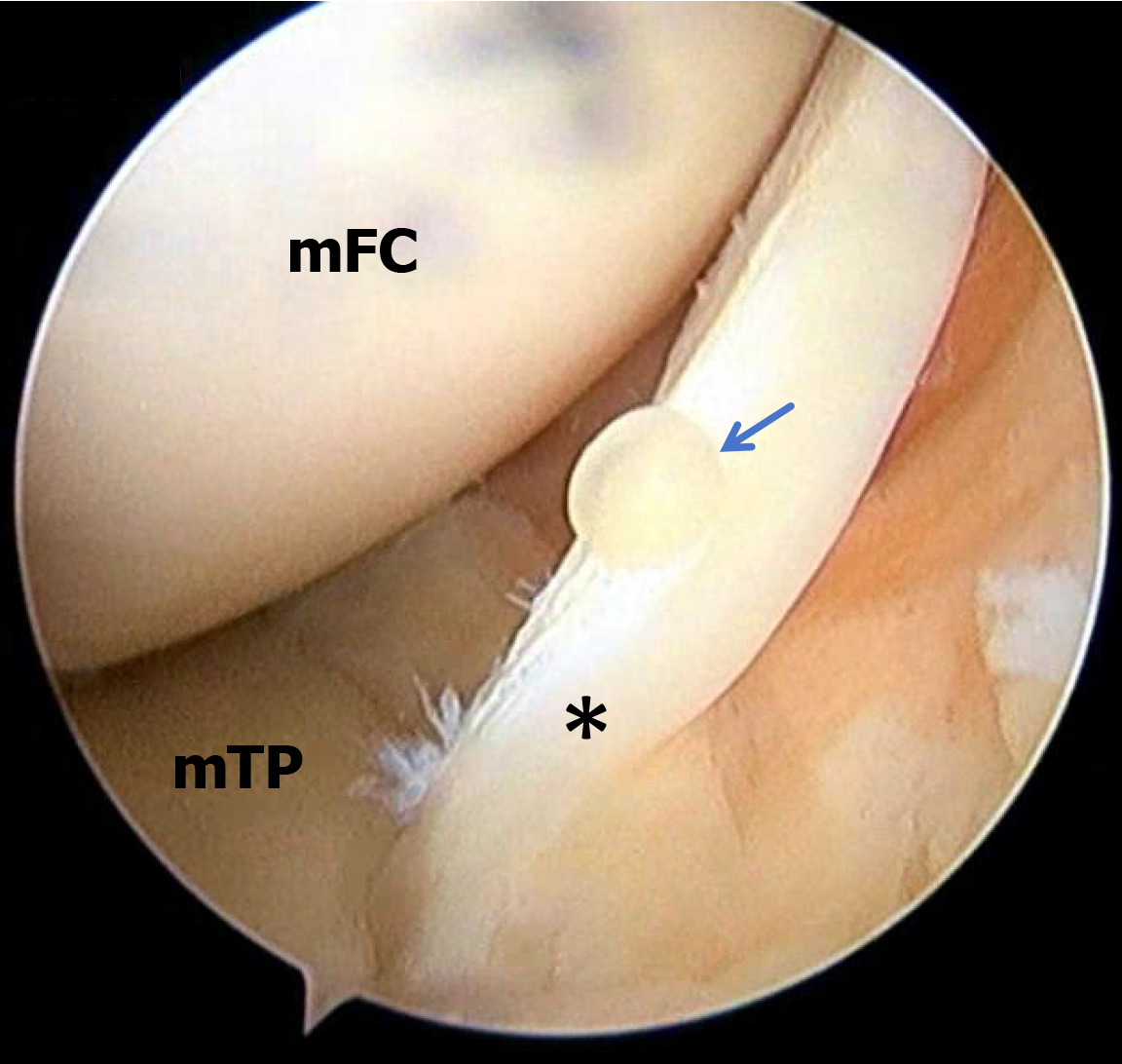
Figure 3 Arthroscopic view through the anterolateral approach of the left knee reveal that the medial meniscus has an oblique tear forming a flipped flap (indicated by the black asterisks), combined with a free-edge cyst (indicated by the blue arrow).
mFC: Medial femoral condyle; mTP: Medial tibial plateau.
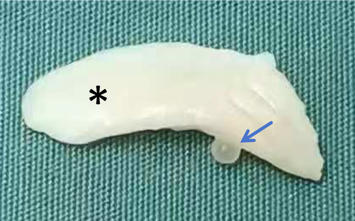
Figure 4 The resected portion of the meniscus during surgery.
The black asterisks indicates an oblique tear of the meniscus, and the blue arrow points to a cyst at the free edge of the meniscus.
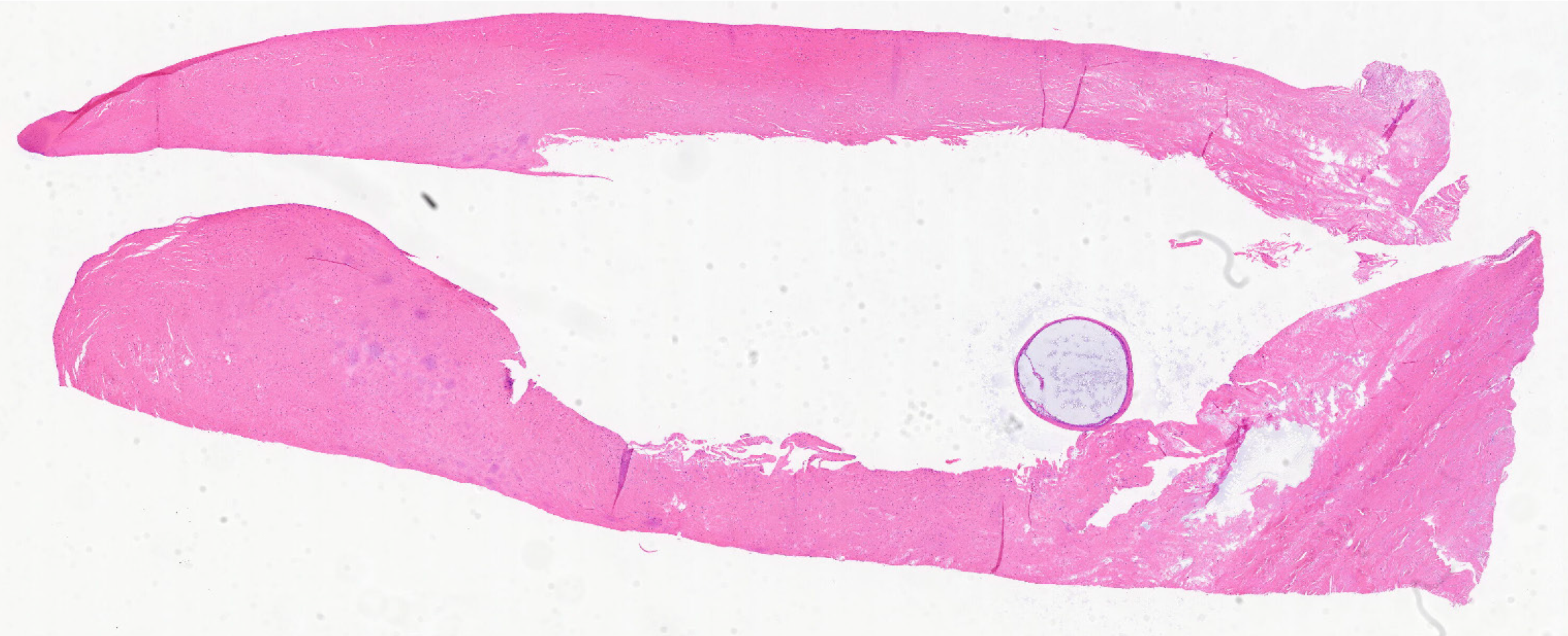
Figure 5
Pathological hematoxylin and eosin staining of the cyst tissue section reveals a typical cyst wall.
- Citation: Ding M, Liao BH, Shangguan L, Wang YC, Xu H. Arthroscopic management of a rare free-edge medial meniscal cyst: A case report. World J Orthop 2025; 16(6): 108454
- URL: https://www.wjgnet.com/2218-5836/full/v16/i6/108454.htm
- DOI: https://dx.doi.org/10.5312/wjo.v16.i6.108454


