©The Author(s) 2025.
World J Orthop. Jun 18, 2025; 16(6): 106458
Published online Jun 18, 2025. doi: 10.5312/wjo.v16.i6.106458
Published online Jun 18, 2025. doi: 10.5312/wjo.v16.i6.106458
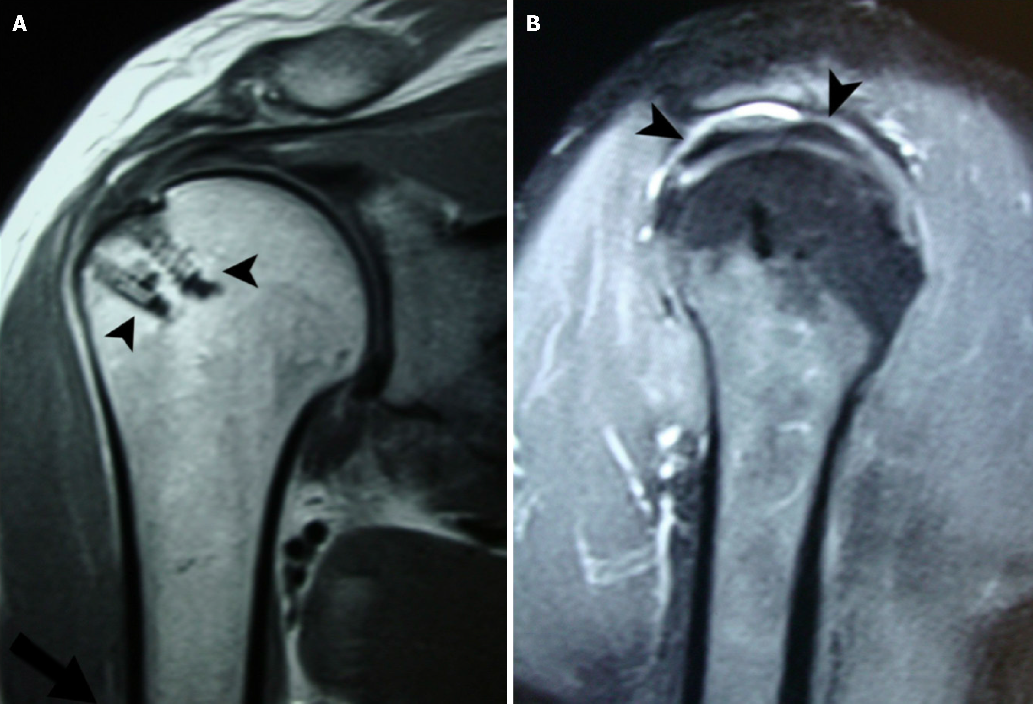
Figure 1 Magnetic resonance imaging evaluation of a repaired rotator cuff 11 months after the first operation in a 63-year-old patient.
A: Coronal magnetic resonance imaging (MRI) of the repaired rotator cuff. The rotator cuff tendons were reattached using a double row technique and absorbable suture anchors (arrowheads). Complete healing with restoration of tendon continuity is observed; B: Sagittal MRI showing restoration of the signal intensity of the rotator cuff tendons (arrowheads).
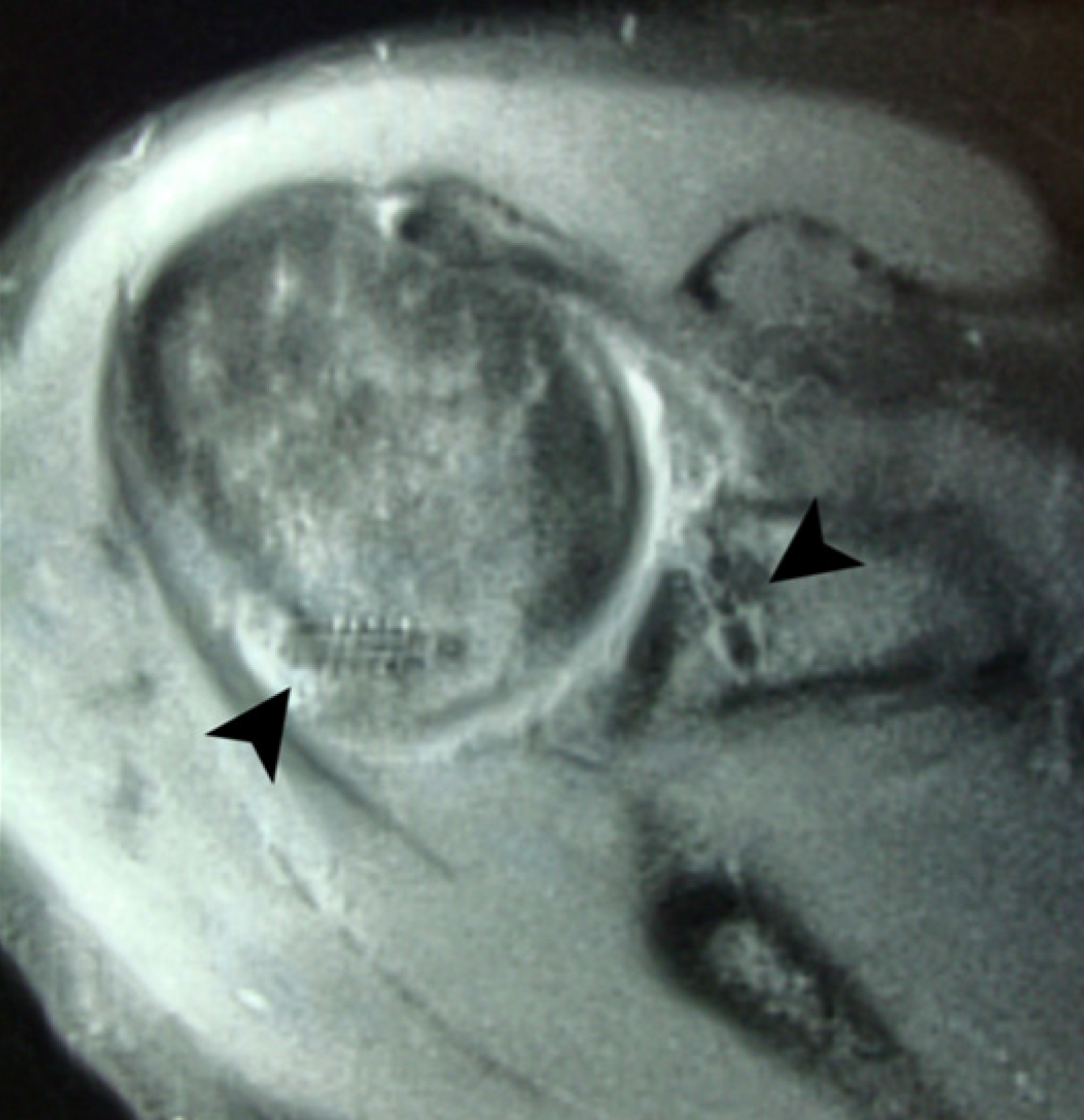
Figure 2 Transverse magnetic resonance imaging of the shoulder in a patient who underwent revision Bankart repair 5 months earlier.
The anterior capsulolabral structures were reattached on the anterior glenoid rim using suture anchors (arrowhead). Remplissage was performed to cover a sizeable engaging Hill-Sachs lesion using another suture anchor (arrowhead).
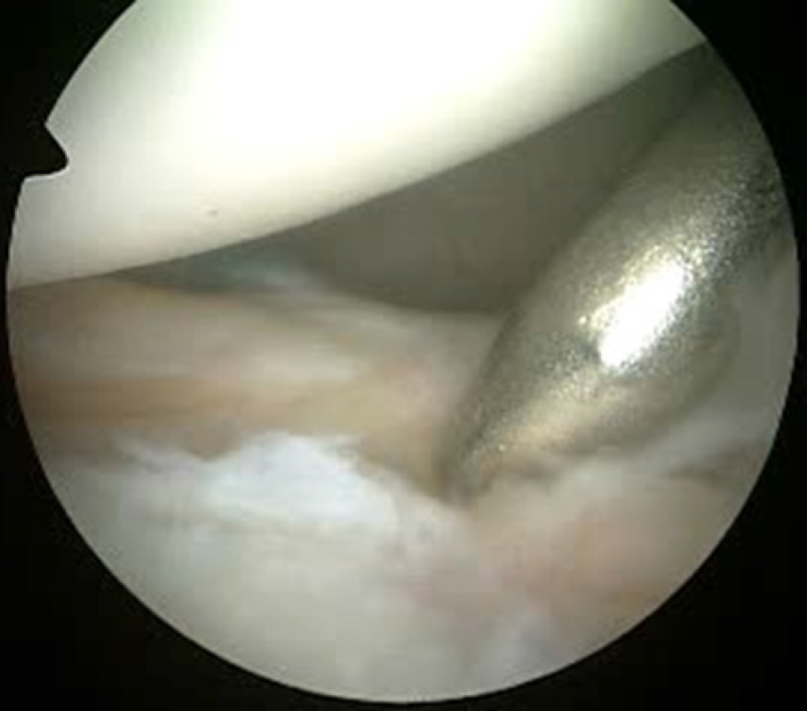
Figure 3 Arthroscopic view of a left shoulder from the posterior viewing portal.
A switching stick is inserted through the anterosuperior portal and used to palpate the area of capsule reattachment.
Figure 4 Arthroscopic evaluation of the glenohumeral joint 14 months after the first operation.
A: The anterior capsule is viewed from the anterosuperior portal revealing complete, watertight healing to the glenoid rim; B: The knots are covered with synovium without causing articular surface fraying.
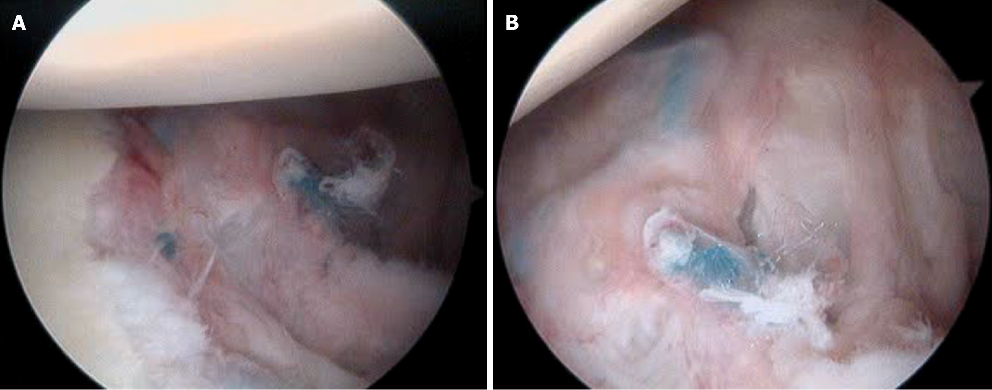
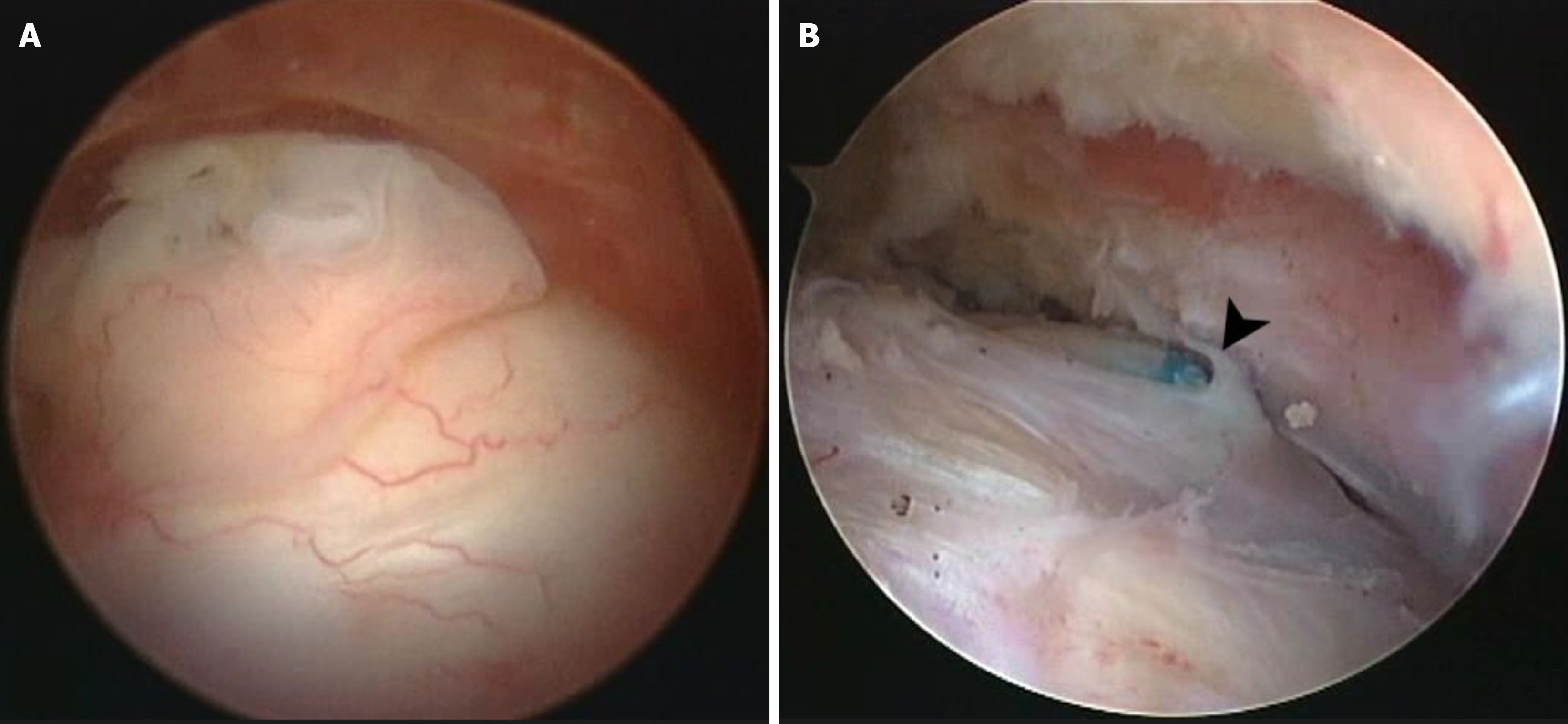
Figure 5 Arthroscopic view of the repaired rotator cuff tendons in the right shoulder from the posterior portal.
A: The repaired rotator cuff is covered with synovium without evidence of synovitis or fraying. This patient was re-operated due to persistent acromioclavicular joint pain; B: The repaired rotator cuff is covered with synovium and an arthroscopic knot is evident (arrowhead). This patient underwent re-operation for stiffness.
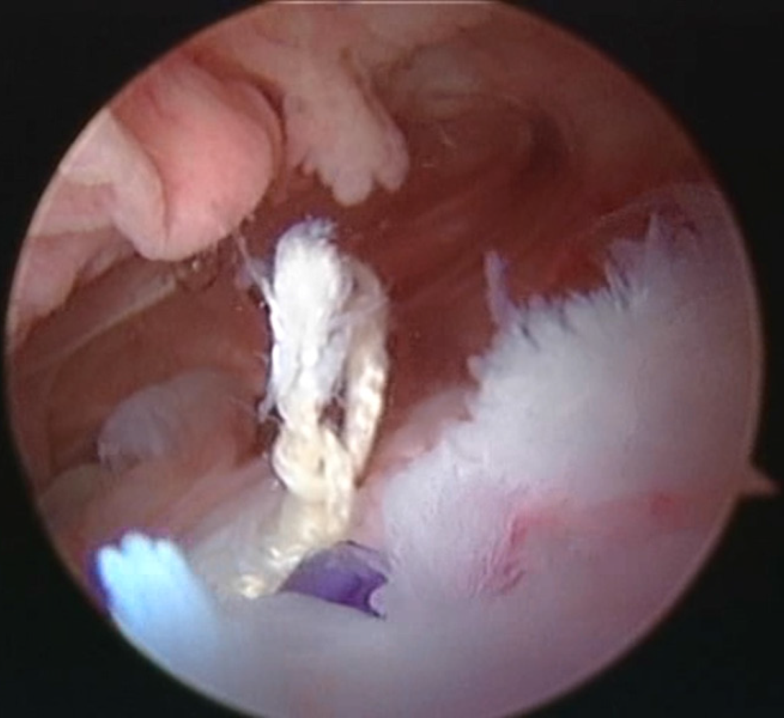
Figure 6 Partial rotator cuff healing failure in the left shoulder viewed from the posterior portal.
The suture cut through the tendon and the anchor is seen uncovered. The orthocord suture appears white because its purple-colored phytoene desaturase coating has been absorbed.
- Citation: Yiannakopoulos C, Koukos C, Habipis A, Apostolou C. Rotator cuff and capsule healing after shoulder arthroscopy: A second look arthroscopic study. World J Orthop 2025; 16(6): 106458
- URL: https://www.wjgnet.com/2218-5836/full/v16/i6/106458.htm
- DOI: https://dx.doi.org/10.5312/wjo.v16.i6.106458


