Copyright
©The Author(s) 2025.
World J Orthop. Apr 18, 2025; 16(4): 103795
Published online Apr 18, 2025. doi: 10.5312/wjo.v16.i4.103795
Published online Apr 18, 2025. doi: 10.5312/wjo.v16.i4.103795
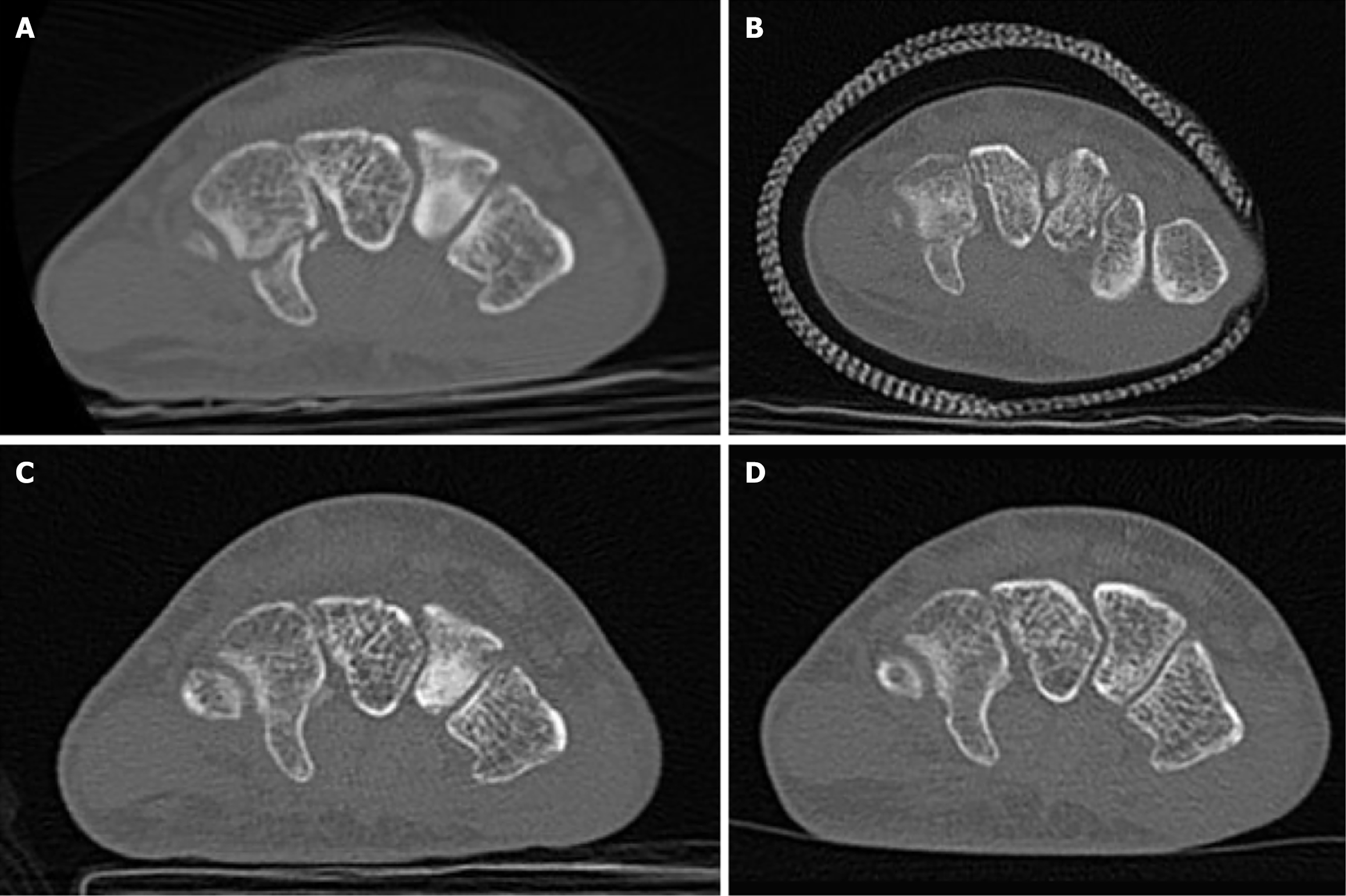
Figure 1 Result of case 6.
A: At the time of injury - type III fracture with small bone fragments; B: Four weeks after the start of treatment - in the cast, showing continuity; C: Eight weeks after the start of treatment - callus is visible, showing continuity between bone fragments; D: Twelve weeks after the start of treatment - bone healing is complete.
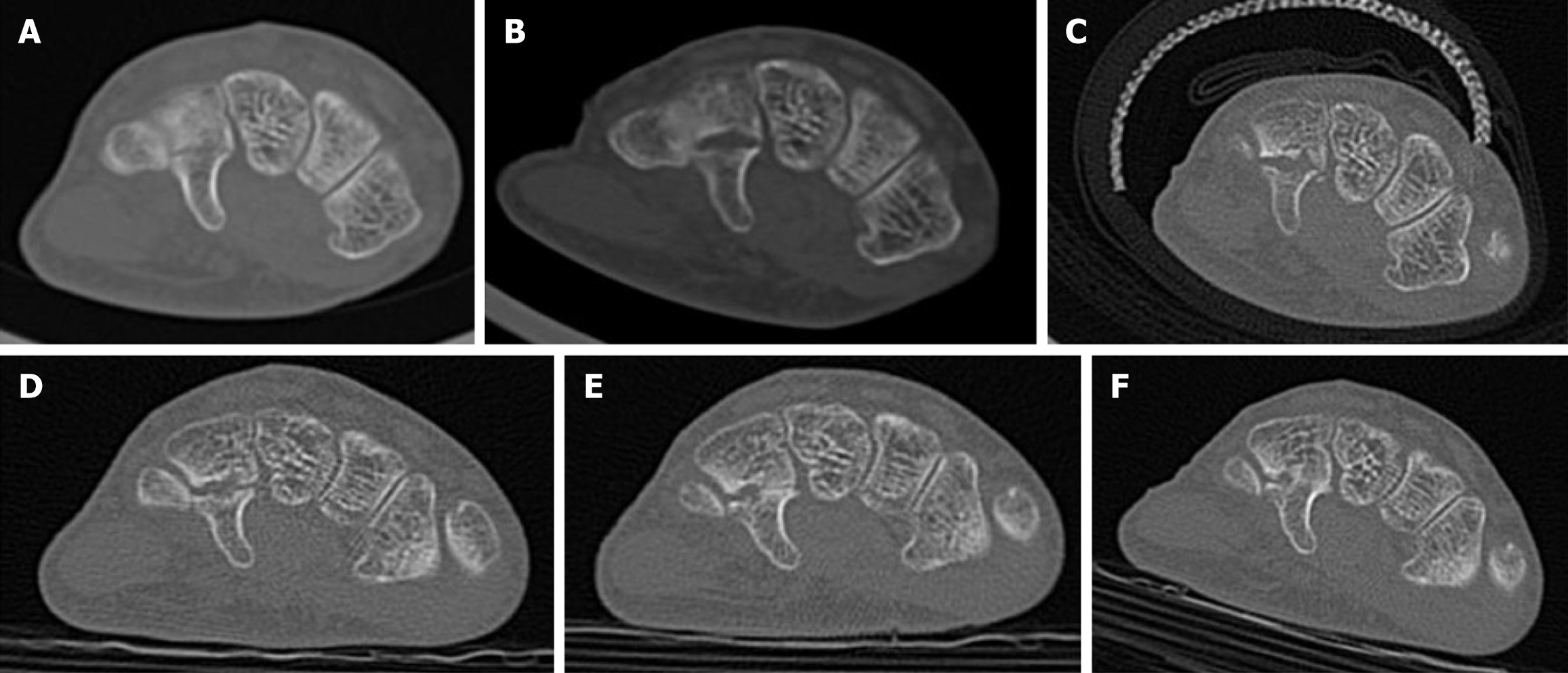
Figure 2 Result of case 11.
A: At the time of injury - type III fracture, a slight fracture line at the base of hook of hamate; B: Six weeks post-injury, before treatment - separation between bone fragments is observed; C: Ten weeks post-injury, 4 weeks after the start of treatment - small bone fragments surround the area, and callus formation is slightly visible; D: Twenty-two weeks post-injury, 12 weeks after the start of treatment - the gap between bone fragments is shortened, and a shadow suggestive of callus is observed; E: Thirty weeks post-injury, 24 weeks after the start of treatment - partial callus formation and continuity between bone fragments are visible; F: Thirty-six weeks post-injury, 42 weeks after the start of treatment - further bone formation between bone fragments is observed.
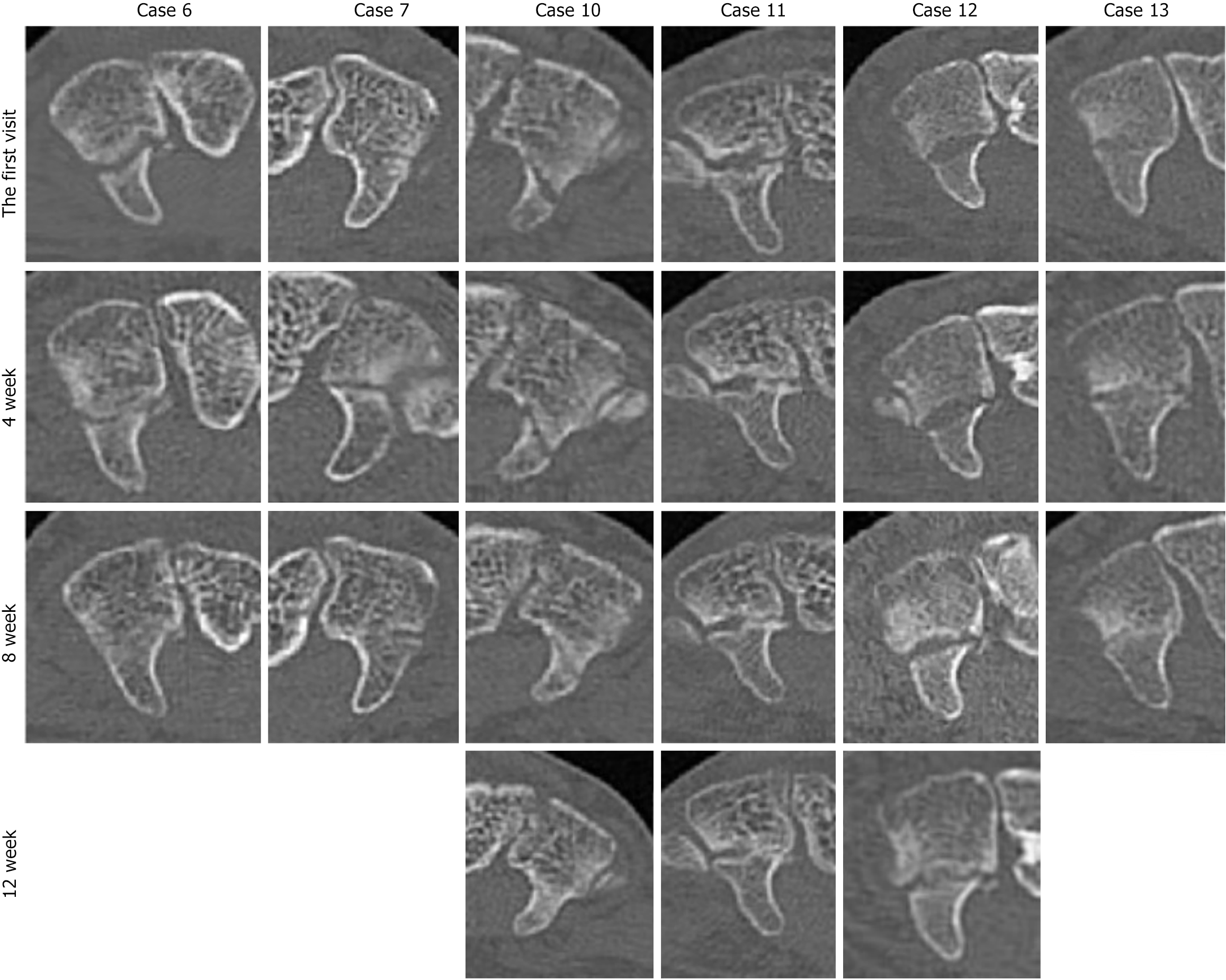
Figure 3 Progression of bone healing assessed trough computed tomography.
Bone healing progresses from the central trabecular side, with the cortical side healing later.
Figure 4 Case 13.
Wrist range of motion after 12 weeks (4 weeks of cast immobilization and 8 weeks of splint immobilization) following the removal of the fixation. There were no restrictions in range of motion or pain observed on the injured left side.
- Citation: Tanaka T, Yoshii Y. Sixteen patients regarding the conservative treatment for hook of hamate fracture. World J Orthop 2025; 16(4): 103795
- URL: https://www.wjgnet.com/2218-5836/full/v16/i4/103795.htm
- DOI: https://dx.doi.org/10.5312/wjo.v16.i4.103795


