©The Author(s) 2024.
World J Orthop. Jul 18, 2024; 15(7): 675-682
Published online Jul 18, 2024. doi: 10.5312/wjo.v15.i7.675
Published online Jul 18, 2024. doi: 10.5312/wjo.v15.i7.675
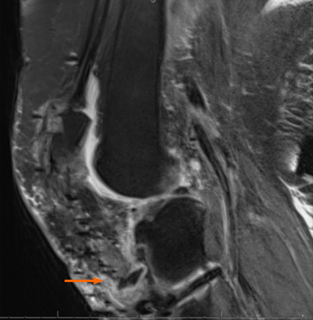
Figure 1 Preoperative sagittal magnetic resonance imaging.
(Proton Density weighted image fat saturation)-demonstrates avulsion fracture of the tibial tuberosity with patellar tendon rupture and patella alta.
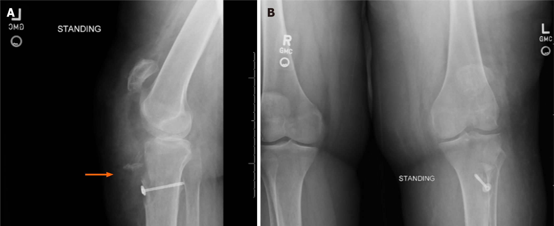
Figure 2 Preoperative plain radiographs.
A: Left knee lateral radiograph demonstrating patella alta and tibial tubercle avulsion fracture; B: Standing Anterior-Posterior radiograph bilateral knees. Note patella alta of left knee with normal patellar height of right side.
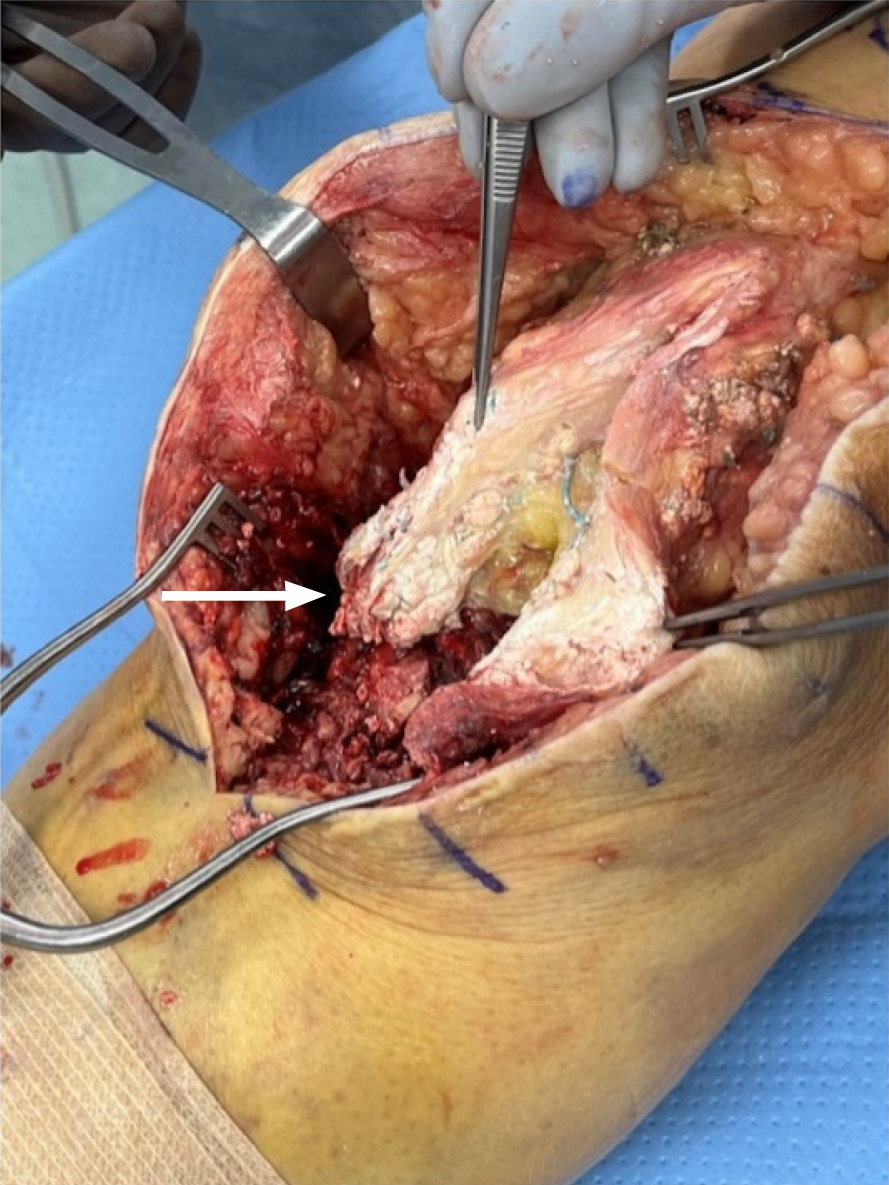
Figure 3 Intraoperative visualization of gouty infiltration of patellar tendon from prior allograft reconstruction.
Gouty tophus noted within surrounding soft tissues with bony destruction of tibial tubercle.
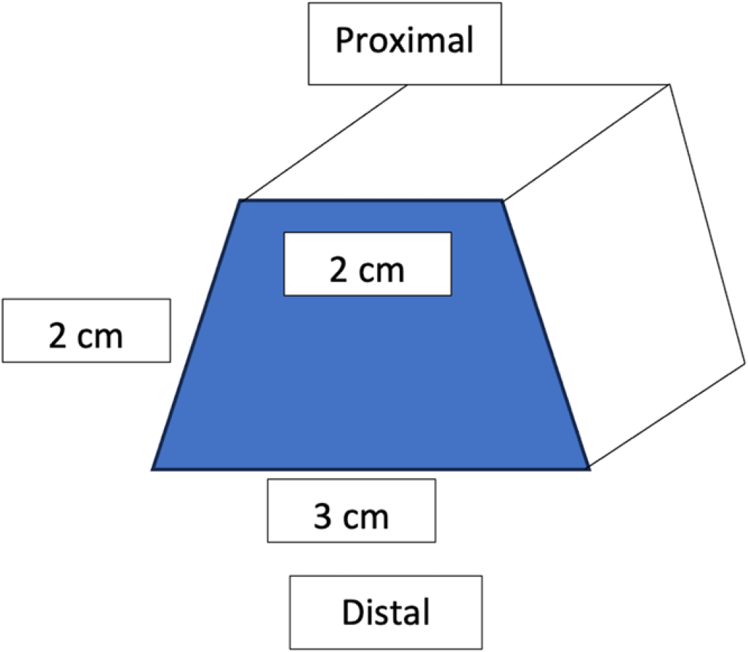
Figure 4 Three-dimensional representation of bone block after contouring.
A corresponding notch in the tibial tubercle was made to create a dovetail joint for bone block placement. This was done to prevent proximal migration a reinforce stability of the reconstruction.
Figure 5 Intraoperative images of reconstructive technique.
A: Achilles bone block secured with screws with tensioning of graft proximally by transosseous tunnel technique; B: Dermal allograft placed overlying Achilles allograft using transverse slit to pass excess proximal tendon graft. Dermal allograft secured proximal to inferior pole patella with knotless fiber tack anchors; C: Dermal allograft secured distally with four swivel lock anchors with speed bridge technique and fiber tape; D: Extra proximal Achilles allograft reinforced to quadriceps tendon.
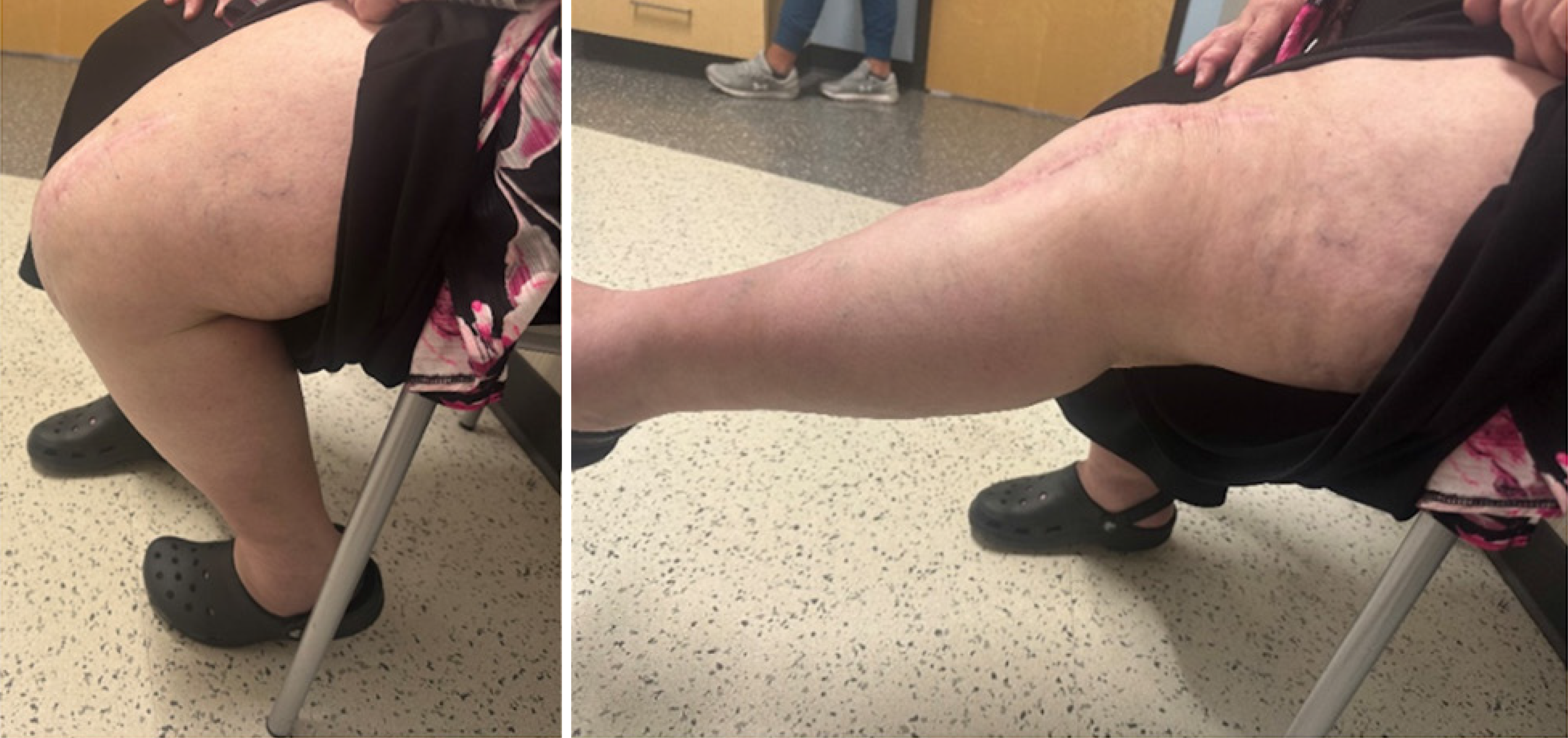
Figure 6
Active knee range of motion from 5 to 115 degrees at 1 year follow up.
- Citation: Edge CC, Widmeyer J, Protzuk O, Johnson M, O’Connell R. Gouty destruction of a patellar tendon reconstruction and novel revision reconstruction technique: A case report. World J Orthop 2024; 15(7): 675-682
- URL: https://www.wjgnet.com/2218-5836/full/v15/i7/675.htm
- DOI: https://dx.doi.org/10.5312/wjo.v15.i7.675


