©The Author(s) 2024.
World J Orthop. Jul 18, 2024; 15(7): 618-626
Published online Jul 18, 2024. doi: 10.5312/wjo.v15.i7.618
Published online Jul 18, 2024. doi: 10.5312/wjo.v15.i7.618
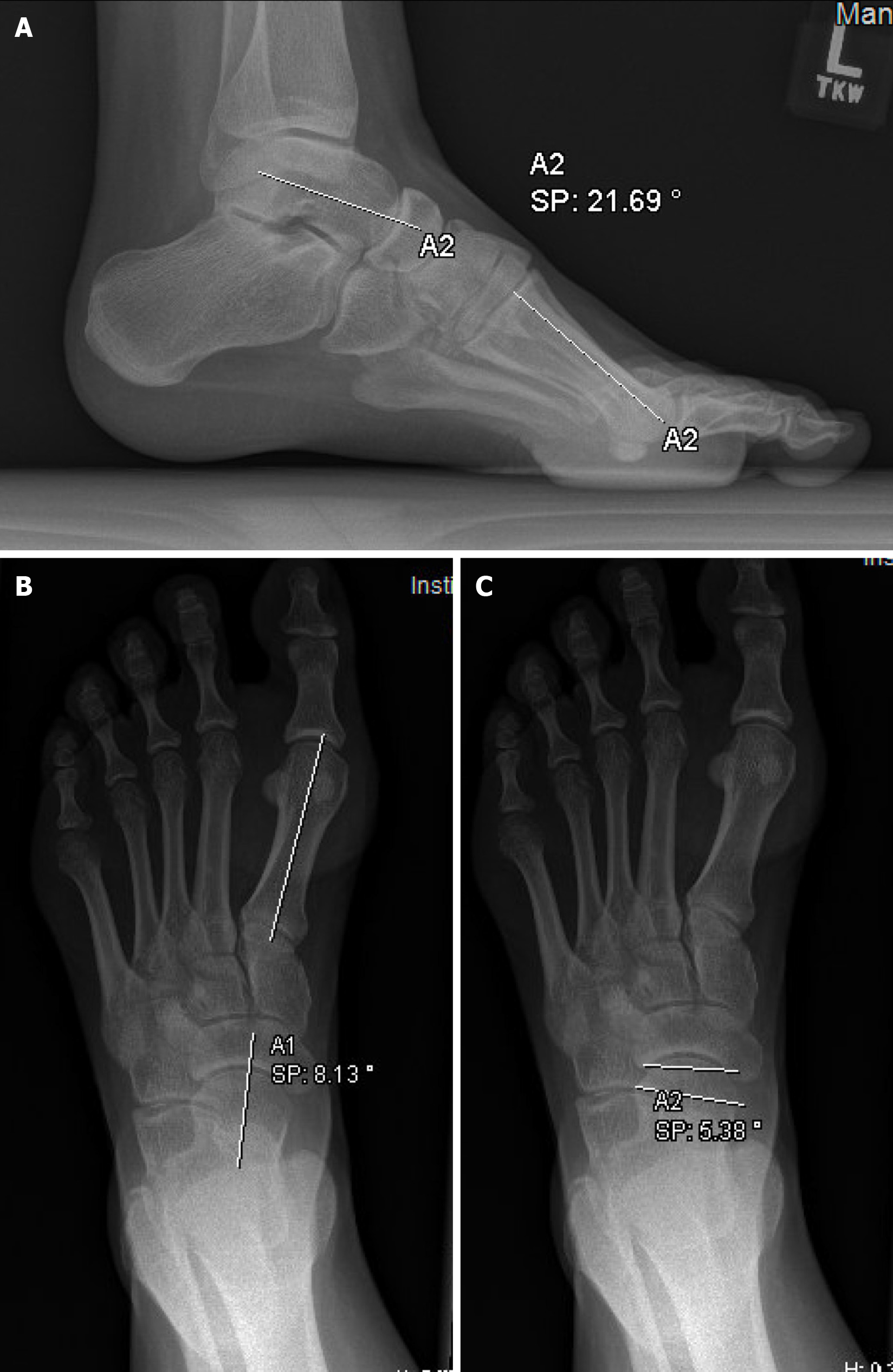
Figure 1 Preoperative lateral and anteroposterior radiographs showing Meary’s angle, anteroposterior talo-first metatarsal angle, and talonavicular coverage.
A: Meary’s angle (lateral talus-first metatarsal angle); B: Anteroposterior talo-first metatarsal angle; C: Talonavicular coverage.
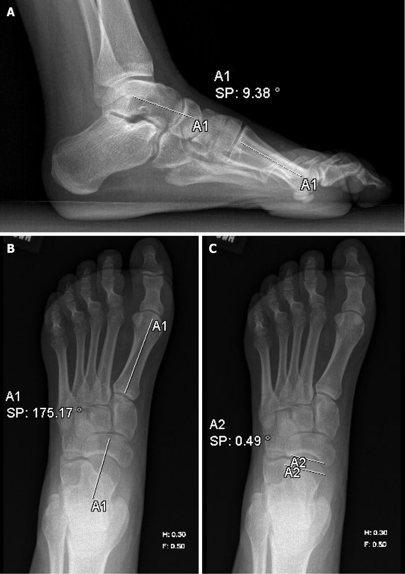
Figure 2 Lateral, and anteroposterior radiographs obtained after first stage radical plantar fascia release demonstrating improvement in Meary’s angle, anteroposterior talo-first metatarsal angle angle, and talonavicular coverage.
A: Meary’s angle (lateral talus-first metatarsal angle); B: Anteroposterior talo-first metatarsal angle; C: Talonavicular coverage.
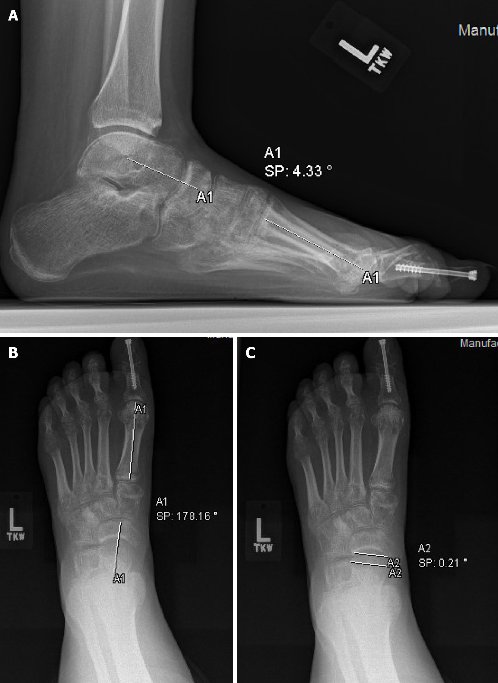
Figure 3 Postoperative lateral and anteroposterior radiographs obtained after second-stage cuneiform osteotomy demonstrating further improvement in Meary’s angle, anteroposterior talo-first metatarsal angle angle, and talonavicular coverage.
A: Meary’s angle (lateral talus-first metatarsal angle); B: Anteroposterior talo-first metatarsal angle; C: Talonavicular coverage.
- Citation: Padgett AM, Kothari E, Conklin MJ. Two-stage corrective operation for the treatment of pes cavovarus in patients with spina bifida. World J Orthop 2024; 15(7): 618-626
- URL: https://www.wjgnet.com/2218-5836/full/v15/i7/618.htm
- DOI: https://dx.doi.org/10.5312/wjo.v15.i7.618


