©The Author(s) 2024.
World J Orthop. May 18, 2024; 15(5): 404-417
Published online May 18, 2024. doi: 10.5312/wjo.v15.i5.404
Published online May 18, 2024. doi: 10.5312/wjo.v15.i5.404
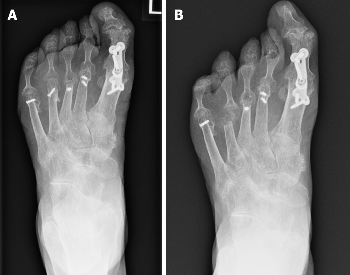
Figure 1 Radiograph demonstrating osteomyelitis affecting the 4th distal metatarsal and base of proximal phalanx.
A: Before conservative surgery; B: Post conservative surgery.
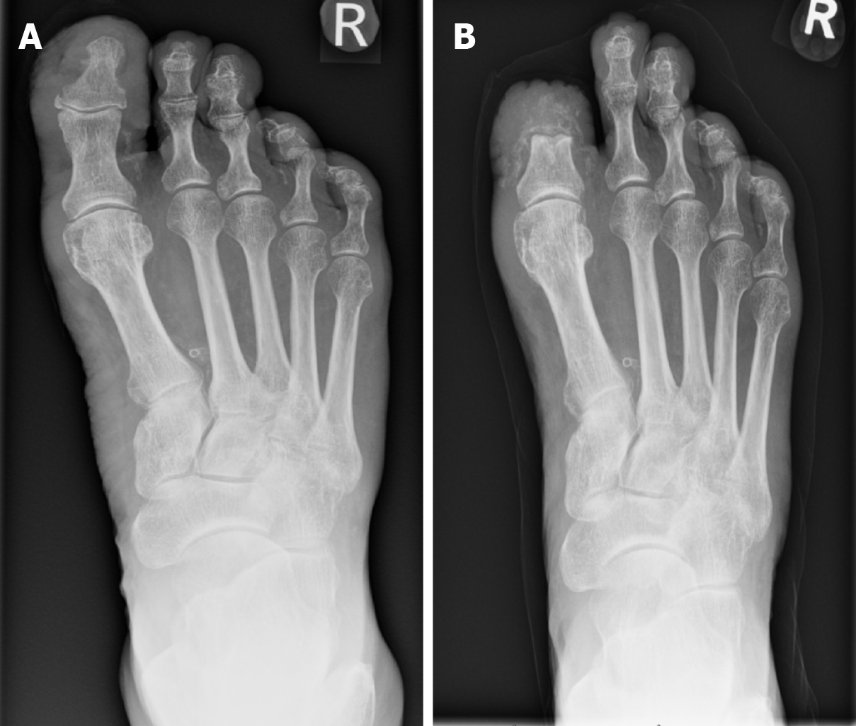
Figure 2 Radiograph demonstrating osteomyelitis affecting the right hallux distal phalanx and proximal phalanx.
A: Before minor amputation; B: Post minor amputation.
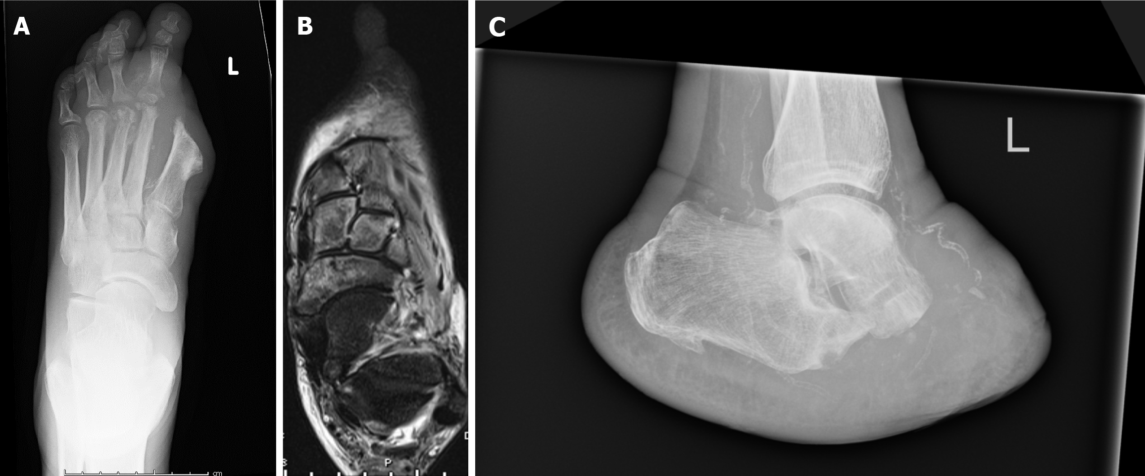
Figure 3 Imaging demonstrating recurrent osteomyelitis post-minor amputation (left hallux amputation).
A: Radiograph demonstrating the involvement of the 2nd and 3rd metatarsals; B: An MRI Scan showing involvement to just before the talo-navicular and calcaneo-cuboid joints; C: A radiograph demonstrating result of a Choparts's Amputation.
Figure 4 A radiograph demonstrating left midfoot charcot arthropathy.
A: Before Charcot reconstruction surgery; B: Post Charcot reconstruction surgery.
- Citation: Roberts RHR, Davies-Jones GR, Brock J, Satheesh V, Robertson GA. Surgical management of the diabetic foot: The current evidence. World J Orthop 2024; 15(5): 404-417
- URL: https://www.wjgnet.com/2218-5836/full/v15/i5/404.htm
- DOI: https://dx.doi.org/10.5312/wjo.v15.i5.404


