©The Author(s) 2024.
World J Orthop. Jan 18, 2024; 15(1): 61-72
Published online Jan 18, 2024. doi: 10.5312/wjo.v15.i1.61
Published online Jan 18, 2024. doi: 10.5312/wjo.v15.i1.61
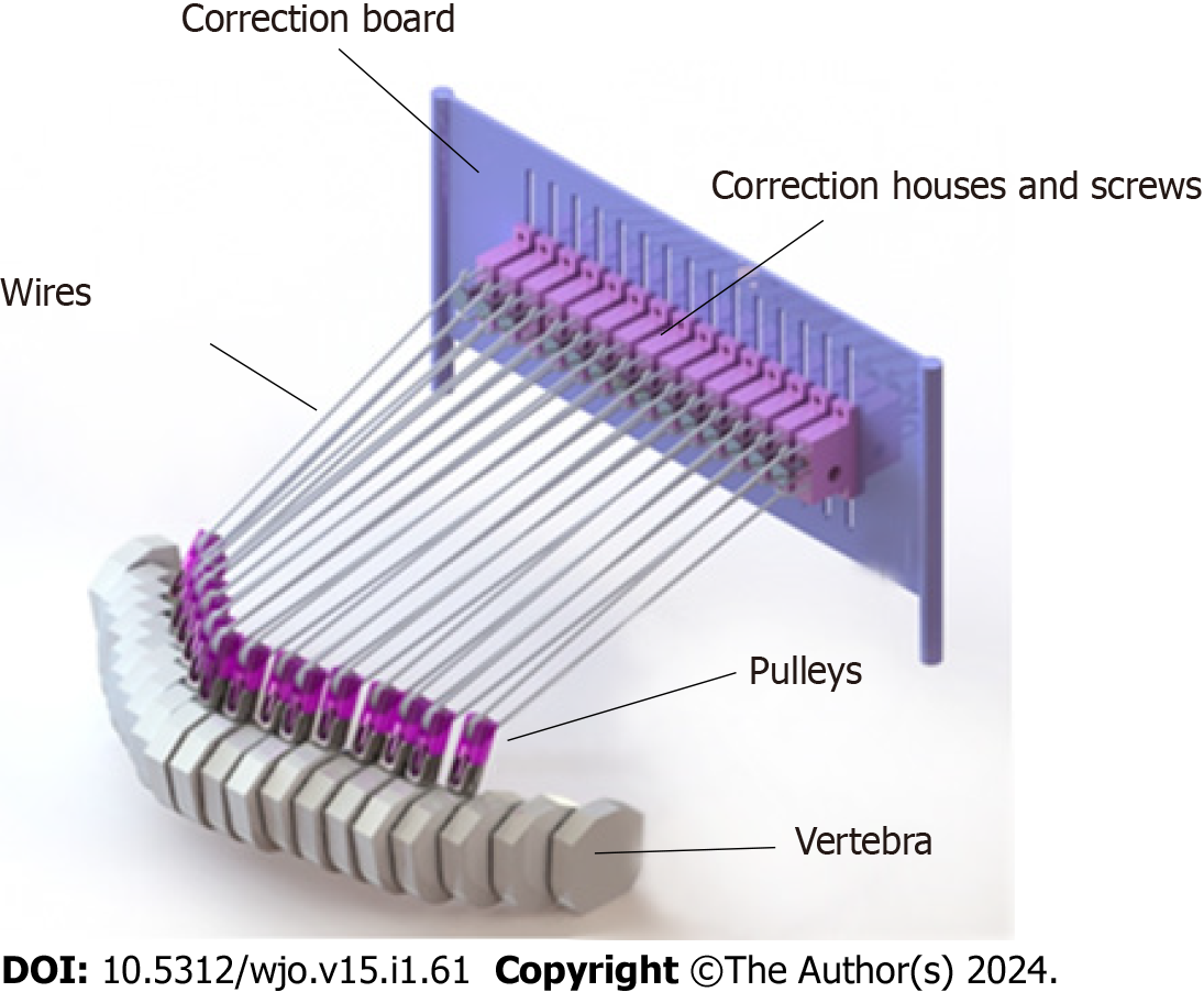
Figure 1 Three-dimensional design of Scoliocorrector Fatma-UI.
Scoliocorrector Fatma-UI consists of a board, pulling screws, screw housings, pulleys, and wire.
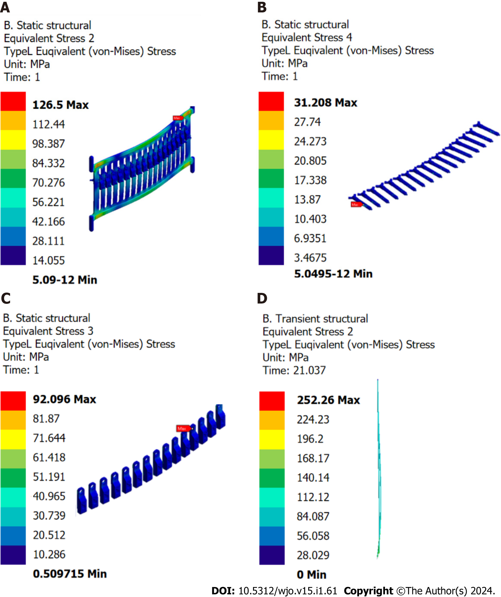
Figure 2 Finite element analysis of Scoliocorrector Fatma-UI components.
A: Correction board; B: Correction screw; C: Correction house; D: Wire. No deformation was observed in any of the components.
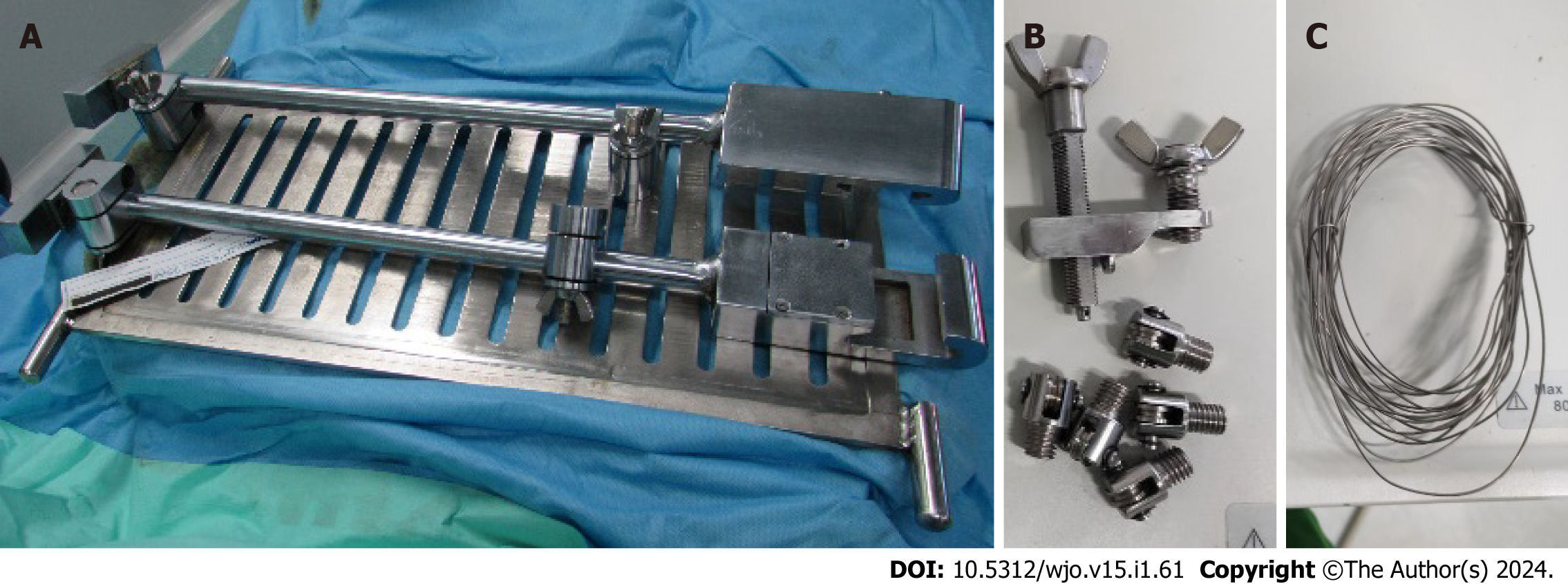
Figure 3 Prototype of Scoliocorrector Fatma-UI.
A: Correction board; B: Pulleys, correction house and correction screws; C: Wire.
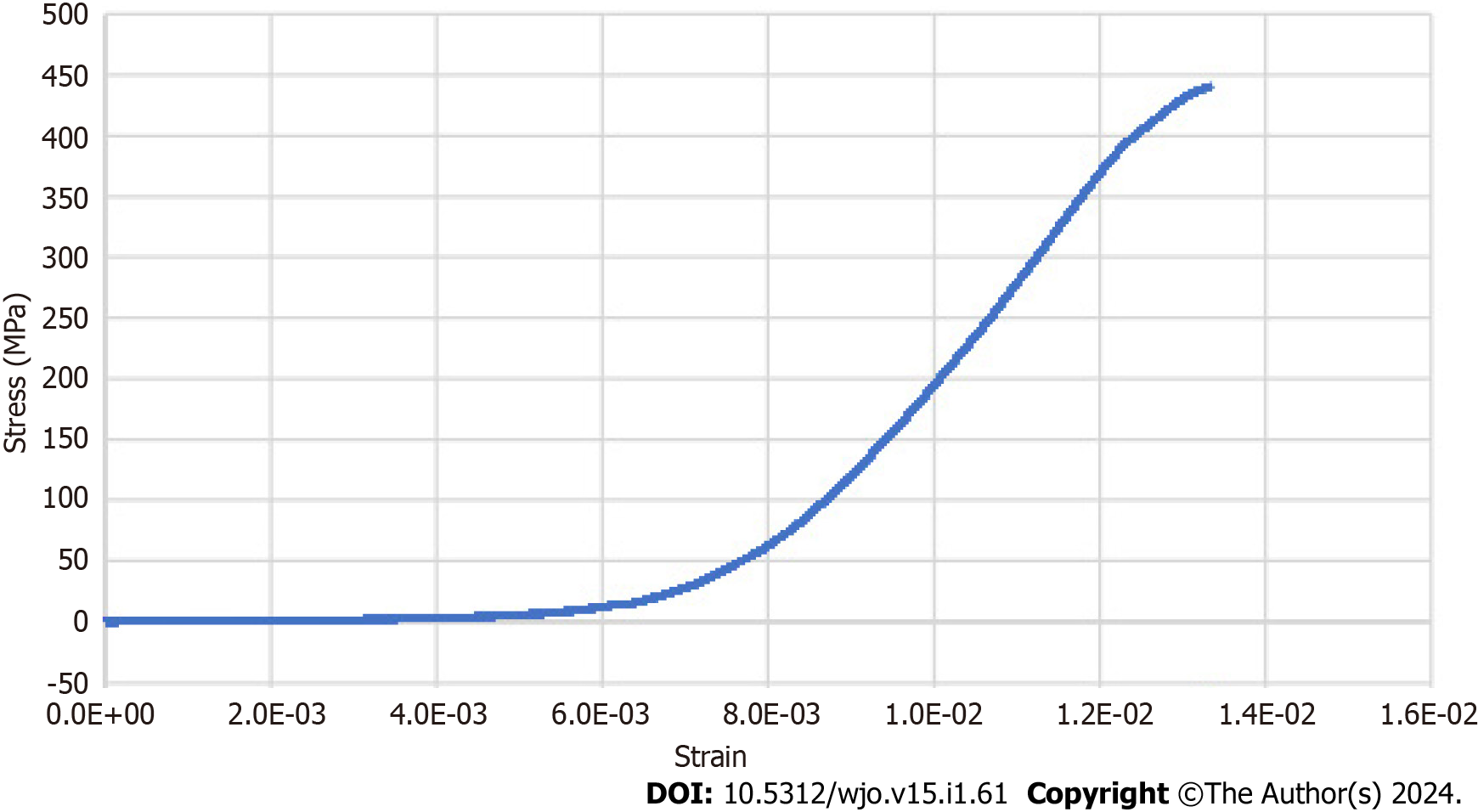
Figure 4 Stress-strain curve of Scoliocorrector Fatma-UI.
No breaking point was observed in this curve.
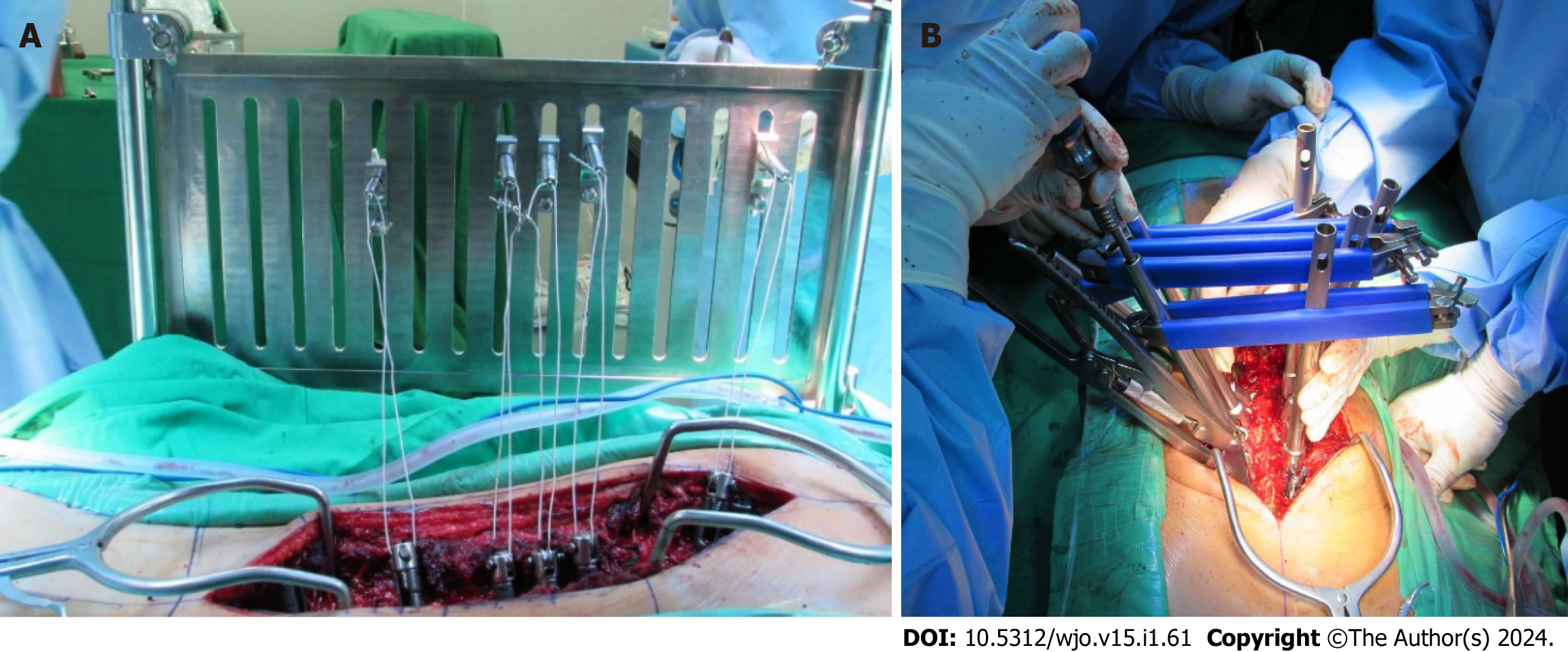
Figure 5 Deformity correction using Scoliocorrector Fatma-UI and direct vertebral rotation.
A: Scoliocorrector Fatma-UI (SCFUI); B: Direct vertebral rotation. Correction using SCFUI was done gradually.
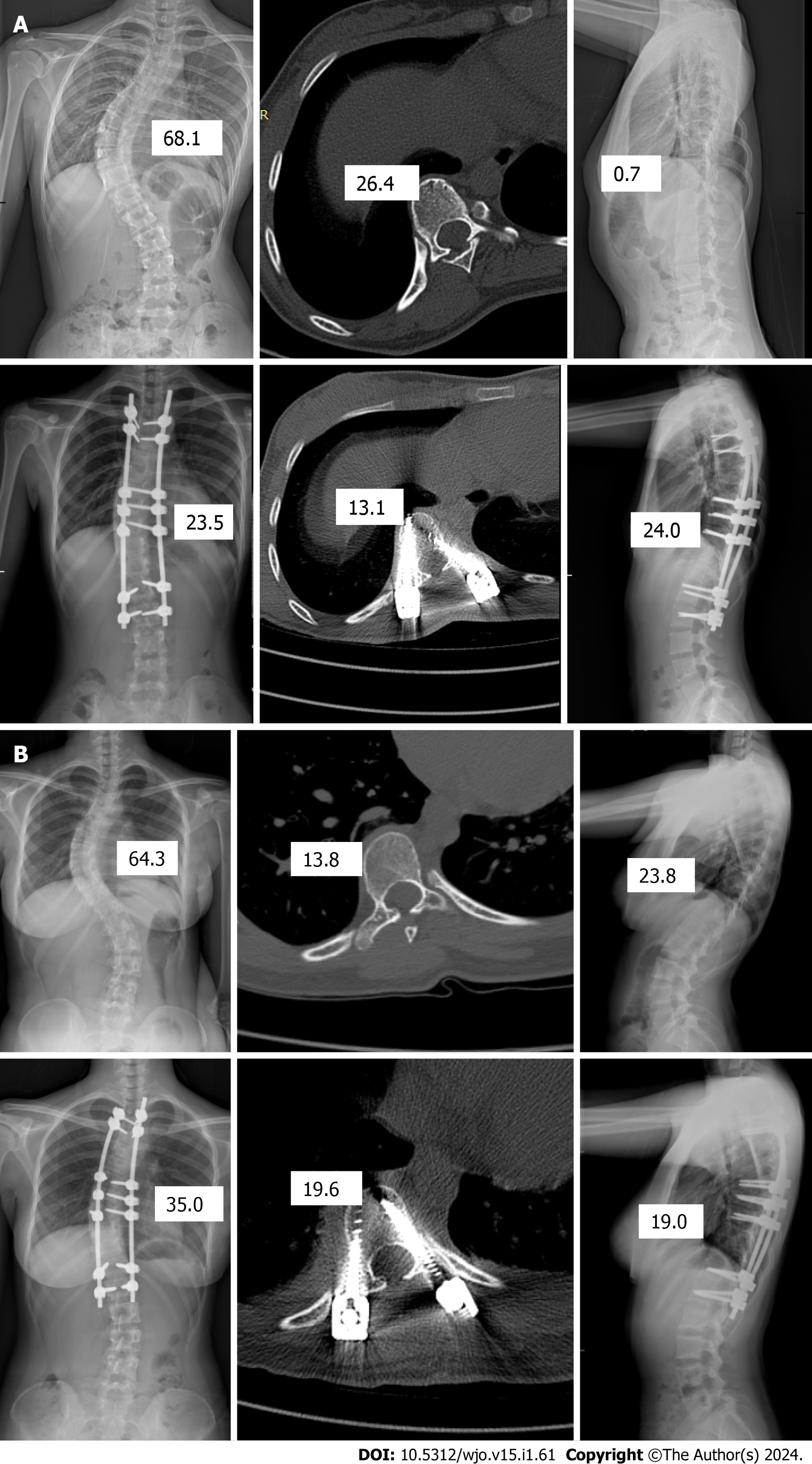
Figure 6 Radiographical finding of intervention and control group.
A: Intervention group; B: Control group. Both groups improved in coronal, rotational, and sagittal angles although they were more profound in the intervention group.
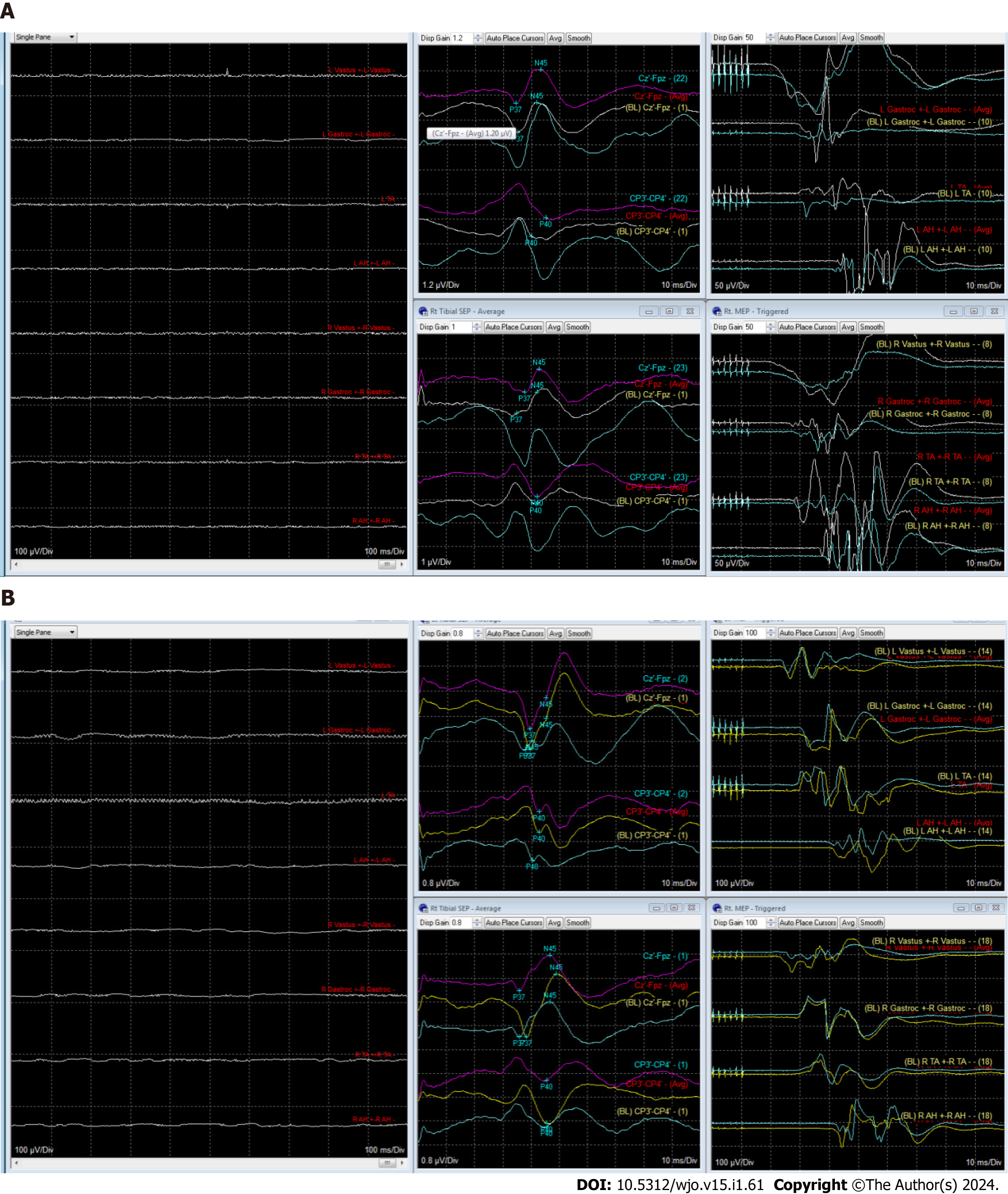
Figure 7 Intraoperative neuromonitoring finding in intervention and control group patient.
A: Intervention group patient; B: Control group patient. Blue graph indicated baseline recording and magenta graph indicated after correction recording. There was not any significant change of somatosensory evoked potential and motor-evoked potential during surgery in both groups.
- Citation: Phedy P, Dilogo IH, Indriatmi W, Supriadi S, Prasetyo M, Octaviana F, Noor Z. Scoliocorrector Fatma-UI for correction of adolescent idiopathic scoliosis: Development, effectivity, safety and functional outcome. World J Orthop 2024; 15(1): 61-72
- URL: https://www.wjgnet.com/2218-5836/full/v15/i1/61.htm
- DOI: https://dx.doi.org/10.5312/wjo.v15.i1.61


