Copyright
©The Author(s) 2023.
World J Orthop. Sep 18, 2023; 14(9): 698-706
Published online Sep 18, 2023. doi: 10.5312/wjo.v14.i9.698
Published online Sep 18, 2023. doi: 10.5312/wjo.v14.i9.698
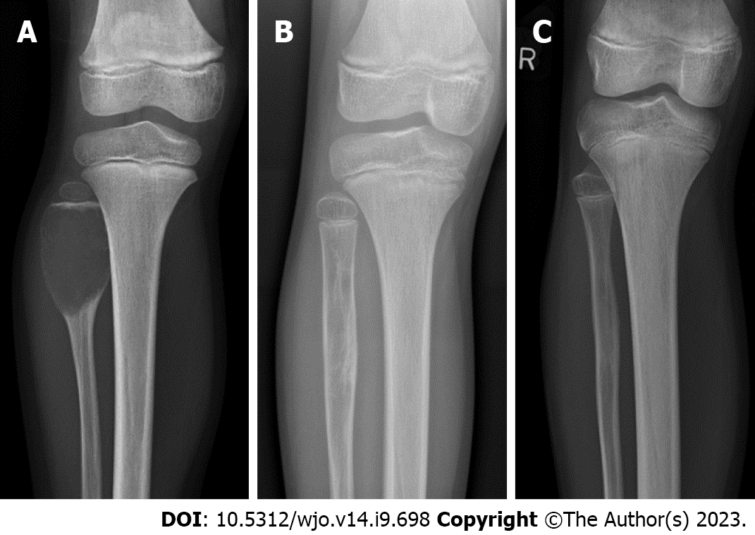
Figure 1 Radiographs of an aneurysmal bone cyst in the proximal fibula of a 5-year-old girl.
A: At presentation; B: After three injections of polidocanol; C: At 38 mo follow-up
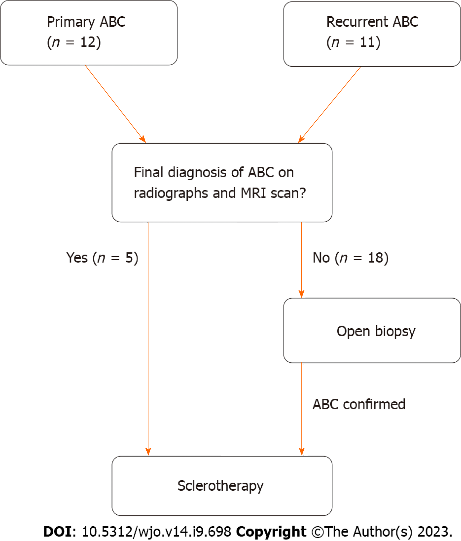
Figure 2 Flowchart of the algorithm from initial diagnosis to final management.
ABC: Aneurysmal bone cysts; MRI: Magnetic resonance imaging.
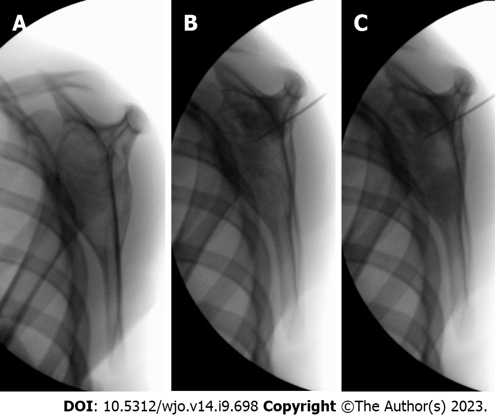
Figure 3 Intraoperative fluoroscopy images showing sclerotherapy of an aneurysmal bone cyst in the scapula.
A: Before injection; B: During injection with polidocanol and contrast agent; C: After injection
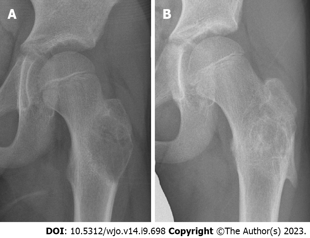
Figure 4 Radiographs of an aneurysmal bone cyst in the proximal femur showing complete ossification.
A: At presentation; B: At 26 mo follow-up after sclerotherapy.
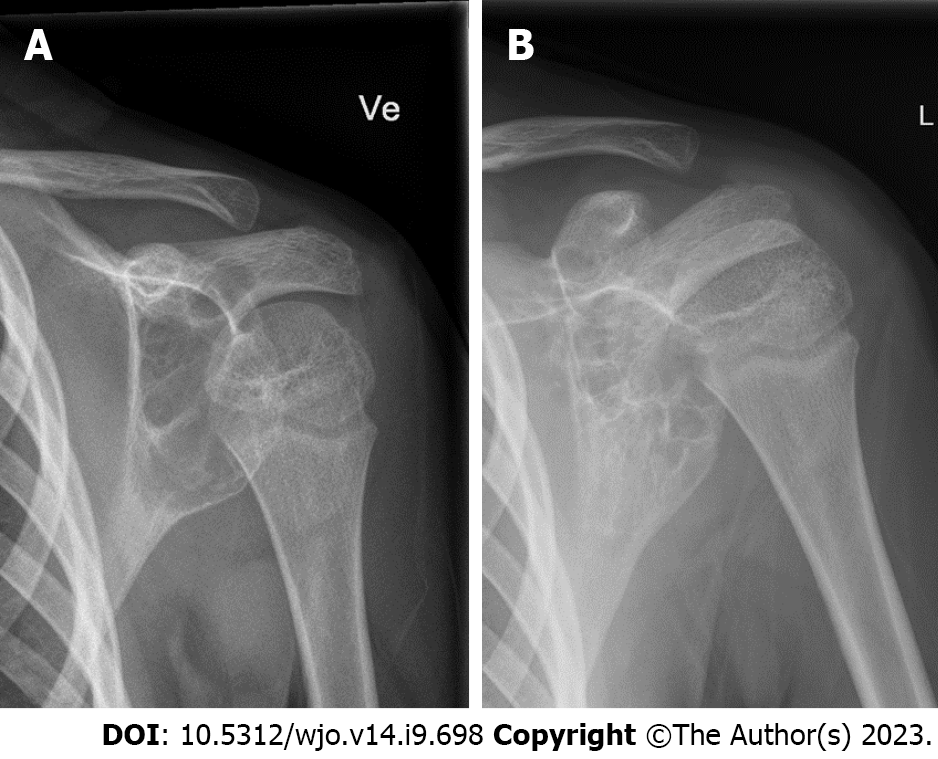
Figure 5 Radiographs of an aneurysmal bone cyst in the scapula showing partial ossification.
A: At presentation; B: At 24 mo follow-up after sclerotherapy.
- Citation: Weber KS, Jensen CL, Petersen MM. Sclerotherapy as a primary or salvage procedure for aneurysmal bone cysts: A single-center experience. World J Orthop 2023; 14(9): 698-706
- URL: https://www.wjgnet.com/2218-5836/full/v14/i9/698.htm
- DOI: https://dx.doi.org/10.5312/wjo.v14.i9.698


