©The Author(s) 2023.
World J Orthop. Dec 18, 2023; 14(12): 843-852
Published online Dec 18, 2023. doi: 10.5312/wjo.v14.i12.843
Published online Dec 18, 2023. doi: 10.5312/wjo.v14.i12.843
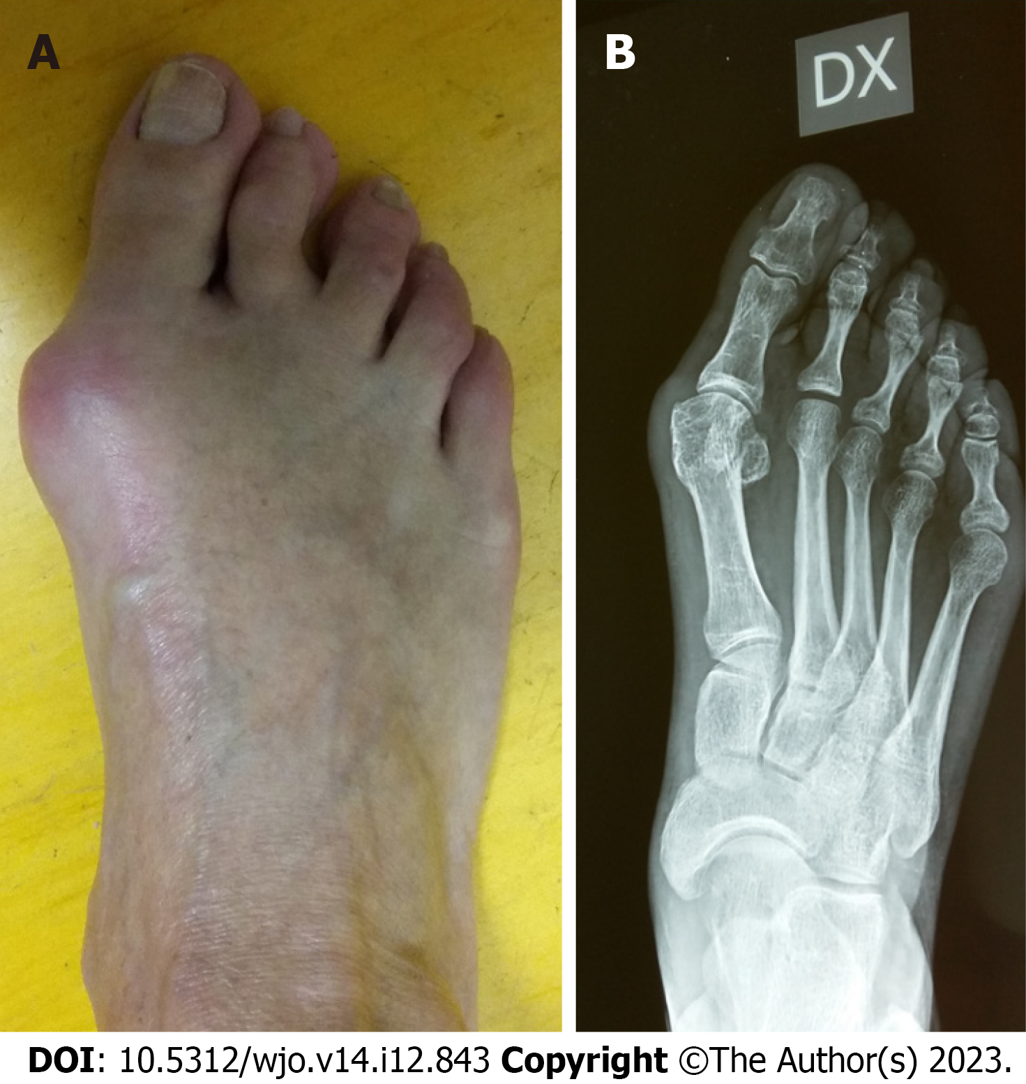
Figure 1 Preoperative clinical and radiological features of a 42 years female patient with hallux valgus.
A: Preoperative clinical; B: Radiological features.
Figure 2 Postoperative fluoroscopic picture of the patient treated with percutaneous hallux valgus correction without fixation (bandages) and follow-up.
A: Postoperative fluoroscopic picture of the patient treated with percutaneous hallux valgus correction without fixation (bandages); B: Clinical and postoperative X-rays of the previous patients at 6 mo of follow-up.
- Citation: Zanchini F, Catani O, Sergio F, Boemio A, Sieczak A, Piscopo D, Risitano S, Colò G, Fusini F. Role of lateral soft tissues release in percutaneous hallux valgus correction: A medium term retrospective study. World J Orthop 2023; 14(12): 843-852
- URL: https://www.wjgnet.com/2218-5836/full/v14/i12/843.htm
- DOI: https://dx.doi.org/10.5312/wjo.v14.i12.843


