Copyright
©The Author(s) 2022.
World J Orthop. May 18, 2022; 13(5): 538-543
Published online May 18, 2022. doi: 10.5312/wjo.v13.i5.538
Published online May 18, 2022. doi: 10.5312/wjo.v13.i5.538
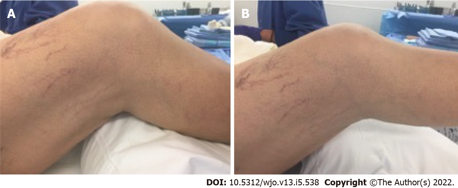
Figure 1 Preoperative flexion and extension of the right knee.
A: Preoperative flexion of the right knee; B: Preoperative extension of the right knee.
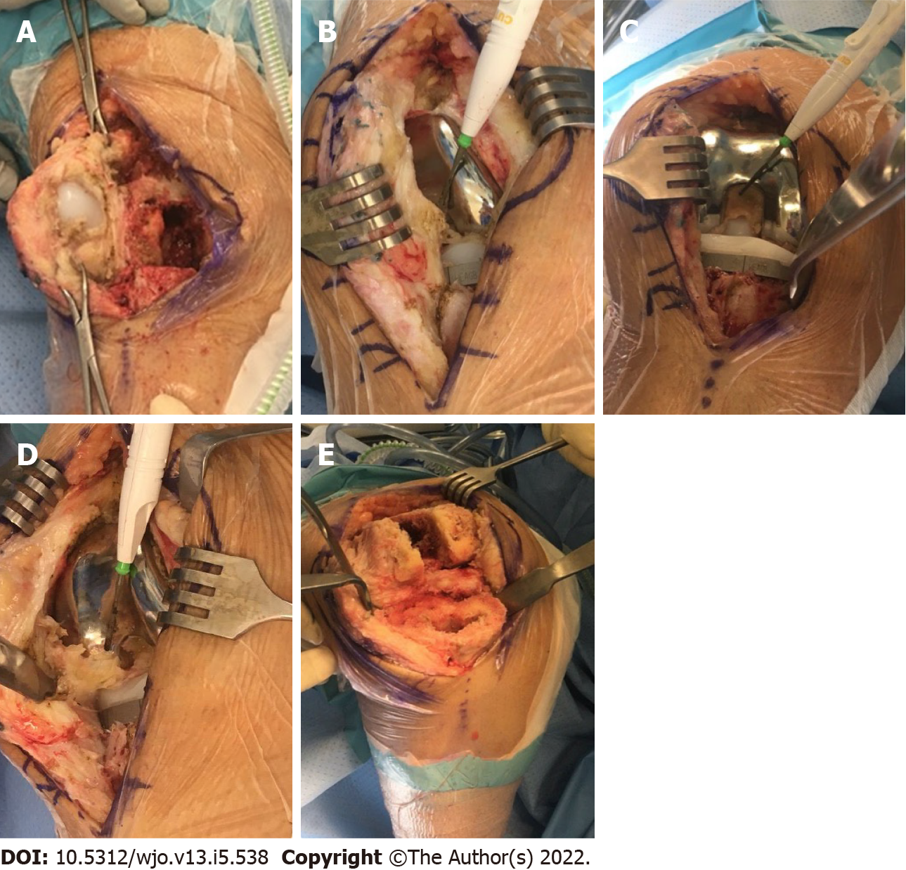
Figure 2 Images during the surgery.
A: Imaging depicting parapatellar adhesions with loose patellar component; B: Intraoperative adhesions from patellar tendon into intercondylar notch; C: Intercondylar scar/fibrous tissue and tibial component; D: Tendo-notch to posterior capsular fibrous adhesions being divided with electrocautery; E: Imaging depicting femur and tibia after removal of components and subtotal synovectomy and excision of adhesions.
- Citation: Mitchell S, Lee A, Stenquist R, Yatsonsky II D, Mooney ML, Shendge VB. Extensive adhesion formation in a total knee replacement in the setting of a gastrointestinal stromal tumor: A case report. World J Orthop 2022; 13(5): 538-543
- URL: https://www.wjgnet.com/2218-5836/full/v13/i5/538.htm
- DOI: https://dx.doi.org/10.5312/wjo.v13.i5.538


