©The Author(s) 2022.
World J Orthop. Nov 18, 2022; 13(11): 993-1005
Published online Nov 18, 2022. doi: 10.5312/wjo.v13.i11.993
Published online Nov 18, 2022. doi: 10.5312/wjo.v13.i11.993
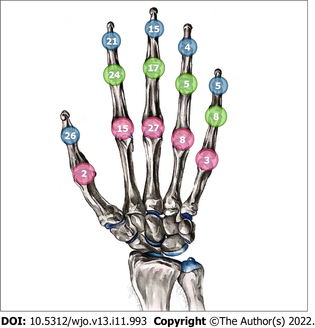
Figure 1 Distribution of 180 small joints involved in 170 episodes of septic arthritis.
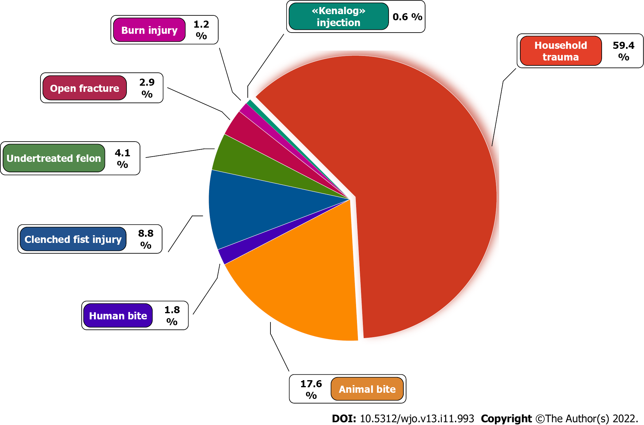
Figure 2 Causative factors of septic arthritis of the hand.
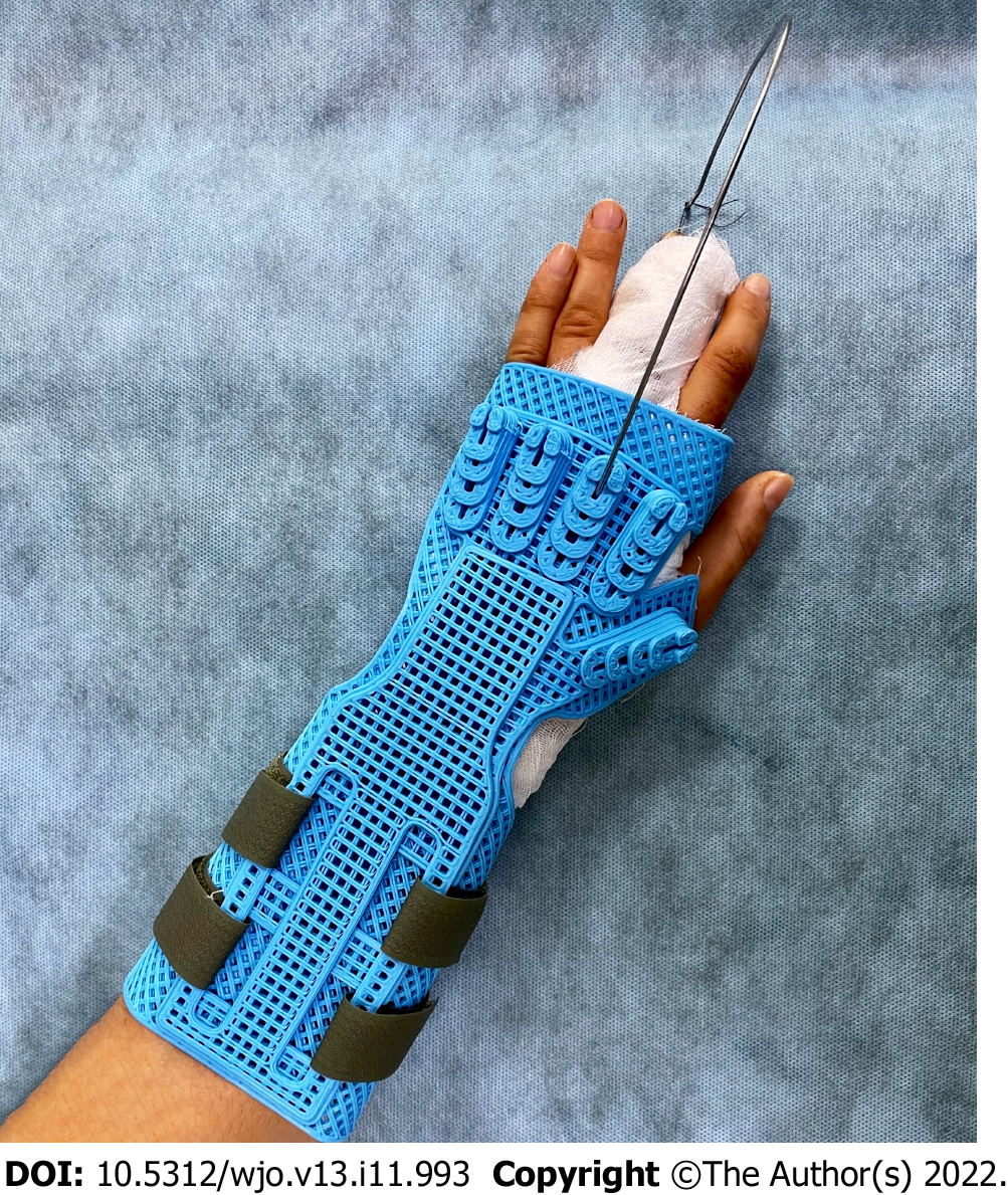
Figure 3 Immobilization of the hand using a developed design that allows axial extension of the affected finger.
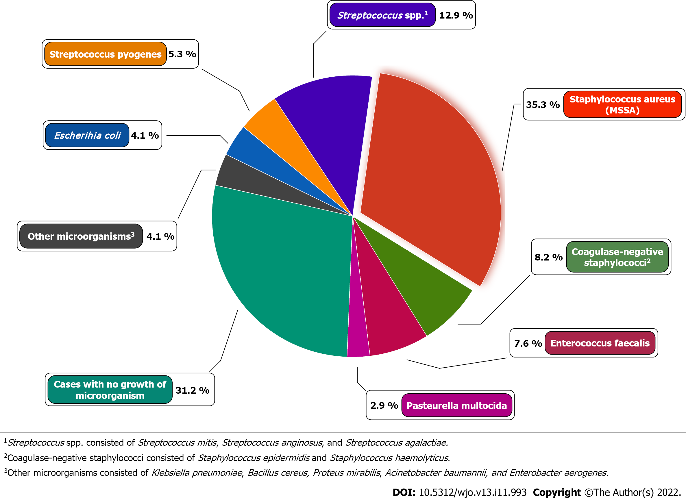
Figure 4 Microorganisms cultured from patients with septic arthritis of the hand.
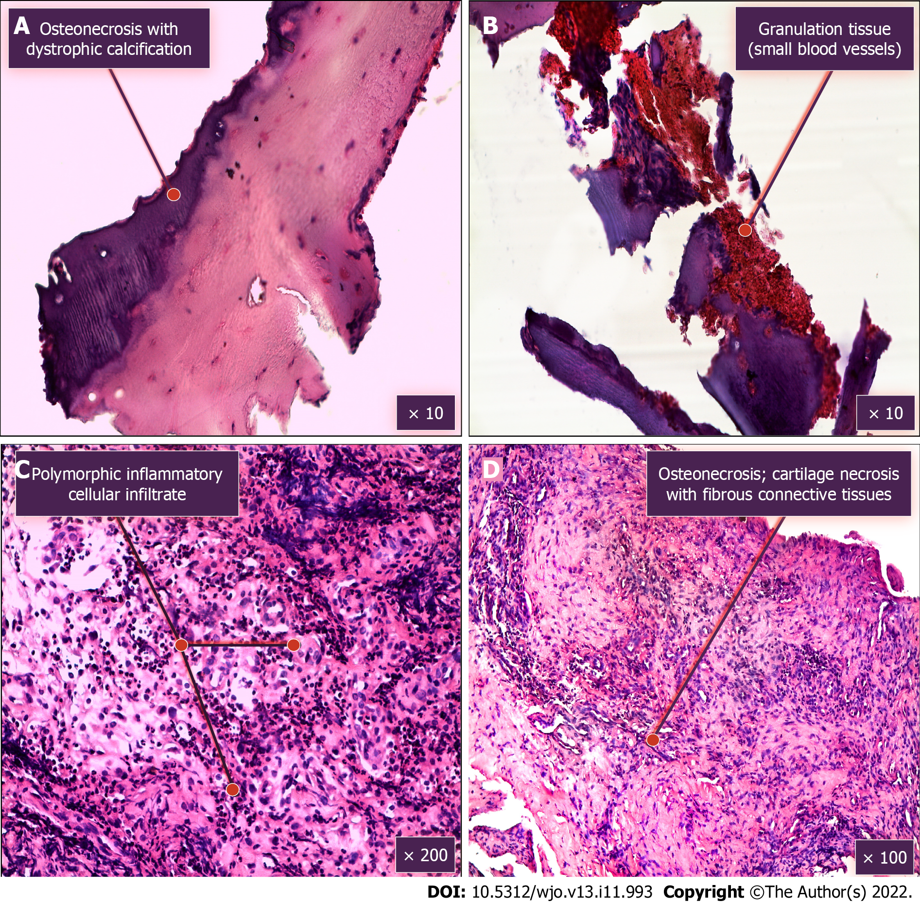
Figure 5 Morphological examination of tissues removed during surgical treatment (hematoxylin and eosin staining).
A: Osteonecrosis with dystrophic calcification; B: Granulation tissue (small blood vessels); C: Polymorphic inflammatory cellular infiltrate; D: Osteonecrosis; cartilage necrosis with fibrous connective tissues.
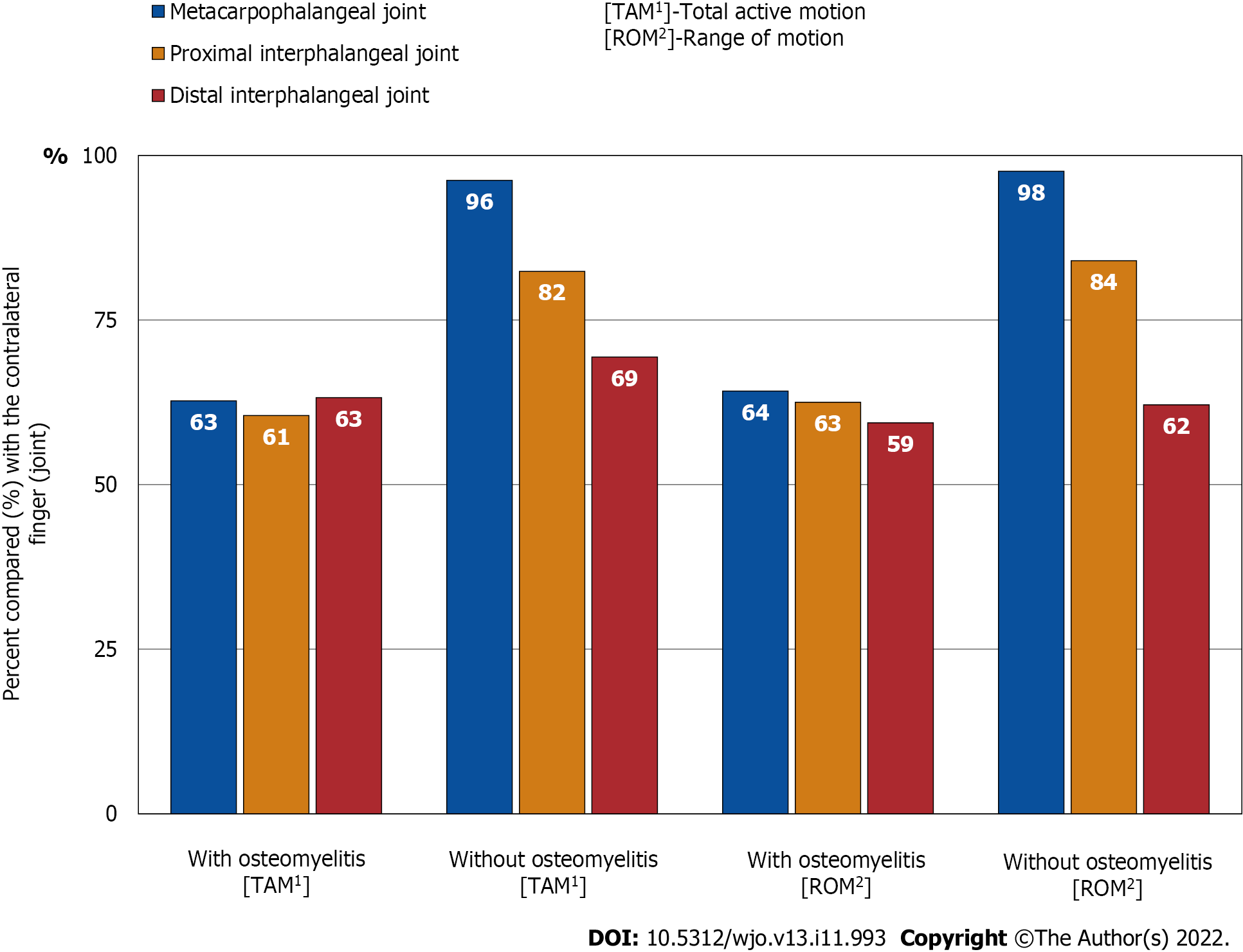
Figure 6 Summary of total active motion and range of motion.
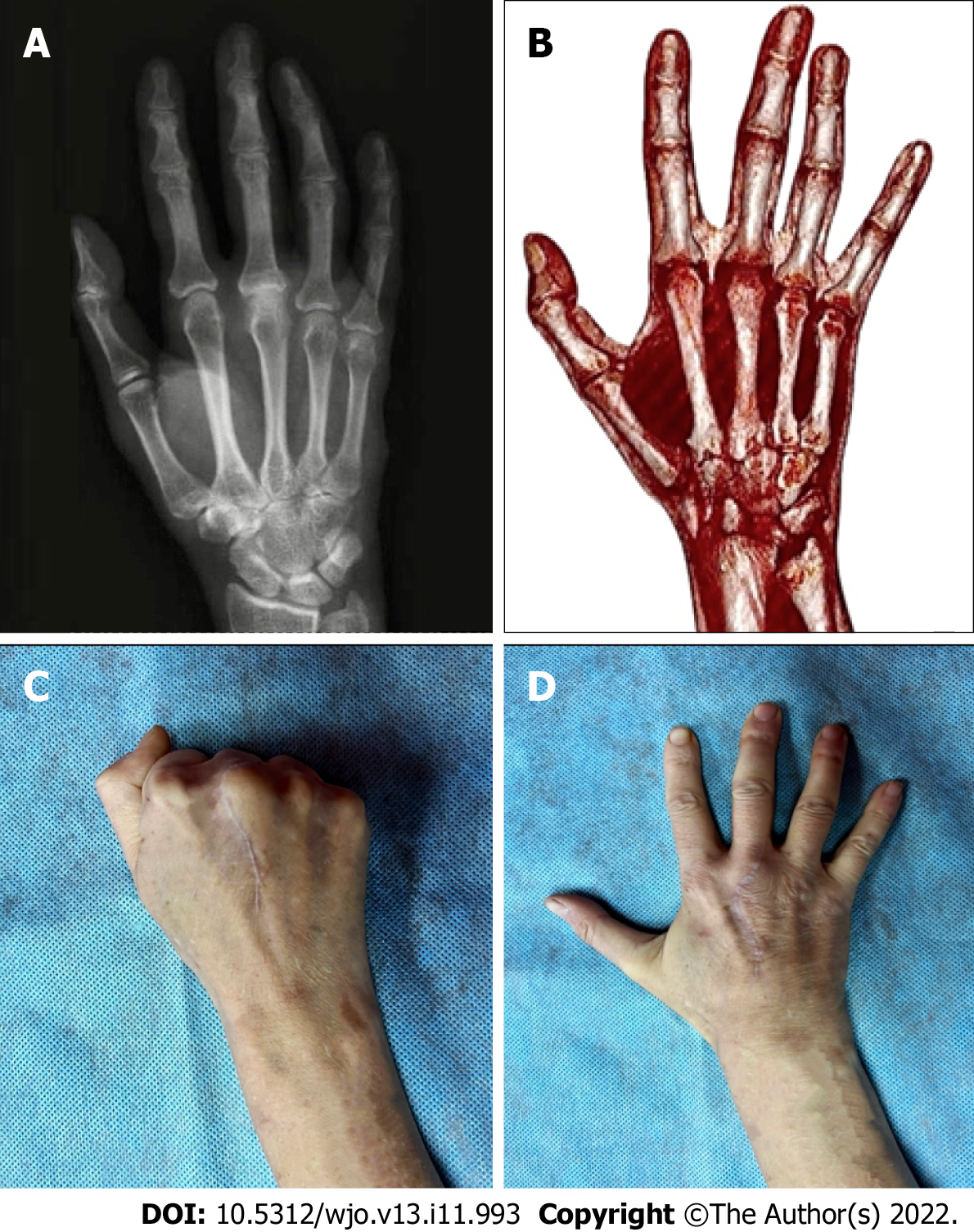
Figure 7 Outcomes of septic arthritis with osteomyelitis of the metacarpophalangeal joint, third finger (right hand).
A: Preoperative radiography; B: 3D-reconstruction (CT) 5 mo after operative intervention; C and D: Long-term outcomes of the function recovery of the metacarpophalangeal joint.
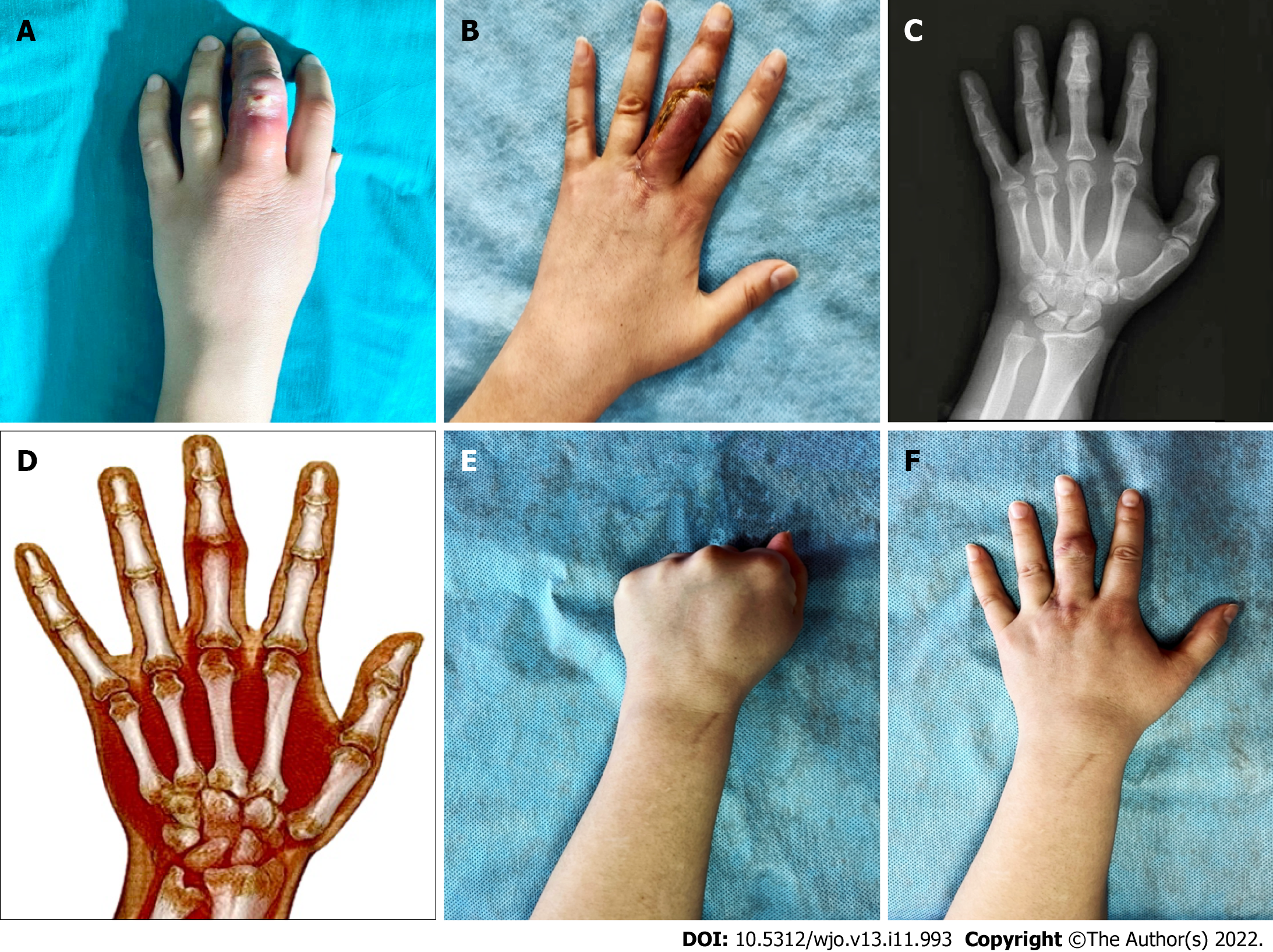
Figure 8 Treatment of septic arthritis with osteomyelitis of the proximal interphalangeal joint, third finger (left hand).
A: Clinical presentation at admission; B: Short-term outcomes after operative intervention; C: Preoperative radiography; D: 3D-reconstruction (CT) 4 mo after operative intervention; E and F: Long-term outcomes of the function recovery of the proximal interphalangeal joint.
- Citation: Lipatov KV, Asatryan A, Melkonyan G, Kazantcev AD, Solov’eva EI, Gorbacheva IV, Vorotyntsev AS, Emelyanov AY. Septic arthritis of the hand: From etiopathogenesis to surgical treatment. World J Orthop 2022; 13(11): 993-1005
- URL: https://www.wjgnet.com/2218-5836/full/v13/i11/993.htm
- DOI: https://dx.doi.org/10.5312/wjo.v13.i11.993


