Copyright
©The Author(s) 2022.
World J Orthop. Oct 18, 2022; 13(10): 940-948
Published online Oct 18, 2022. doi: 10.5312/wjo.v13.i10.940
Published online Oct 18, 2022. doi: 10.5312/wjo.v13.i10.940
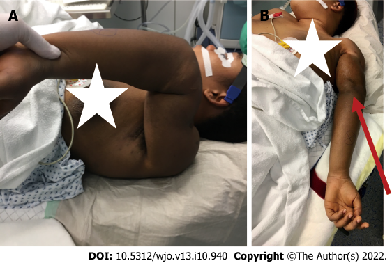
Figure 1 Severe malalignment of the patient’s left arm.
A: The patient in the operating room demonstrating the extreme deformity of the left upper extremity; B: In the supine position the palm of her hand and her olecranon (red narrow) were facing up.
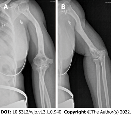
Figure 2 Preoperative X-ray of the patient’s left upper extremity.
A: Preoperative X-rays of the left shoulder and upper arm of the patient revealed a deformed humerus and a complete consolidation; B: In addition, the X-ray displayed classic signs of post-traumatic osteoarthritis of the shoulder.
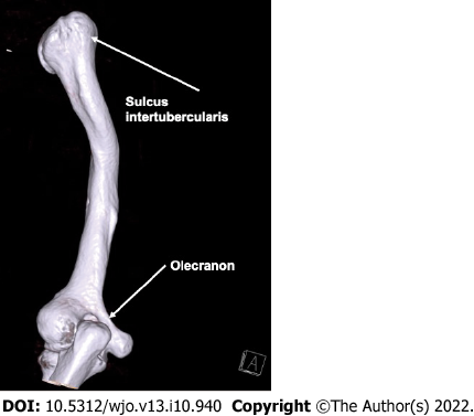
Figure 3 Preoperative three-dimensional computed tomography of the patient’s humerus.
Extreme malrotation of 180° was detectable. The sulcus intertubercularis and olecranon served as landmarks.
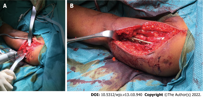
Figure 4 Intraoperative photographs.
A: Transversal osteotomy of the humerus was performed at the level of the former fracture; B: Internal fixation was done with the use of an eight-hole dynamic compression plate (Limited Contact Dynamic Compression Plate System).
Figure 5 Postoperative X-rays of the patient’s left humerus after corrective osteotomy and internal fixation.
Figure 6 X-rays at the patient’s 2-year follow-up appointment.
There was complete consolidation of the osteotomy zone.
Figure 7 Postoperative cosmetic and functional outcome.
- Citation: Wenning KE, Schildhauer TA, Jones CB, Hoffmann MF. Derotational osteotomy and internal fixation of a 180° malrotated humerus: A case report. World J Orthop 2022; 13(10): 940-948
- URL: https://www.wjgnet.com/2218-5836/full/v13/i10/940.htm
- DOI: https://dx.doi.org/10.5312/wjo.v13.i10.940


