Copyright
©The Author(s) 2021.
World J Orthop. Jun 18, 2021; 12(6): 445-455
Published online Jun 18, 2021. doi: 10.5312/wjo.v12.i6.445
Published online Jun 18, 2021. doi: 10.5312/wjo.v12.i6.445
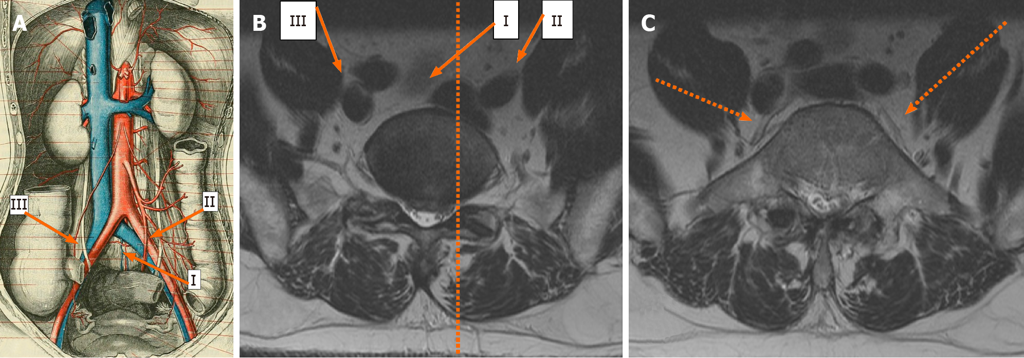
Figure 1 Vascular anatomy as depicted through an illustration showing the frontal aspect (A), and that depicted through magnetic resonance imaging axial sections (B, C).
Image B shows magnetic resonance imaging (MRI) axial section through the L5-S1 disc, and the facet line (dotted line) running anteroposteriorly through the medial border of the left L5-S1 facet joint cutting through the left common iliac vein. The three oblique approaches to L5-S1; namely the left intra-bifurcation (i), the left pre-psoas (ii), and the right pre-psoas (iii) are shown in A and B. Image C shows an MRI axial section through mid-L5 vertebral body and shows the left and right ilio-lumbar veins (dotted arrows).
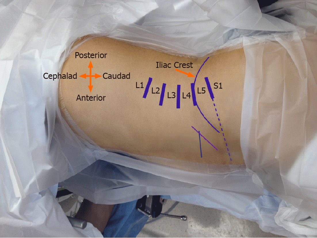
Figure 2 Clinical photograph of a patient in the right lateral decubitus position with superimposed skin markings.
Disc levels from L1 to S1 are marked. L5-S1 disc marking is extended anteriorly (dotted line) to guide the incision. A longer oblique line (purple) somewhat parallel to the anterior iliac crest marks the suggested incision for a left intra-bifurcation approach for L3-S1 oblique lumbar interbody fusion (OLIF). The shorter transverse line (green) is the suggested incision (along skin lines) for a left pre-psoas approach for L3-S1 OLIF.
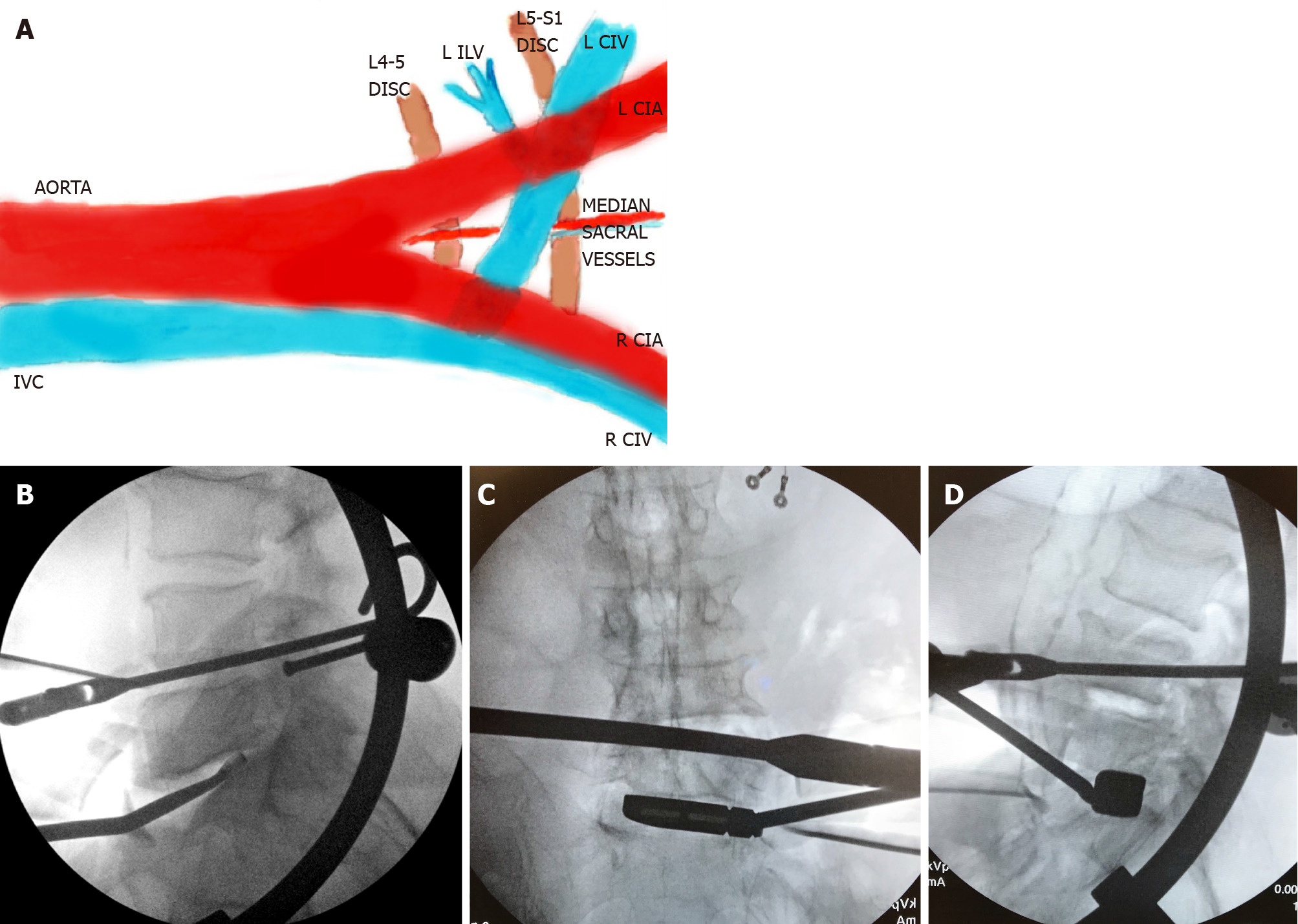
Figure 3 Illustration showing left-sided oblique anterolateral approaches (A), and intraoperative fluoroscopy images (B-D).
Image A shows the median sacral vessels that are often encountered in the left intra-bifurcation approach, and the left ilio-lumbar vein (ILV), which may need ligation in a left pre-psoas approach. Image B shows a lateral fluoroscopy image with a double-bent curette which may be helpful in preparing the portions of the L5 inferior endplate that may not be under direct visualization. Images C and D show intraoperative anteroposterior and lateral fluoroscopy images during a left pre-psoas approach with specialized trial instrument bent in two planes. IVC: Inferior vena cava; CIV: Common iliac vein; CIA: Common iliac artery; ILV: Ilio-lumbar vein; R: Right; L: Left.
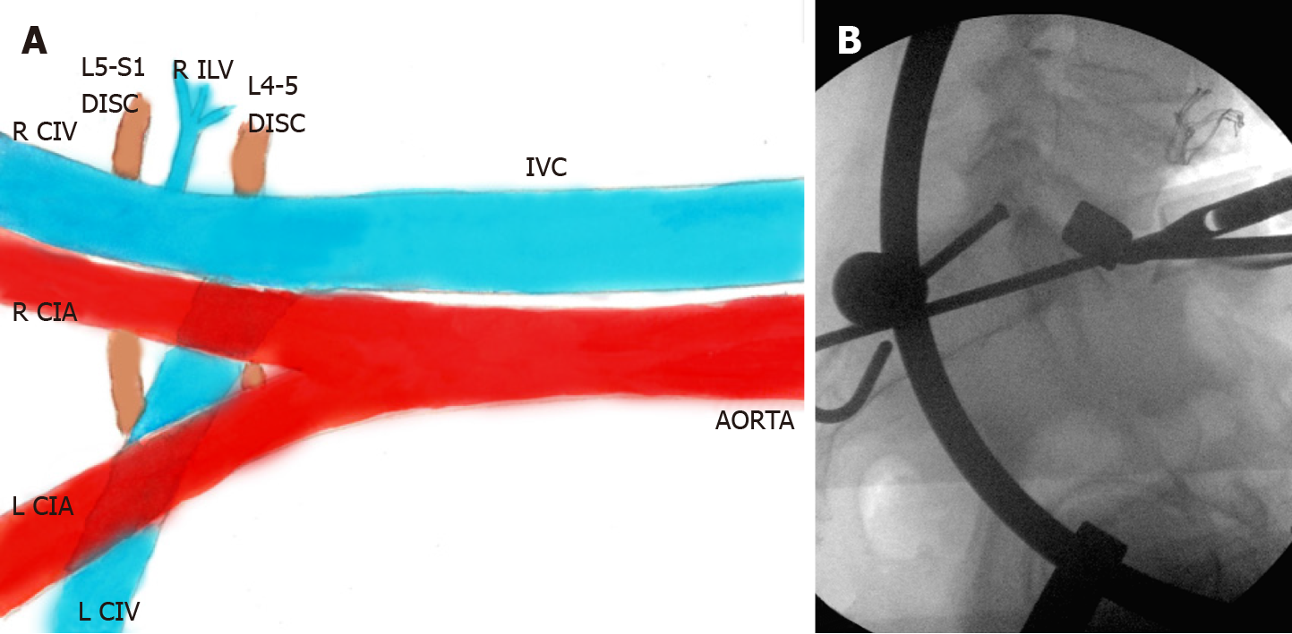
Figure 4 Illustration showing right-sided oblique anterolateral approach (A), and a lateral fluoroscopy image (B) during a right pre-psoas approach.
The right ilio-lumbar vein (ILV) is longer in length and smaller in caliber (A) when compared to the left ILV (Figure 3A). Also note that the right common iliac vein (CIV) is visible throughout in the right pre-psoas approach, unlike the left CIV, which for the most part, is covered by the accompanying left common iliac artery. Image B again identifies specialized trial instrument bent in two planes (similar to Figure 3C and D). IVC: Inferior vena cava; CIV: Common iliac vein; CIA: Common iliac artery; ILV: Ilio-lumbar vein; R: Right; L: Left.
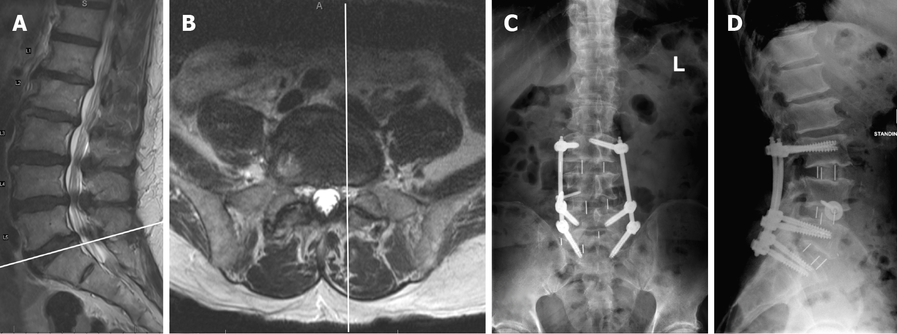
Figure 5 Preoperative magnetic resonance imaging sagittal (A) and axial (B) images and postoperative anteroposterior (C) and lateral (D) upright radiographs for Case 1.
Image A shows the scout line corresponding to the axial image B. The anteroposterior facet line through the medial border of the left L5-S1 facet joint runs medial to the medial border of the left CIV (B). CIV: Common iliac vein.
Figure 6 Preoperative upright anteroposterior (A) and lateral (B) radiographs, preoperative magnetic resonance imaging sagittal (C) and axial (D) images, and postoperative anteroposterior (E) and lateral (F, G) upright radiographs for Case 2.
Image C shows the scout line corresponding to the axial image D. The left common iliac vein is in the midline medial to the anteroposterior facet line (D). CIV: Common iliac vein.
Figure 7 Preoperative magnetic resonance imaging sagittal (A) and axial (B) images and postoperative (C) anteroposterior and (D) lateral upright radiographs for Case 3.
Image A shows the scout line corresponding to the axial image B. The anteroposterior facet line through the medial border of the left L5-S1 facet joint runs lateral to the medial border of the left common iliac vein (CIV) (B). The fat plane is better visualized under the right CIV than under the left CIV (B). CIV: Common iliac vein.
- Citation: Berry CA. Nuances of oblique lumbar interbody fusion at L5-S1: Three case reports. World J Orthop 2021; 12(6): 445-455
- URL: https://www.wjgnet.com/2218-5836/full/v12/i6/445.htm
- DOI: https://dx.doi.org/10.5312/wjo.v12.i6.445


