©The Author(s) 2021.
World J Orthop. Nov 18, 2021; 12(11): 850-858
Published online Nov 18, 2021. doi: 10.5312/wjo.v12.i11.850
Published online Nov 18, 2021. doi: 10.5312/wjo.v12.i11.850
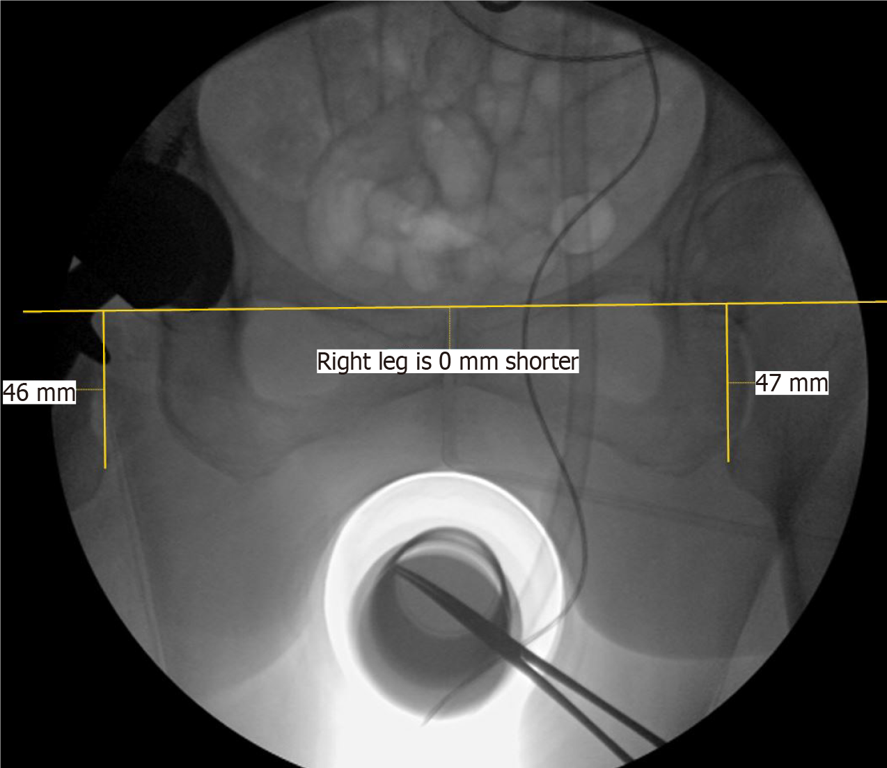
Figure 1 Intra-operative fluoroscopy image capture.
Representative image of observer obtained leg length discrepancy measurements on a saved intra-operative fluoroscopic view of the pelvis. Image capture was performed by the OEC image intensifier intra-operatively as described. Shown is a line drawn through bilateral radiographic teardrops with perpendicular lines to the medial prominence of bilateral lesser trochanters.
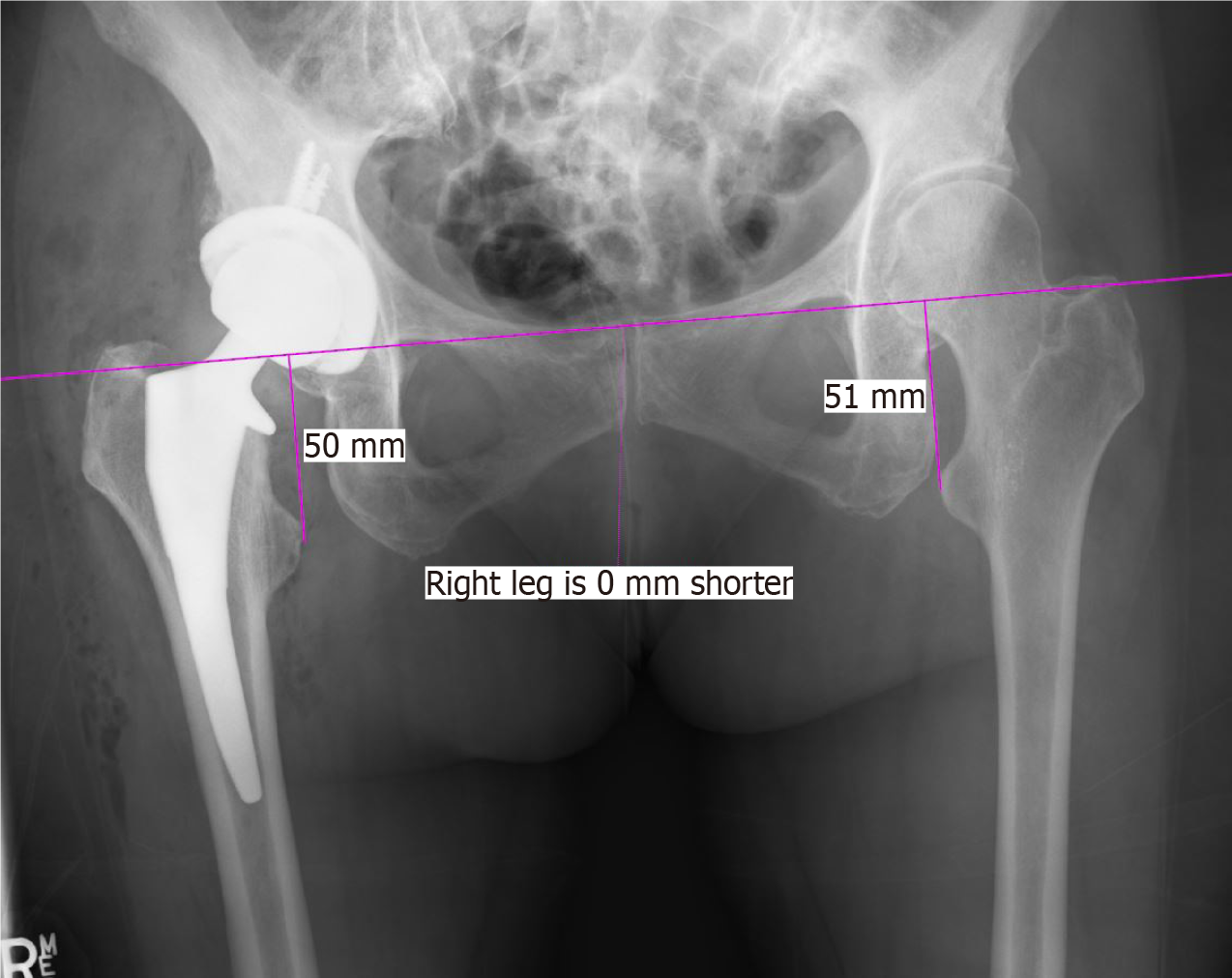
Figure 2 Post-operative x-ray image capture.
Representative image of observer obtained leg length discrepancy measurements on a corresponding post-operative anterior-posterior x-ray of the pelvis. Single line drawn through bilateral radiographic teardrops with perpendicular lines to the medial prominence of bilateral lesser trochanters.
Figure 3 Measured leg length discrepancy.
A: Measured leg length discrepancy based on teardrop reference point. Comparison showing mean x-ray leg length discrepancy (LLD) and fluoroscopic LLD using radiographic teardrop reference points as measured by two independent observers. Corresponding linear regression r2 value of 0.56; B: Measured leg length discrepancy (LLD) based on ischium reference point. Comparison showing mean x-ray and fluoroscopic LLD using ischium references points as measured by two independent observers. Corresponding linear regression r2 value of 0.87. LLD: Leg length discrepancy.
- Citation: Caus S, Reist H, Bernard C, Blankstein M, Nelms NJ. Reliability of a simple fluoroscopic image to assess leg length discrepancy during direct anterior approach total hip arthroplasty. World J Orthop 2021; 12(11): 850-858
- URL: https://www.wjgnet.com/2218-5836/full/v12/i11/850.htm
- DOI: https://dx.doi.org/10.5312/wjo.v12.i11.850


