Copyright
©The Author(s) 2020.
World J Orthop. Sep 18, 2020; 11(9): 411-417
Published online Sep 18, 2020. doi: 10.5312/wjo.v11.i9.411
Published online Sep 18, 2020. doi: 10.5312/wjo.v11.i9.411
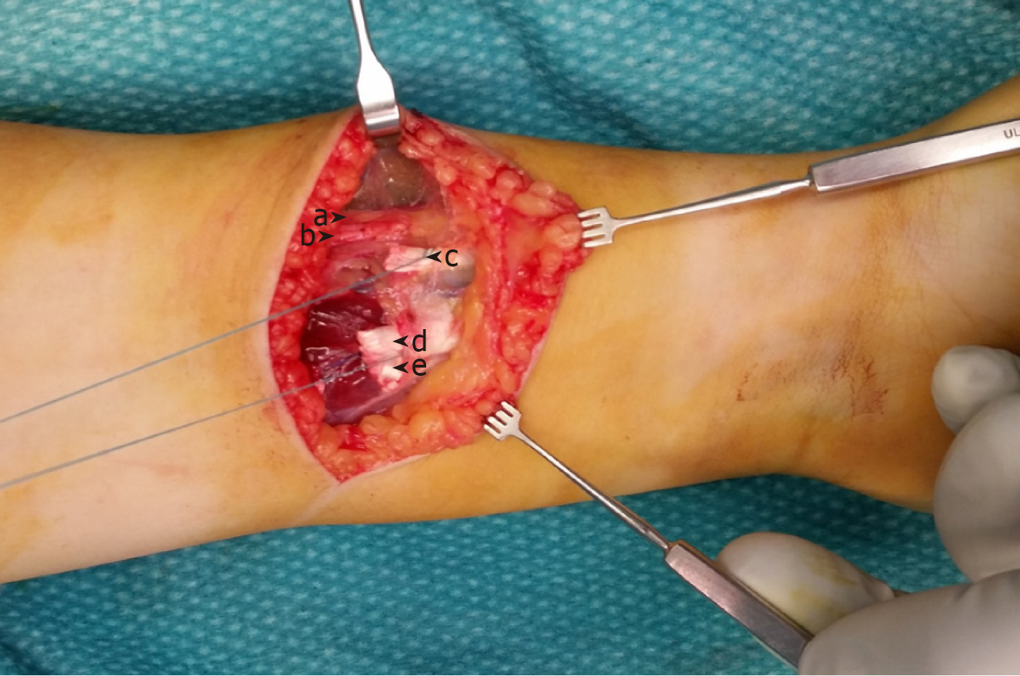
Figure 1 Intraoperative findings.
a: Proprius brachioradialis tendon; b: Superficial branch of radial nerve; c: Supernumerary brachioradialis tendon with tendon loop; d: Flexor carpi radialis tendon with tendon loop; e: Palmaris longus tendon.
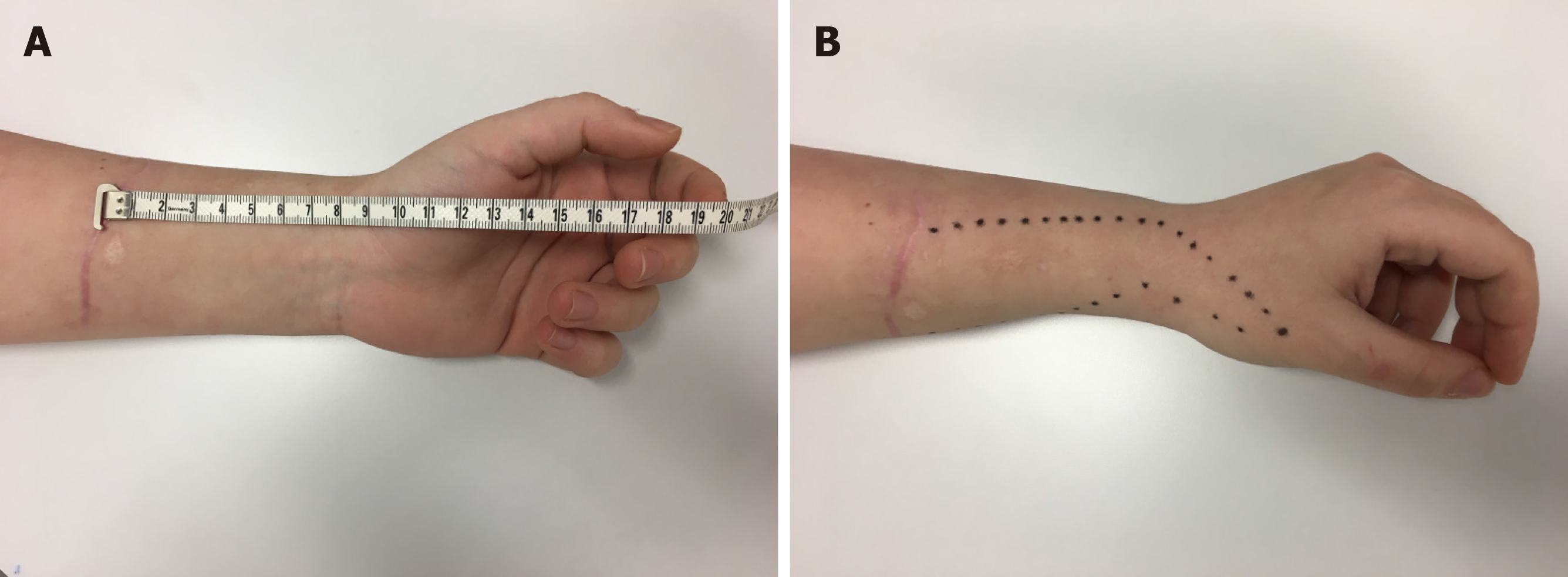
Figure 2 Clinical presentation at 5 mo postoperatively.
A: Scarification and distance of the scar to the flexion crease; B: The area of loss of sensibility.
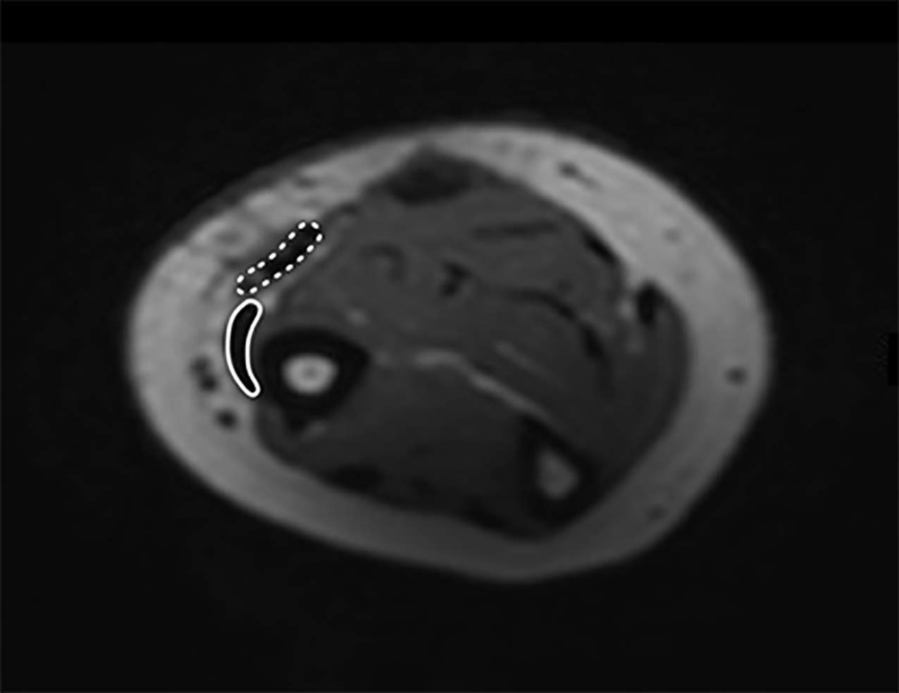
Figure 3 Magnetic resonance imaging series at 5 mo postoperatively, 85 mm proximal to the radius styloid tip at the level of injury and tendon suture with relationship of proprius brachioradialis (full line) and supernumerary brachioradialis (interrupted line).
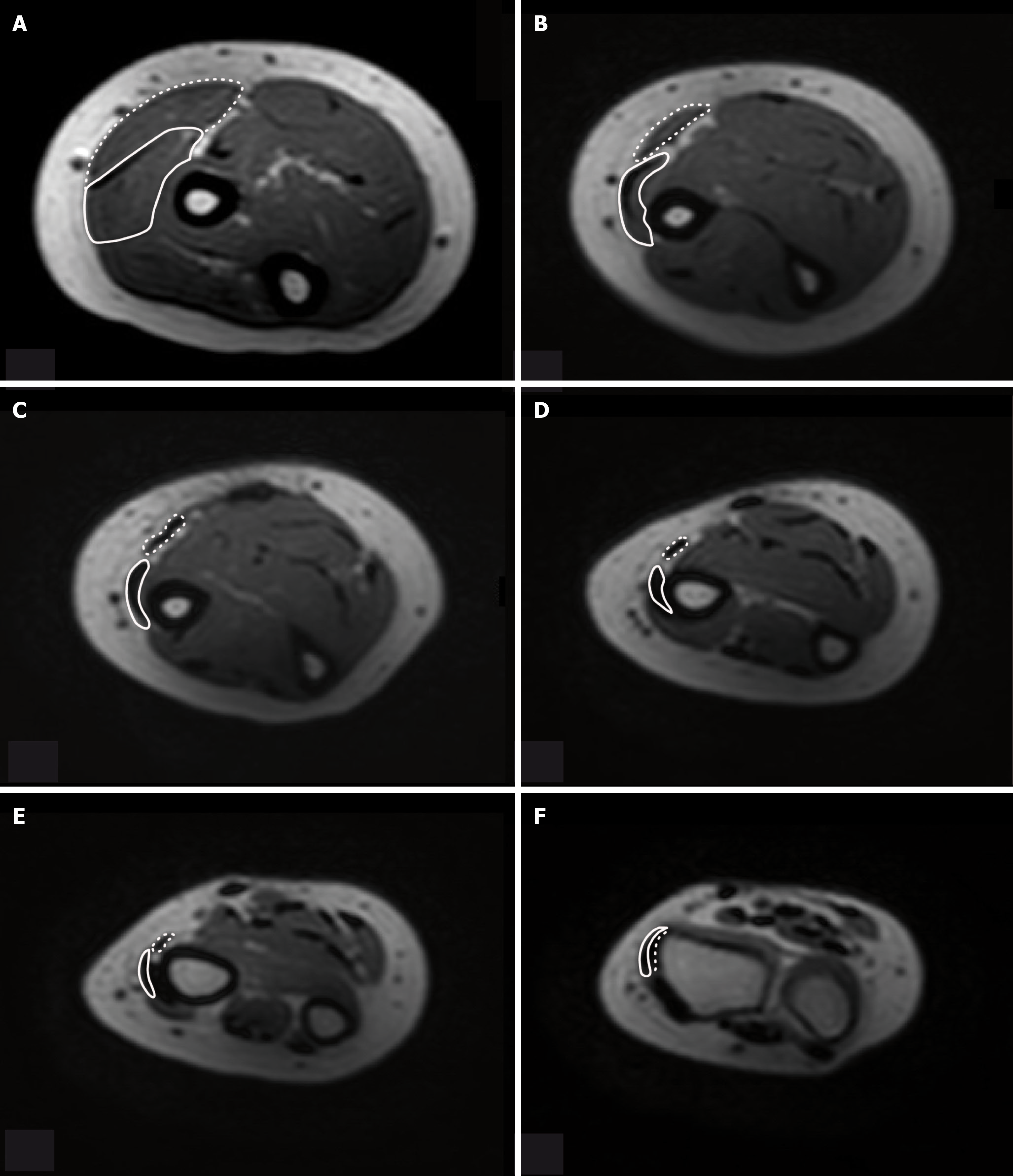
Figure 4 Magnetic resonance imaging series of forearm at 5 mo postoperatively with relationship of proprius brachioradialis (full line) and supernumerary brachioradialis (interrupted line).
- Citation: Kravarski M, Goerres GW, Antoniadis A, Guenkel S. Supernumerary brachioradialis - Anatomical variation with magnetic resonance imaging findings: A case report. World J Orthop 2020; 11(9): 411-417
- URL: https://www.wjgnet.com/2218-5836/full/v11/i9/411.htm
- DOI: https://dx.doi.org/10.5312/wjo.v11.i9.411


