©The Author(s) 2020.
World J Orthop. Feb 18, 2020; 11(2): 137-144
Published online Feb 18, 2020. doi: 10.5312/wjo.v11.i2.137
Published online Feb 18, 2020. doi: 10.5312/wjo.v11.i2.137
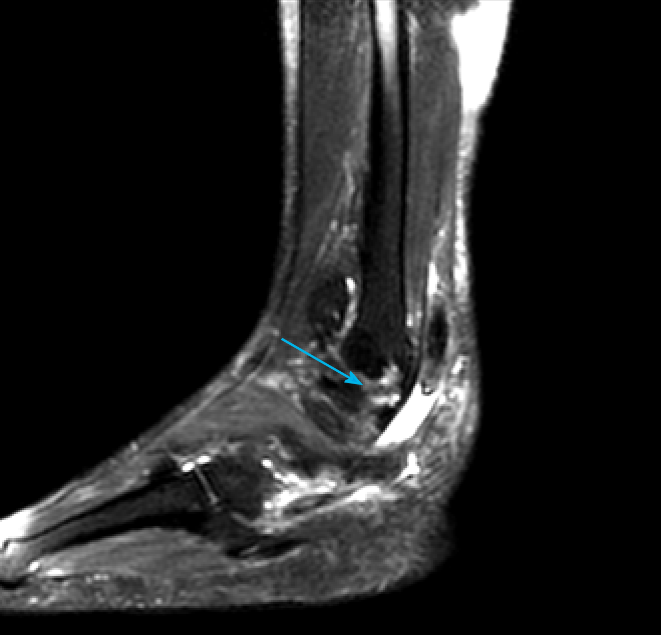
Figure 1 Preoperative ankle magnetic resonance imaging for diagnosis and evaluation of the type of peroneal tendon injuries.
Ankle magnetic resonance imaging T2-weighted sagittal image showed complete rupture of the peroneus longus tendon (blue arrow).
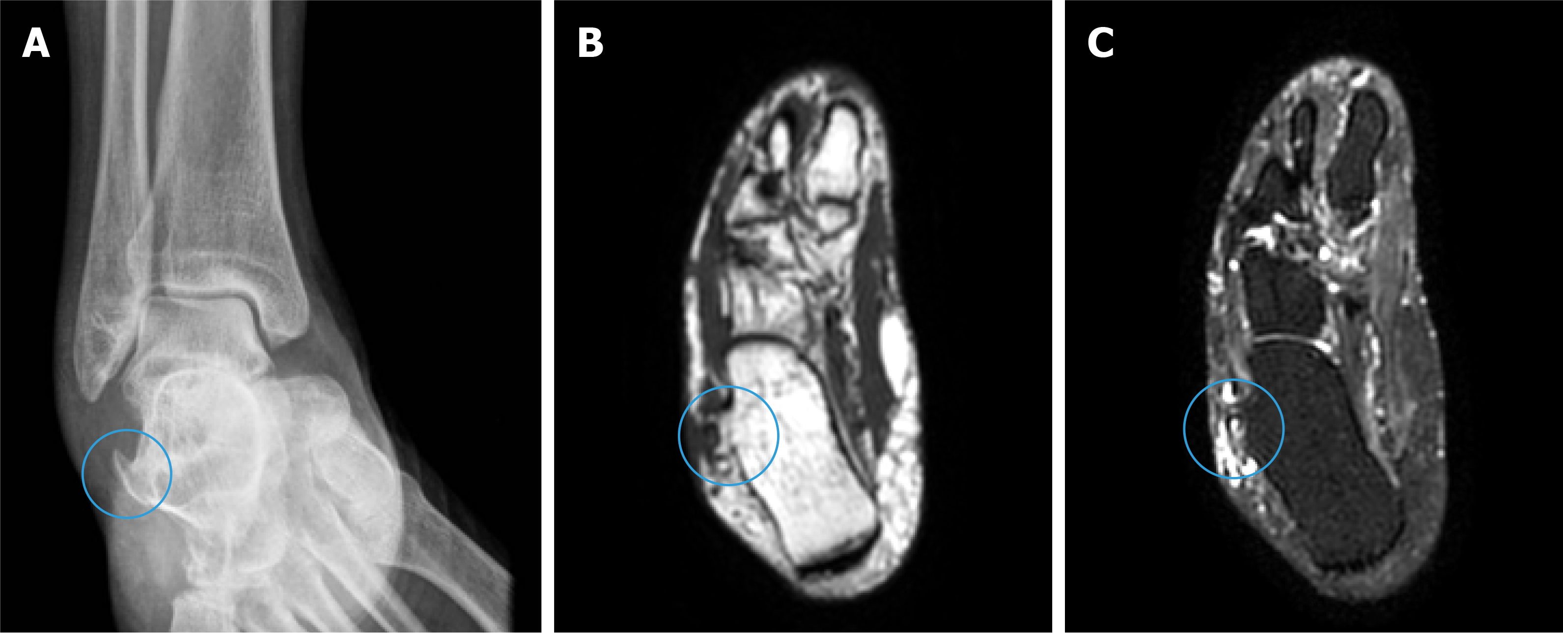
Figure 2 Preoperative plain radiograph and magnetic resonance imaging evaluation for surgical planning.
A: Ankle plain radiograph anteroposterior view showing a sharp hypertrophic peroneal tubercle (blue circle); B and C: Ankle magnetic resonance imaging T1- and T2-weighted axial images (B and C respectively) demonstrated the hypertrophic peroneal tubercle with underlying bone edema (blue circles).
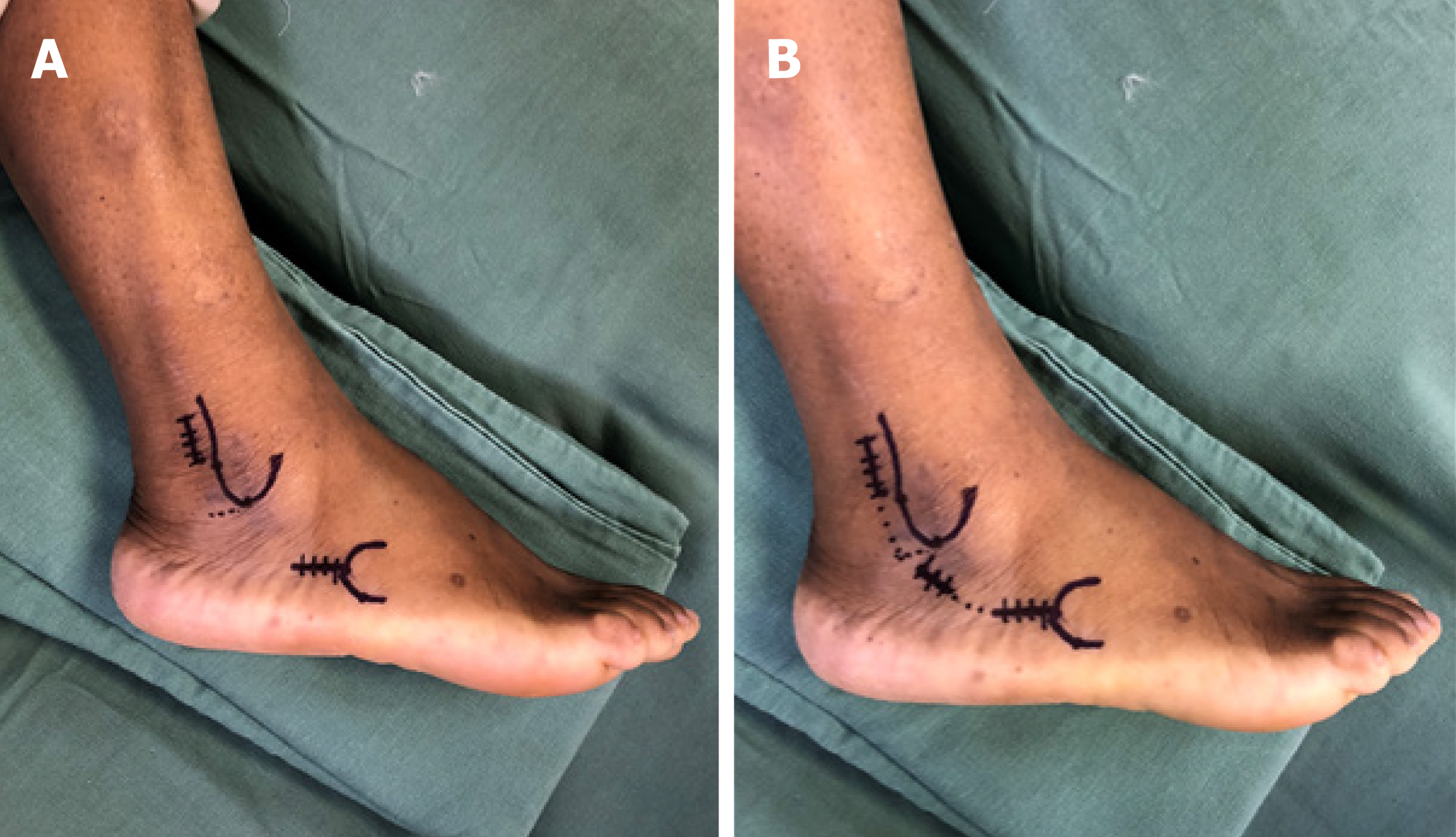
Figure 3 Preoperative skin marking of the proximal and distal incisions.
A: The proximal incision was 3 cm in length, starting 1.5 cm above the tip of the lateral malleolus and 1 cm posterior to the distal fibula. Distally, a longitudinal incision of 3 cm in length was made parallel to the ground, going backwards from the tip of the base of the fifth metatarsal base. In the distal incision, the distal stump of the remaining peroneal longus tendon was dissected and released at the cuboid groove; B: Due the presence of a hypertrophic peroneal tubercle, a short middle incision of 2 cm was performed for its resection.
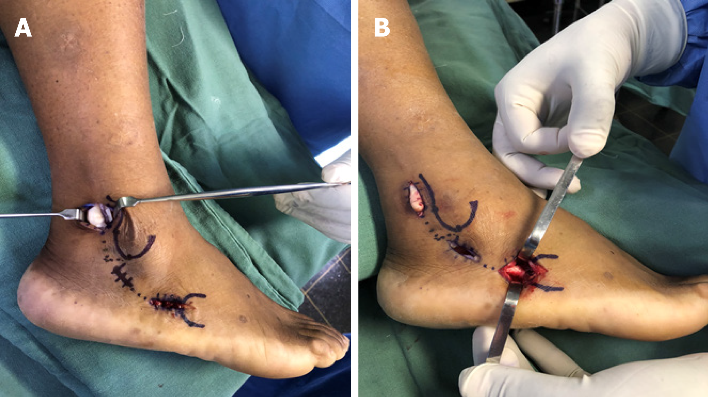
Figure 4 Intraoperative images.
A: The peroneal tendon sheath was opened, preserving the superior peroneal retinaculum, and both tendons were exposed; B: The peroneal tubercle was resected through the middle incision, and the peroneus longus tendon was identified and released in the distal incision.
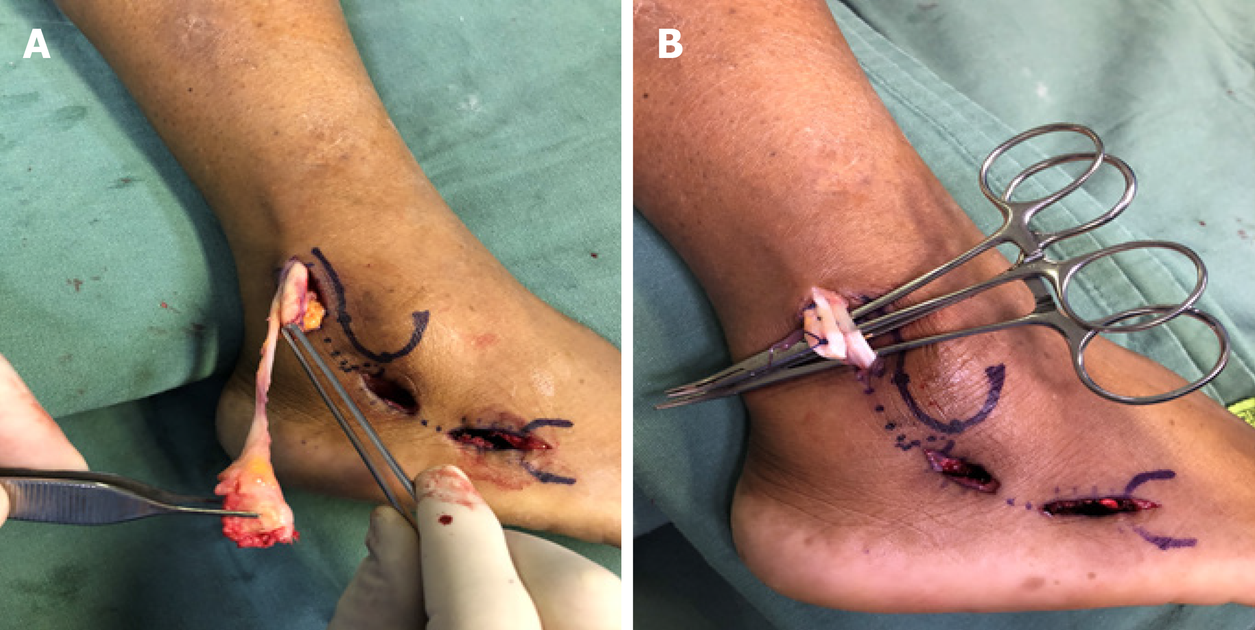
Figure 5 Tenodesis of the peroneus longus tendon to the peroneus brevis tendon.
A: The peroneus longus tendon was brought out of its sheath through the proximal incision, and its nonviable portion was identified and resected; B: Side-to-side tenodesis of the peroneus longus tendon to the peroneus brevis tendon was carried out with two U-shaped lateral sutures using No. 1 Vicryl.
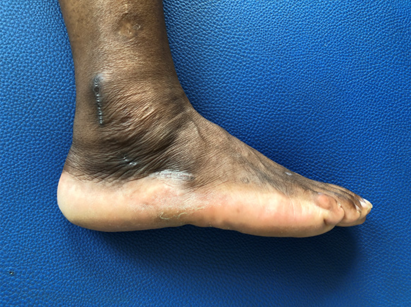
Figure 6 Clinical aspect at 12 wk after surgery.
The surgical incisions were fully healed and swelling had decreased.
- Citation: Nishikawa DRC, Duarte FA, Saito GH, de Cesar Netto C, Fonseca FCP, Miranda BR, Monteiro AC, Prado MP. Minimally invasive tenodesis for peroneus longus tendon rupture: A case report and review of literature. World J Orthop 2020; 11(2): 137-144
- URL: https://www.wjgnet.com/2218-5836/full/v11/i2/137.htm
- DOI: https://dx.doi.org/10.5312/wjo.v11.i2.137


