Copyright
©The Author(s) 2019.
World J Orthop. Jun 18, 2019; 10(6): 247-254
Published online Jun 18, 2019. doi: 10.5312/wjo.v10.i6.247
Published online Jun 18, 2019. doi: 10.5312/wjo.v10.i6.247
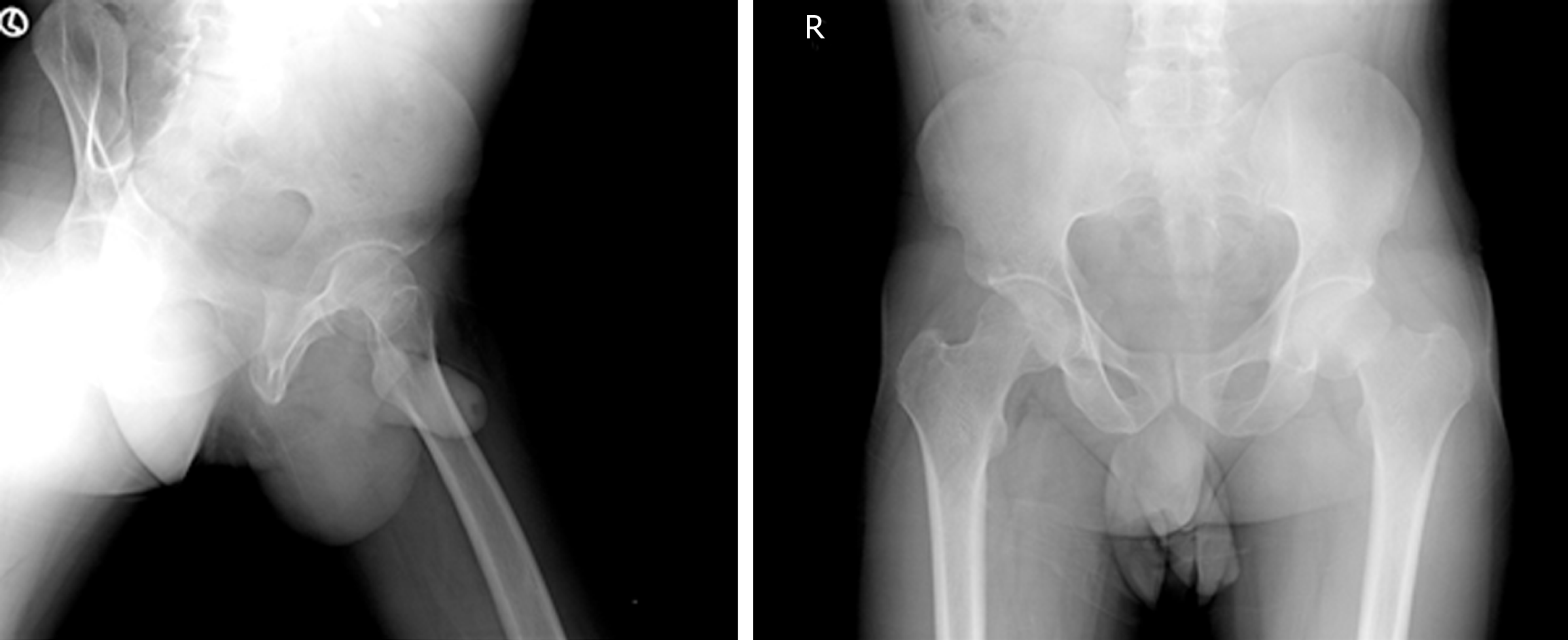
Figure 1 Preoperative anteroposterior and lateral X-rays demonstrating transcervical stress fracture of left femoral neck, with Singh’s grade III osteopenia noted on the right proximal femur.
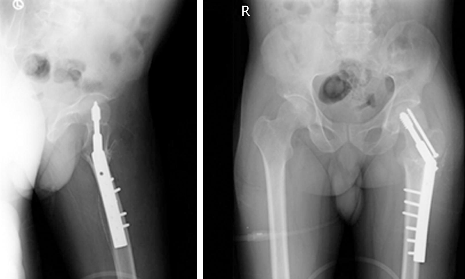
Figure 2 Postoperative AP and lateral radiographs of left proximal femur valgus Osteotomy demonstrating sliding hip screw fixation.
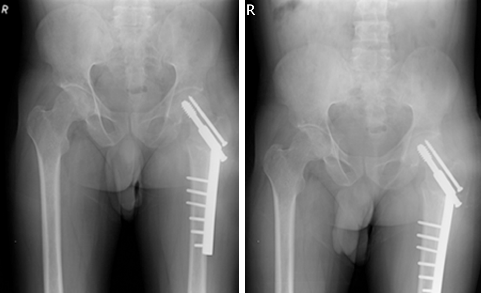
Figure 3 Three months postop follow up with sound union
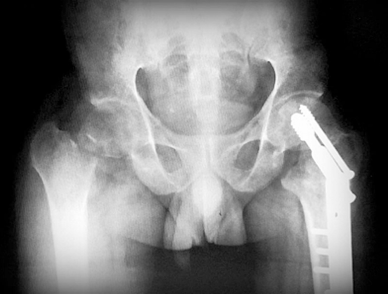
Figure 4 Six months post operative follow up with stress fracture non union of the Right femur neck
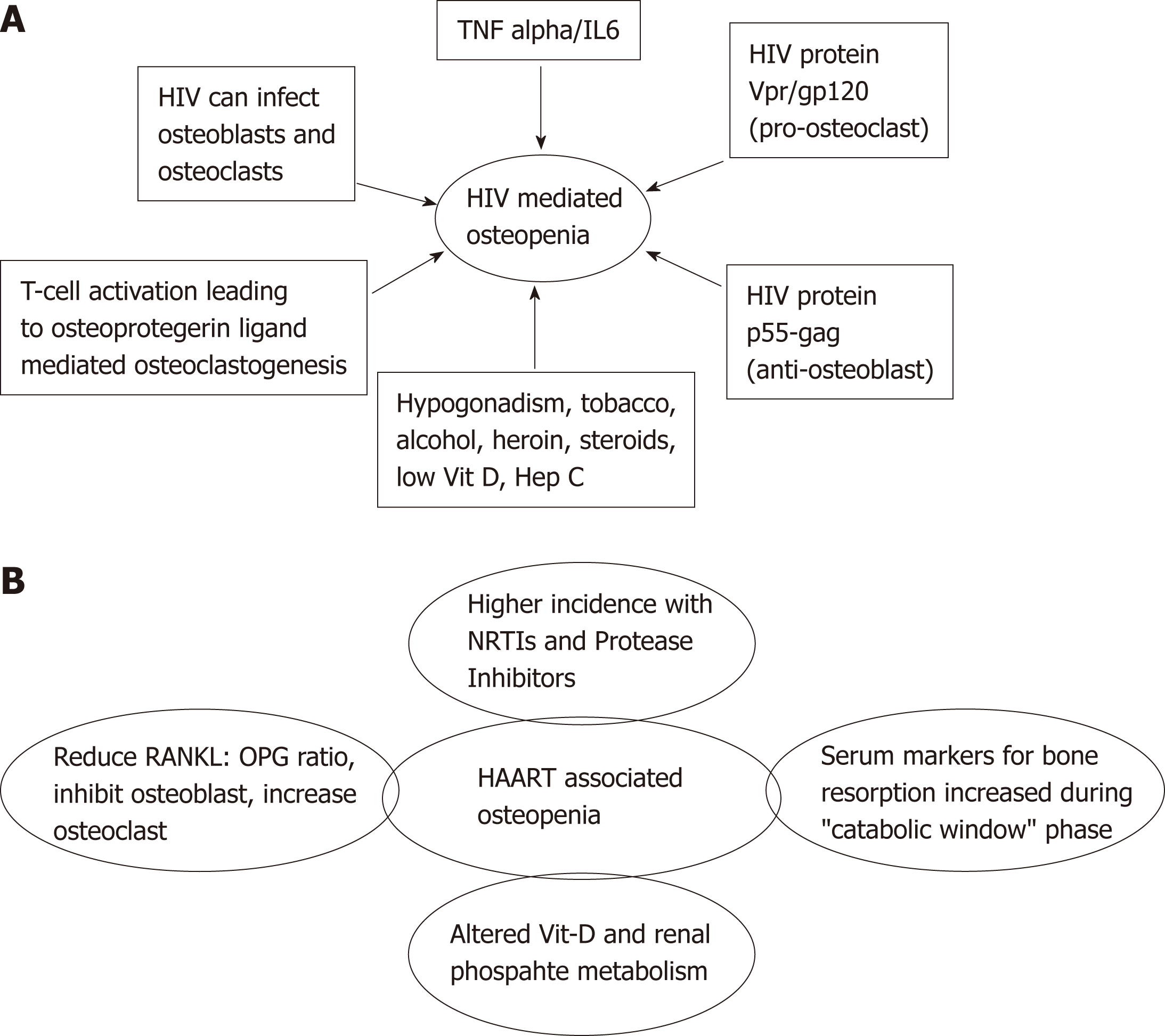
Figure 5 Suspected mechanisms.
A: Human immunodeficiency virus and mediated osteopenia; B: Highly active antiretroviral therapy associated osteopenia. HIV: Human immunodeficiency virus; TNF alpha: Tumor necrosis factor alpha; HAART: Highly active antiretroviral therapy; Vit D: Vitamin D; Hep C: Hepatitis C; NRTIs: Nucleoside reverse transcriptase inhibitors ;OPG: Osteoprotegerin.
- Citation: Chaganty SS, James D. Bilateral sequential femoral neck stress fractures in young adult with HIV infection on antiretroviral therapy: A case report. World J Orthop 2019; 10(6): 247-254
- URL: https://www.wjgnet.com/2218-5836/full/v10/i6/247.htm
- DOI: https://dx.doi.org/10.5312/wjo.v10.i6.247


