©The Author(s) 2019.
World J Orthop. Jan 18, 2019; 10(1): 45-53
Published online Jan 18, 2019. doi: 10.5312/wjo.v10.i1.45
Published online Jan 18, 2019. doi: 10.5312/wjo.v10.i1.45
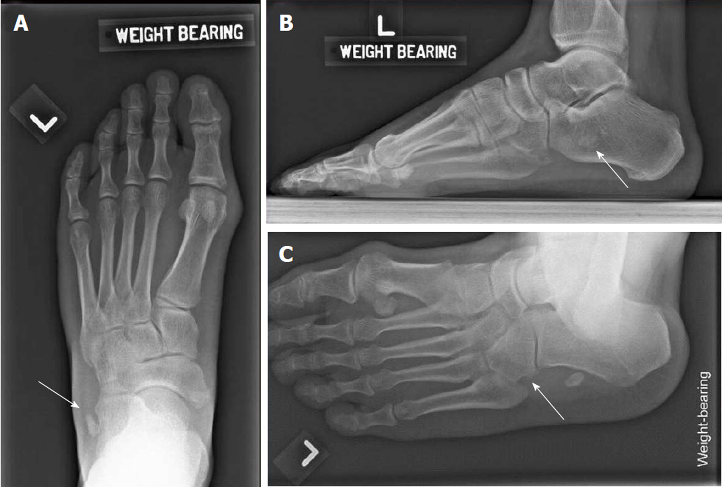
Figure 1 Initial weight bearing radiographs of the injured foot.
A: Weight-bearing dorsoplantar view of left foot showing well-defined bony fragment (white arrow) lateral to the anterior left calcaneum; B: Left lateral weight-bearing views shows a bony fragment at the level of the calcaneum; C: Left oblique view. White arrow shows the absence of the os peronuem at its usual anatomical. Instead, it was displaced proximally to the level of the anterior calcaneum process.
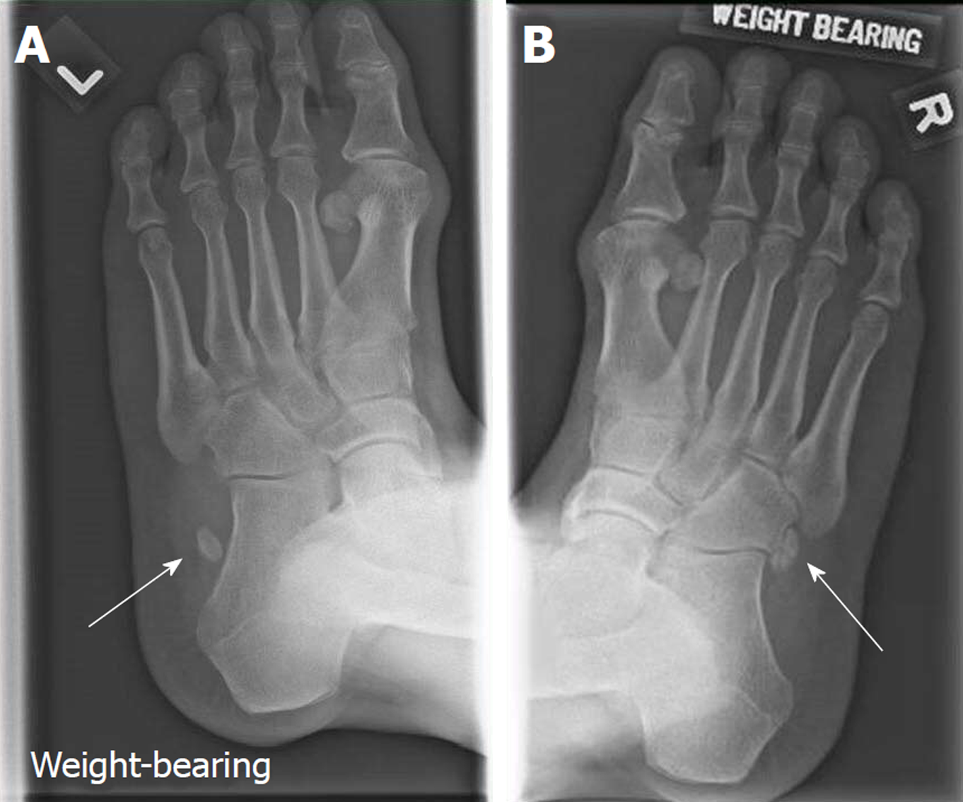
Figure 2 Comparing radiographs of bilateral oblique foot views.
A: Weight-bearing oblique view of left foot show a bony fragment lateral to the anterior left calcaneum; B: Shows the position of the undisplaced os peroneum on the right foot. The displaced os peroneum over the left foot is better appreciated when compared against the contralateral foot.
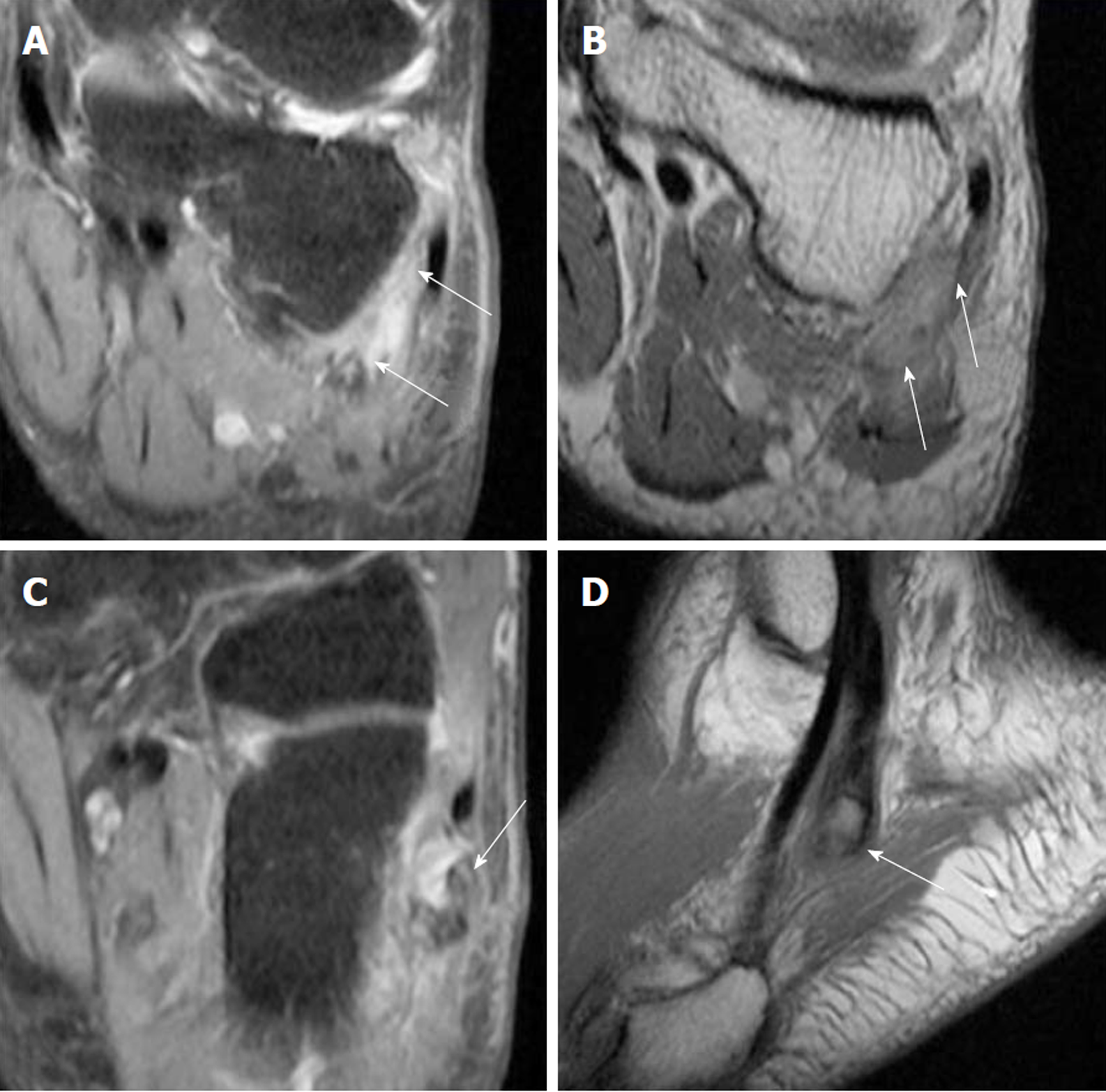
Figure 3 Magnetic resonance imaging of left foot.
A: (Coronal) Peroneal tenosynovitis; B: (Coronal) Both peroneal tendons run within a flat peroneal groove with intact superior peroneal retinaculum. No fixed lateral subluxation of the peroneal tendons; C, D: (Sagittal view). Five millimeter os peroneum within the peroneus longus tendon (PLT), with full-thickness rupture of the PLT. The tear measures 2 cm in length, with proximal migration of the tendon at the level of the cuboid bone.
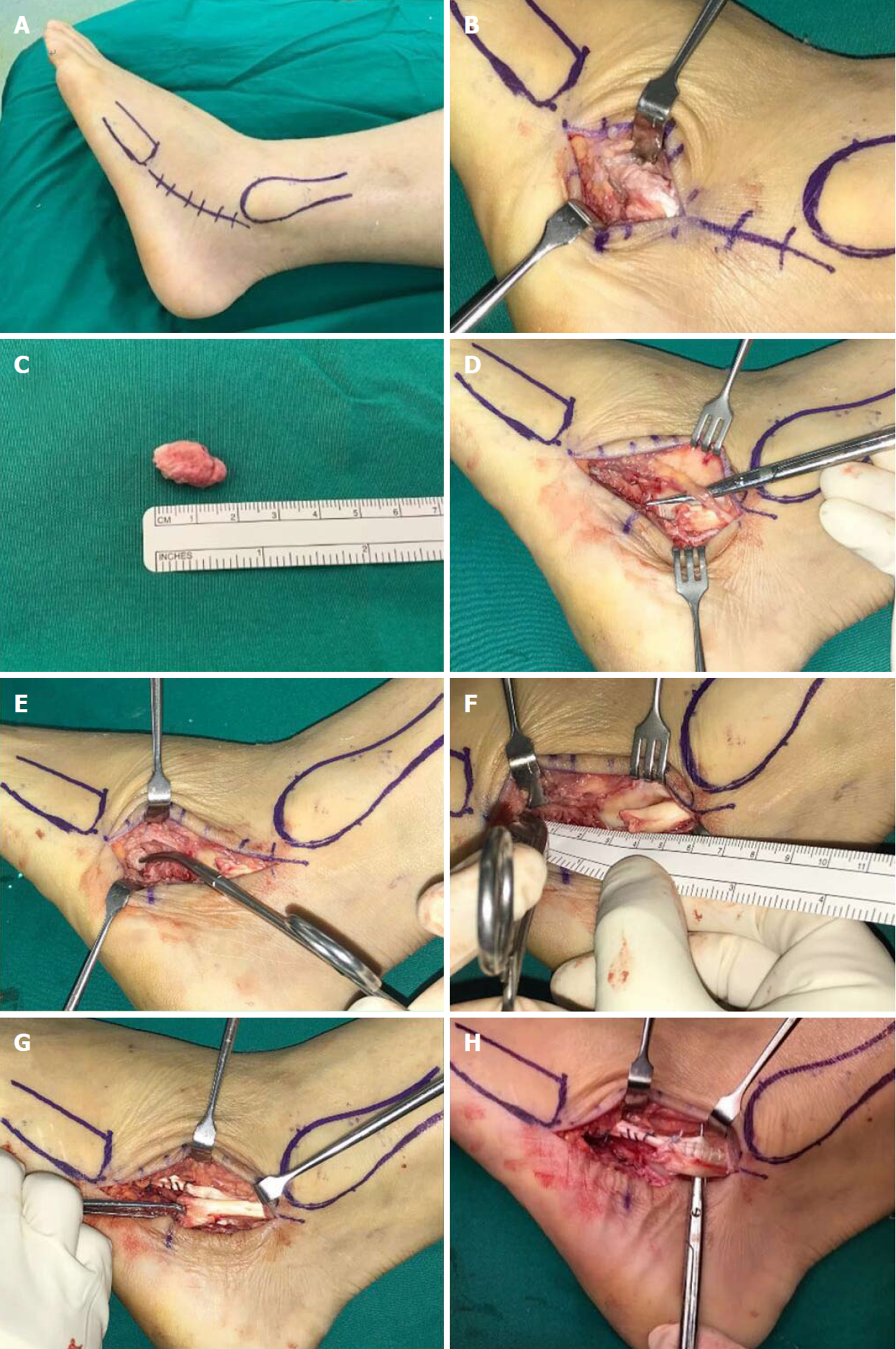
Figure 4 Intra-operative images.
A: Lateral position adopted with a thigh tourniquet applied; B: Incision was made from the left fibula tip to the base of the fifth metatarsal - centred around the os peroneum; C: The os peroneum was identified and excised and unhealthy tendon and devitalised synovium were debrided; D: The sural nerve was identified and protected during the surgery; E and F: Proximal and distal ends of the peroneus longus tendon (PLT) was mobilised and defect gap measured; G: The longitudinal split tear in the peroneus brevis was repaired; H: Side-to-side tenodesis of the PLT to the peroneus brevis tendon was performed.
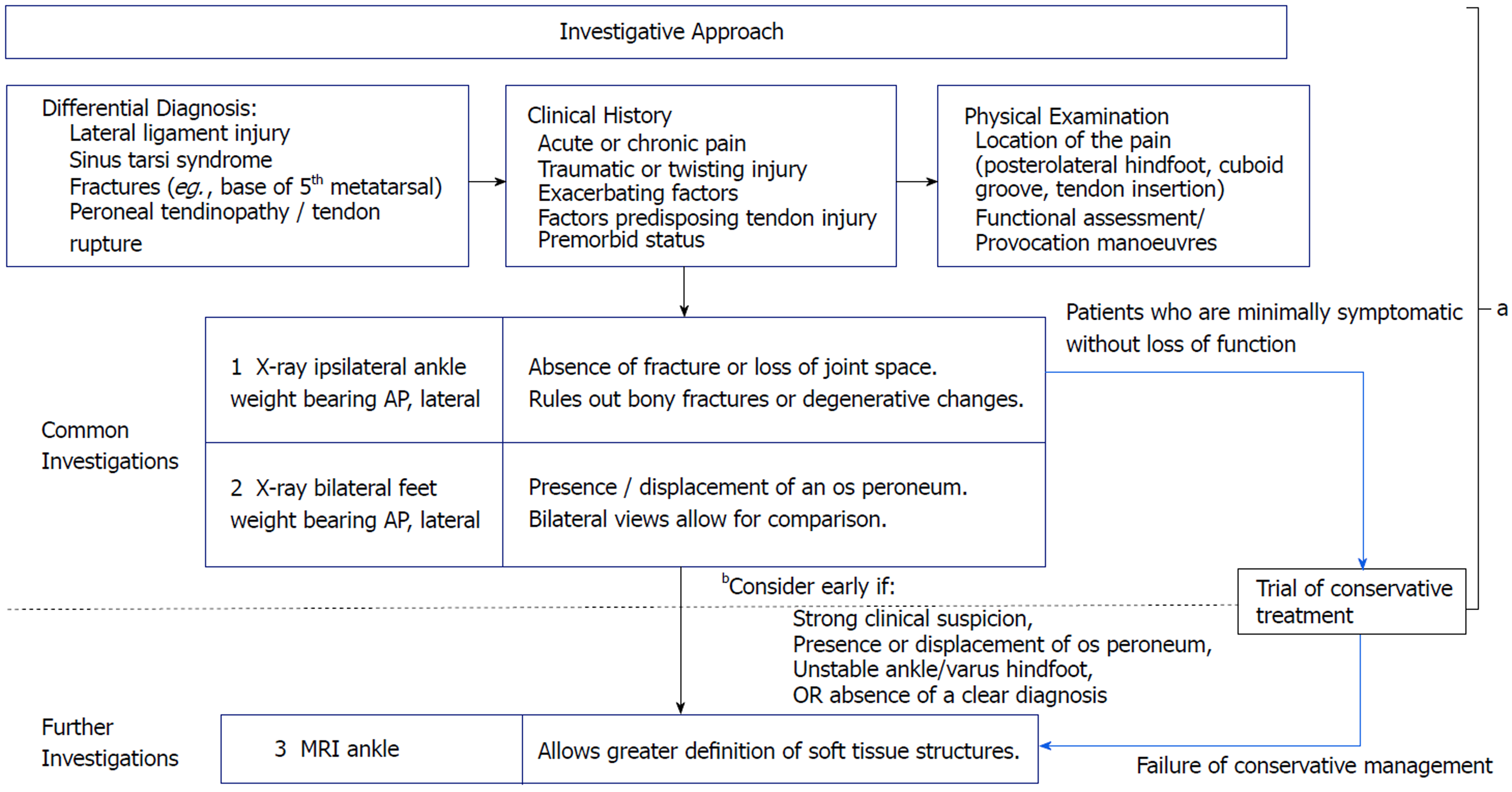
Figure 5 Investigative pathway when approaching patients with lateral ankle discomfort.
aAt this juncture, most differential diagnosis can be ruled out with astute clinical and radiological findings; bEarly magnetic resonance imaging ankle is warranted should there remain a strong clinical suspicion, persistently symptomatic patient and even the absence of a definitive diagnosis.
- Citation: Koh D, Liow L, Cheah J, Koo K. Peroneus longus tendon rupture: A case report. World J Orthop 2019; 10(1): 45-53
- URL: https://www.wjgnet.com/2218-5836/full/v10/i1/45.htm
- DOI: https://dx.doi.org/10.5312/wjo.v10.i1.45


