Copyright
©The Author(s) 2015.
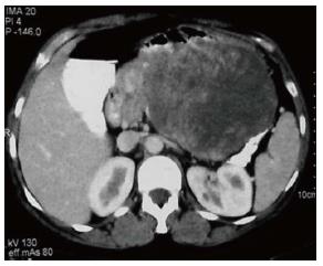
Figure 1 Axial computed tomography scan shows a well defined heterogeneous mass lesion arising from body and tail of pancreas with marked central necrosis which was proven as solid pseudo-papillary epithelial neoplasm after surgery.
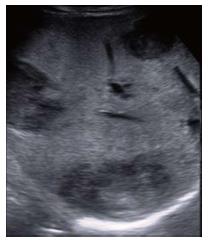
Figure 2 Ultrasonography image showing multiple well defined hypoechoic lesions in the background of fatty liver, suggestive of metastases.
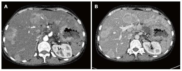
Figure 3 Arterial (A) and venous (B) phases of multiphase contrast enhanced axial computed tomography images showing multiple arterial enhancing focal lesions in liver with no significant wash out in venous phase.
Figure 4 DSA: (A) Common hepatic artery and (B) right hepatic artery angiography showing multiple tumor blush in the liver, (C) post embolisation spot image showing lipiodol retention within the lesions.
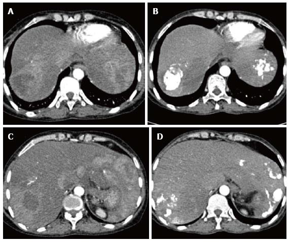
Figure 5 Pre and Post transarterial chemoembolization.
A and C showing arterial enhancing lesions in pre transarterial chemoembolisation (TACE) images; B and D are corresponding post TACE images showing lipiodol retention with significant reduction in enhancing component of the lesions.
- Citation: Prasad T, Madhusudhan K, Srivastava DN, Dash NR, Gupta AK. Transarterial chemoembolization for liver metastases from solid pseudopapillary epithelial neoplasm of pancreas: A case report. World J Radiol 2015; 7(3): 61-65
- URL: https://www.wjgnet.com/1949-8470/full/v7/i3/61.htm
- DOI: https://dx.doi.org/10.4329/wjr.v7.i3.61


