Published online Jun 26, 2017. doi: 10.4330/wjc.v9.i6.558
Peer-review started: October 29, 2016
First decision: January 14, 2017
Revised: February 10, 2017
Accepted: April 6, 2017
Article in press: April 10, 2017
Published online: June 26, 2017
Processing time: 240 Days and 20.1 Hours
We are reporting a case of a 80-year-old lady with effort angina who underwent coronary angiography through the right radial artery, using a dedicated radial multipurpose 5 French Optitorque Tiger catheter. The catheter was advanced into the left ventricle and a left ventriculogram was obtained, while the catheter appeared optimally placed at the centre of the ventricle and the pressure waveform was normal. A large posterior interventricular vein draining into the right atrium was opacified, presumably because the catheter’s end hole inadvertently cannulated an endocardial opening of a small thebesian vein, with subsequent retrograde filling of the epicardial vein. Our case suggests that caution is needed when a dedicated radial catheter with both an end-hole and a side hole is used for a ventriculogram, as a normal left ventricular pressure waveform does not exclude malposition of the end-hole against the ventricular wall.
Core tip: Use of a dedicated radial catheter with both an end-hole and a side hole to perform a left ventriculogram, can result in inadvertent cannulation of a small Thebesian vein and subsequent opacification of a large epicardial vein. When such catheters are used for ventriculogram, a normal ventricular pressure waveform does not exclude malposition of the end-hole against the ventricular wall and extra caution is needed in order to prevent iatrogenic myocardial injury. We review current literature on myocardial injury induced by end-hole catheters used for left ventriculograms.
- Citation: Aznaouridis K, Masoura C, Kastellanos S, Alahmar A. Inadvertent cardiac phlebography. World J Cardiol 2017; 9(6): 558-561
- URL: https://www.wjgnet.com/1949-8462/full/v9/i6/558.htm
- DOI: https://dx.doi.org/10.4330/wjc.v9.i6.558
In the last years, the use of radial artery as an arterial access site for coronary procedures has gained increasing popularity, as it is considered safer compared to transfemoral procedures. Recent data from large randomized trials suggest that the radial access is associated with a reduction of major adverse events in patients with acute coronary syndrome undergoing invasive management[1]. Furthermore, there is expanding use of dedicated “multipurpose” radial catheters, which enable the operator to cannulate both coronary arteries, and also to perform chamber injection and left ventriculogram. For transradial procedures, this single-catheter approach has been shown to decrease radiation exposure, fluoroscopy time, contrast volume and total procedure time compared with standard Judkins catheters[2]. Using a single catheter also reduces the risk of spasm of the radial artery. On the other hand, those dedicated radial catheters may rarely cause myocardial injury when used for a left ventriculogram. We present a rare angiographic finding in a patient who underwent cardiac catheterization through the radial artery using a dedicated radial catheter.
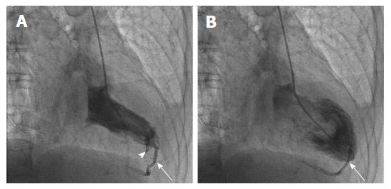
A 80-year-old lady with effort angina underwent coronary angiography through the right radial artery, using a dedicated radial “multipurpose” 5 French Optitorque Tiger catheter (Terumo, Somerset, New Jersey). Coronary angiogram of the left and right coronary arteries demonstrated a significant stenosis in the ostium of a modest sized intermediate artery. The catheter was then advanced into the left ventricle (LV) and appeared optimally placed at the centre of the LV, while the LV pressure waveform was normal. A left ventriculogram was obtained after delivering 10 mL of contrast with a vigorous hand injection. Few ectopics were noticed at the beginning of the injection, followed by a small deflection of the catheter’s tip, a minor subendocardial staining (arrowhead in panel A) and visualization of a large posterior interventricular vein, which seemed to drain directly into the right atrium (arrows in Figure 1 and supplementary Video 1).
We observed no persistent staining of the myocardium and the patient did not experience any discomfort, arrhythmia or electrocardiographic changes. The patient was monitored and was discharged few hours later.
Dedicated radial “multipurpose” catheters such as the Tiger catheter have been specifically designed to minimise catheter exchange with ability to access both coronary ostia from the radial approach, and also provide ability to perform chamber injection with the presence of both an end hole and a single side hole. In our case, we assume that the catheter’s end hole cannulated an endocardial opening of a small Thebesian vein, with subsequent retrograde filling of the epicardial vein. Venae cordis minimae (Thebesian veins) are small valveless venous conduits that connect the coronary arteries, veins or capillaries with the cardiac chambers. Most Thebesian veins of the ventricles are connected to the cardiac venous system[3], as was the case in our patient.
We screened the available literature and we identified a total of 7 reports with 8 cases of myocardial injury following contrast injection with end-hole catheters for left ventriculogram[4-10]. The characteristics of the patients and procedures and the type of myocardial injury and outcome are shown in Table 1. In 5 of those cases, a dedicated transradial end-hole catheter with one side hole near the tip (radial Tiger catheter) or two side holes (radial Jacky catheter) was used[4,6,7,9,10]. Traditional multipurpose (MPA) catheters with an end-hole and 2 side holes near the tip were used in 2 cases[4,5], whereas a catheter with a single end-hole (Judkins right 4) was used from femoral access in 1 case[8]. The Thebesian venous network and/or cardiac veins were visualized in 4 cases[4,5,8,9]. A high-pressure power injection had been performed in most cases describing myocardial laceration/dissection and persistent myocardial staining[6,7,9,10]. Complete myocardial “perforation” with presence of contrast in the pericardial space was confirmed in 2 of those cases[6,10], and emergency pericardiocentesis due to tamponade was performed in one patient[6]. No fatalities were reported (Table 1).
| Case | Ref. | Demographics | Catheter, access | Injection characteristics | Complication | Clinical findings/outcome |
| 1 | Judkins et al[4] | 72 yr, woman, aortic stenosis | Multipurpose-1 (right radial access) | Not provided | Opacification of Thebesian veins, coronary veins and coronary sinus | Not provided |
| 2 | Judkins et al[4] | 77 yr, woman, chest pain | Optitorque Tiger (right radial access) | Not provided | Opacification of Thebesian veins and coronary veins | Not provided |
| 3 | Singhal et al[5] | 46 yr, man, hypertrophic cardiomyopathy | Multipurpose-2 (femoral access) | Power injection, 25 mL of contrast, 10 mL/s | Opacification of Thebesian veins, coronary veins and coronary sinus | Ventricular tachycardia requiring cardioversion/uneventful recovery and next day discharge |
| 4 | Frizzell et al[6] | 76 yr, woman, myocardial infarction | Optitorque Tiger (radial access) | Power injection, 30 mL of contrast over 10 s | Laceration/dissection of anterolateral myocardium and pericardial staining | Chest discomfort, pericardial effusion and cardiac tamponade/pericardiocentesis, uneventful recovery |
| 5 | Rossington et al[7] | 71 yr, woman, angina | Optitorque Tiger (right radial access) | Power injection, 25 mL of contrast, 8 mL/s, 600 psi | Laceration/dissection of anterolateral myocardium | Chest discomfort, transient bundle branch block/uneventful course and next day discharge |
| 6 | Aqel et al[8] | 50 yr, woman, chest pain | Judkins right 4 (femoral access) | Hand injection | Opacification of Thebesian veins, coronary veins and coronary sinus | Not provided |
| 7 | Kang et al[9] | 66 yr, woman, angina | Optitorque Jacky radial (radial access) | Power injection, 30 mL of contrast over 2 s, 600 psi | Laceration/dissection of anterior myocardium and opacification of anterior interventricular vein and coronary sinus | Not provided |
| 8 | Basit et al[10] | 69 yr, man, inferior wall ischemia | Optitorque Jacky radial (radial access) | Not provided | Laceration/dissection of myocardium with pericardial opacification | Chest pain, trivial pericardial effusion/uneventful recovery |
Even a pigtail catheter can rarely cause severe myocardial injury during ventriculography when its tip is inappropriately positioned[11]. However, apposition of the pigtail catheter’s end-hole against the endocardium or cannulation of Thebesian veins is extremely unlikely, and therefore this catheter should be the preferred option for ventriculograms.
Caution is needed when a dedicated radial catheter with both an end-hole and side holes is used for a ventriculogram, as a normal left ventricular pressure waveform (likely from the side hole) does not exclude unsafe positioning of the end-hole against the ventricular wall[6,7]. This malposition of the catheter may result in cannulation and injection in a Thebesian vein[4,5,8], or injection against the endocardial layer. In our case, only a small volume of contrast was delivered in an endocardial opening of the Thebesian network with a hand injection, and this may partly explain the relatively “benign” outcome of opacifying an epicardial vein without causing any major myocardial injury. In this scenario, it seems that the injected contrast drains through the Thebesian network into the cardiac veins and therefore no significant intramyocardial shearing forces are generated. However, serious complications such as laceration/dissection of the myocardium or even catastrophic “perforation” of the ventricular wall[6,7,9,10] may occur when the contrast is injected against the endocardium, or when of a large contrast volume is injected in a Thebesian vein with high-pressure (with an automated power injector), as in this case the small Thebesian network would likely be unable to accommodate the forcefully injected large volume of contrast. Hence, we believe that our case indirectly supports the common practice of avoiding the use of radial multipurpose catheters with automated high-pressure power injectors and large volume of contrast for ventriculograms. Therefore, additional care should be taken to confirm that the catheter is optimally positioned at the centre of the left ventricular cavity and that the catheter’s tip is free before contrast injection, and the operator should not rely only on a normal waveform of ventricular pressure. Finally, the injection of contrast must stop immediately when subendocardial or myocardial staining or opacification of an epicardial vein occurs during a ventriculogram with a dedicated radial multipurpose catheter, as this invariably indicates myocardial injury due to malposition of the catheter’s end hole against the endocardium.
This case shows that using a dedicated radial catheter with both an end-hole and a side hole for a left ventriculogram can result in inadvertent cannulation of a small Thebesian vein and subsequent opacification of an epicardial cardiac vein. This was not related to any symptoms or adverse outcomes.
Minor catheter-induced endocardial staining and visualization of posterior interventricular vein.
Catheter-induced laceration/dissection of myocardial wall.
Left ventriculogram with a dedicated radial Tiger catheter.
Current literature on myocardial injury induced by end-hole catheters used for left ventriculogram is reviewed.
When dedicated radial end-hole catheters are used for ventriculogram, a normal ventricular pressure waveform does not exclude malposition of the end-hole against the ventricular wall and extra caution is needed in order to prevent iatrogenic myocardial injury.
This is a case report about an unexpected cardiac phlebography. The manuscript is well written and describes an important aspect related to the use of multipurpose radial catheters.
Manuscript source: Invited manuscript
Specialty type: Cardiac and cardiovascular systems
Country of origin: United Kingdom
Peer-review report classification
Grade A (Excellent): A
Grade B (Very good): 0
Grade C (Good): C, C
Grade D (Fair): 0
Grade E (Poor): E
P- Reviewer: Amiya E, Barili F, Farand P, Zhang ZH S- Editor: Song XX L- Editor: A E- Editor: Li D
| 1. | Valgimigli M, Gagnor A, Calabró P, Frigoli E, Leonardi S, Zaro T, Rubartelli P, Briguori C, Andò G, Repetto A. Radial versus femoral access in patients with acute coronary syndromes undergoing invasive management: a randomised multicentre trial. Lancet. 2015;385:2465-2476. [RCA] [PubMed] [DOI] [Full Text] [Cited by in Crossref: 856] [Cited by in RCA: 974] [Article Influence: 88.5] [Reference Citation Analysis (0)] |
| 2. | Chen O, Goel S, Acholonu M, Kulbak G, Verma S, Travlos E, Casazza R, Borgen E, Malik B, Friedman M. Comparison of Standard Catheters Versus Radial Artery-Specific Catheter in Patients Who Underwent Coronary Angiography Through Transradial Access. Am J Cardiol. 2016;118:357-361. [RCA] [PubMed] [DOI] [Full Text] [Cited by in Crossref: 14] [Cited by in RCA: 15] [Article Influence: 1.5] [Reference Citation Analysis (0)] |
| 3. | Jain AK, Smith EJ, Rothman MT. The coronary venous system: an alternative route of access to the myocardium. J Invasive Cardiol. 2006;18:563-568. [PubMed] |
| 4. | Judkins C, Yamen E. Inadvertent thebesian vein cannulation during radial access ventriculography. JACC Cardiovasc Interv. 2013;6:e9-e10. [RCA] [PubMed] [DOI] [Full Text] [Cited by in Crossref: 2] [Cited by in RCA: 2] [Article Influence: 0.2] [Reference Citation Analysis (0)] |
| 5. | Singhal S, Khoury S. Images in clinical medicine. Imaging of thebesian venous system. N Engl J Med. 2008;359:e8. [RCA] [PubMed] [DOI] [Full Text] [Cited by in Crossref: 7] [Cited by in RCA: 7] [Article Influence: 0.4] [Reference Citation Analysis (0)] |
| 6. | Frizzell JD, Alkouz M, Sheldon MW. Left ventricular perforation during ventriculogram using an optitorque tiger catheter. JACC Cardiovasc Interv. 2014;7:1456-1457. [RCA] [PubMed] [DOI] [Full Text] [Cited by in Crossref: 2] [Cited by in RCA: 2] [Article Influence: 0.2] [Reference Citation Analysis (0)] |
| 7. | Rossington JA, Aznaouridis K, Davison B, Oliver RM. A Tear in the Heart: Myocardial Laceration Following Left Ventriculogram With a Dedicated Radial Catheter. Am J Med Sci. 2016;352:533. [RCA] [PubMed] [DOI] [Full Text] [Cited by in Crossref: 1] [Cited by in RCA: 1] [Article Influence: 0.1] [Reference Citation Analysis (0)] |
| 8. | Aqel R, Gupta R, Zoghbi GJ. Images in cardiology: Thebesian venous lake. Heart. 2005;91:1318. [PubMed] |
| 9. | Kang G, Kang K, Jurado J, Byrnes L. Left ventricular angiography with a Jacky radial catheter at high pressure leading to myocardial staining and opacification of the great cardiac vein and coronary sinus. EuroIntervention. 2012;8:410. [RCA] [PubMed] [DOI] [Full Text] [Cited by in Crossref: 2] [Cited by in RCA: 2] [Article Influence: 0.2] [Reference Citation Analysis (0)] |
| 10. | Basit A, Nazir R, Hahn H. Myocardial and pericardial staining by transradial Optitorque Jacky shape catheter during left ventriculogram. J Invasive Cardiol. 2012;24:128. [PubMed] |
| 11. | Gach O, Lempereur M, Eeckhout E, Legrand V. Unintentional “ventriculo-phlebo-myo-pericardiography”. JACC Cardiovasc Interv. 2014;7:577-578. [RCA] [PubMed] [DOI] [Full Text] [Cited by in Crossref: 2] [Cited by in RCA: 2] [Article Influence: 0.2] [Reference Citation Analysis (0)] |


