Published online Jun 26, 2017. doi: 10.4330/wjc.v9.i6.539
Peer-review started: August 30, 2016
First decision: October 8, 2016
Revised: March 12, 2017
Accepted: April 18, 2017
Article in press: April 20, 2017
Published online: June 26, 2017
Processing time: 301 Days and 4 Hours
To evaluate the long-term outcome of catheter ablation of atrial fibrillation (AF) facilitated by preprocedural three-dimensional (3-D) transesophageal echocardiography.
In 50 patients, 3D transesophageal echocardiography (3D TEE) was performed immediately prior to an ablation procedure (paroxysmal AF: 30 patients, persistent AF: 20 patients). The images were available throughout the ablation procedure. Two different ablation strategies were used. In most of the patients with paroxysmal AF, the cryoablation technique was used (Arctic Front Balloon, CryoCath Technologies/Medtronic; group A2). In the other patients, a circumferential pulmonary vein ablation was performed using the CARTO system [Biosense Webster; group A1 (paroxysmal AF), group B (persistent AF)]. Success rates and complication rates were analysed at 4-year follow-up.
A 3D TEE could be performed successfully in all patients prior to the ablation procedure and all four pulmonary vein ostia could be evaluated in 84% of patients. The image quality was excellent in the majority of patients and several variations of the pulmonary vein anatomy could be visualized precisely (e.g., common pulmonary vein ostia, accessory pulmonary veins, varying diameter of the left atrial appendage and its distance to the left superior pulmonary vein). All ablation procedures could be performed as planned and almost all pulmonary veins could be isolated successfully. At 48-mo follow-up, 68.0% of all patients were free from an arrhythmia recurrence (group A1: 72.7%, group A2: 73.7%, group B: 60.0%). There were no major complications.
3D TEE provides an excellent overview over the left atrial anatomy prior to AF ablation procedures and these procedures are associated with a favourable long-term outcome.
Core tip: Three-dimensional (3-D) transesophageal echocardiography has been shown to be a useful tool for analysing the individual left atrial morphology prior to an ablation procedure. The aim of this study was to evaluate whether favourable long-term results can be obtained by catheter ablation of atrial fibrillation after prior pulmonary vein imaging using 3-D transesophageal echocardiography. In 50 patients, 3-D transesophageal echocardiography was performed immediately prior to an ablation procedure. The image quality was excellent in the majority of patients and several variations of the pulmonary vein anatomy could be visualized precisely. At 48-mo follow-up, 68.0% of all patients were free from an arrhythmia recurrence.
- Citation: Kettering K, Gramley F, von Bardeleben S. Catheter ablation of atrial fibrillation facilitated by preprocedural three-dimensional transesophageal echocardiography: Long-term outcome. World J Cardiol 2017; 9(6): 539-546
- URL: https://www.wjgnet.com/1949-8462/full/v9/i6/539.htm
- DOI: https://dx.doi.org/10.4330/wjc.v9.i6.539
Catheter ablation is an important therapeutic option in patients with symptomatic atrial fibrillation (AF)[1-21]. However, these procedures can be quite challenging because of the variability of the individual left atrial anatomy.
Magnetic resonance imaging (MRI) or multi-detector spiral computed tomography (MDCT) are frequently used prior to an ablation procedure. These three-dimensional (3D) imaging systems provide insights into the morphology of the left atrium (LA). Obviously, the precise knowledge of the left atrial anatomy facilitates the ablation procedures and enhances the saftey of these interventions. However, these imaging techniques are associated with significant limitations [e.g., radiation exposure (MDCT), impaired image quality in patients suffering from AF with fast AV-nodal conduction (especially MRI) and addtitional costs]. Three-dimensional transesophageal echocardiography (3D TEE) provides excellent insights into the left atrial anatomy of individual patients and is free from most of the difficulties associated with MRI or MDCT[22-26]. It has been shown to be associated with a favourable short-term outcome after catheter ablation of AF[27].
The target of this study was to analyse the long-term outcome of AF ablation procedures facilitated by preprocedural 3D TEE with regard to success rates and complication rates.
A total of 50 patients [35 men, 15 women; mean age 60.8 years (SD ± 9.2 years)] were enrolled in this study. All of them underwent 3D transesophageal echocardiography immediately before the ablation procedure, so that a 3D TEE reconstruction of the left atrium and the pulmonary veins (PVs) could be generated.
Catheter ablation was performed for paroxysmal AF in 30 patients and for persistent AF in 20 patients. All patients were highly symptomatic and at least one failed attempt of an antiarrhythmic drug therapy was a prerequisite for being accepted for catheter ablation. Table 1 summarizes clinical characteristics of the patients enrolled in our study. For all patients, this was the first AF ablation procedure.
| Group A | Group A1 | Group A2 | Group B | P | |
| Patients | 30 | 11 | 19 | 20 | 0.07 |
| Men:Women | 18:12 | 5:6 | 13:6 | 17:3 | |
| Age (yr), mean (SD) | 60.0 (9.7) | 61.6 (8.0) | 59.1 (10.7) | 62.1 (8.4) | 0.57 |
| Cardiac disease | 0.05 | ||||
| None | 13 | 8 | 5 | 2 | |
| CAD | 3 | 1 | 2 | 9 | |
| DCM | 0 | 0 | 0 | 1 | |
| Valvular heart disease1 | 5 | 1 | 4 | 5 | |
| Arterial hypertension | 9 | 1 | 8 | 2 | |
| Other | 0 | 0 | 0 | 1 | |
| Previous cardiac surgery | 1 | 0 | 1 | 0 | 0.43 |
| Left ventricular ejection fraction, mean (SD) | 58.0% (5.8%) | 59.1% (7.4%) | 57.4% (4.8%) | 52.6% (9.9%) | 0.06 |
| Antiarrhythmic drug therapy prior to the ablation procedure | 0.68 | ||||
| Class Ic (e.g., flecainide, propafenone) | 1 | 0 | 1 | 2 | |
| Class III (e.g., amiodarone, sotalol) | 5 | 0 | 5 | 2 | |
| Beta-blocker in combination with a class Ic or class III antiarrhythmic drug | 16/7 | 7/3 | 9/4 | 3/7 | |
| Beta-blocker | 1 | 1 | 0 | 6 | |
| Digitalis | 0 | 0 | 0 | 0 | |
| Other | 0 | 0 | 0 | 0 |
The ablation procedures were performed at our University Hospital Center between October 2007 and May 2011.
Inclusion criteria were: (1) documented episodes of recurrent AF (≥ 30 s); (2) severe symptoms despite antiarrhythmic drug therapy (including beta-blockers) or prior attemps of electrical cardioversion; (3) ability and willingness to give informed consent; and (4) age between 18 and 85 years. Patients were not accepted for catheter ablation if one of the following conditions was present: Severe valvular heart disease or any other concomitant cardiac disease requiring surgery, severely impaired left ventricular function (left ventricular ejection fraction < 20%), left atrial diameter > 65 mm (parasternal long-axis view), left atrial thrombus, hyperthyroidism, severe renal insufficiency (creatinine ≥ 3 mg/dL) or another severe concomitant illness.
In all patients, a 3D TEE was performed immediately before the ablation procedure (X7-2t, 7 MHz/IE 33; Philips Healthcare, Best, the Netherlands). The images were available throughout the ablation procedures. They were displayed in a synchronised way with the geometry created with the 3D mappig system (if available).
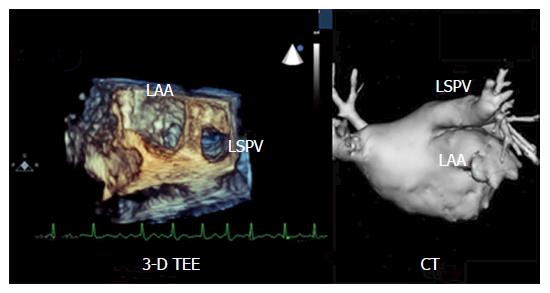
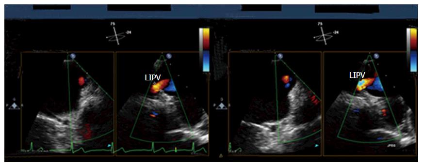
The echocardiographic examination was performed extensively to acquire all relevant information about the left/right atrium, all cardiac valves, the left/right ventricular function and the aorta. In addition, 3D reconstructions of the left atrium and the pulmonary vein ostia were generated. The image quality was classified as: (1) good; (2) acceptable; or (3) not appropriate (for each pulmonay vein ostium). If it was not possible to visualize the right-sided or left-sided PVs at all this was noted as well. The variations of the PV anatomy are summarized in Table 2. A detailed analysis of the 3D TEE findings concerning the left atrial anatomy has been published elsewhere[27].
No other imaging techniques (MDCT or MRI) were used before or after the ablation procedures routinely.
The ablation strategy was depending on the type of AF.
In patients with paroxysmal AF, two strategies were used. In some patients with paroxysmal AF, a cicumferential pulmonary vein ablation was performed in combination with a potential-guided segmental approach in order to achieve complete pulmonary vein isolation [group A1; CARTO system (Biosense Webster, Diamond Bar, CA, United States)]. In most of the patients with paroxysmal AF, the cryoballoon technique (Medtronic, Minneapolis, MN, United States) was used (group A2). We refrained from using the cryoballoon technique if any variations of the pulmonary veins were detected by 3D TEE (e.g., common ostium, accessory pulmonary vein). In patients with persistent AF, a circumferential pulmonary vein ablation was performed in combination with a potential-guided segmental approach to achieve complete pulmonary vein isolation (group B). Furthermore, a linear lesion was created at the roof of the left atrium in some patients. In addition, catheter ablation of the mitral isthmus was performed in selected cases [CARTO system (Biosense Webster)]. The ablation strategies have been described in detail in previous publications[20,27].
In addition, catheter ablation of the right atrial isthmus was performed in patients with inducible or clinically documented episodes of typical atrial flutter. The completeness of the right atrial isthmus lines was confirmed by differential pacing maneuvers in all cases.
After hospital discharge, patients were seen regularly on an outpatient basis. One month after the procedure, a physical examination, a resting electrocardiogram (ECG) and a transthoracic echocardiogram were performed. The patients were questioned whether there was any evidence for an arrhythmia recurrence. In addition, a long-term ECG recording (24-h) was performed.
Three months after the ablation procedure, the patients were re-examined in the same way except for the fact that a 7-d Holter monitoring was performed and that each patient underwent a repeat 3D TEE to rule out a pulmonary vein stenosis. Then, the patients were seen at 3-mo intervals if asymptomatic. If there was an arrhythmia recurrence or other problems occurred, the further follow-up and future strategy (e.g., medical therapy, electrical cardioversion, repeat ablation procedure) were planned on an individual basis.
Twelve months, twenty-four and fourty-eight months after the ablation procedure another 7-d Holter monitoring was performed (or the results of repeated 24-h recordings obtained by the referring physicians were reviewed). A blanking period of 3 mo was employed after ablation when evaluating the follow-up results.
Oral anticoagulation was continued for at least 3 mo after the procedure in all patients and was discontinued only in patients with a CHADS2 score ≤ 1 thereafter. Since October 2010 the CHADS2-VASc score was used for risk assessment and oral anticoagulation was only discontinued in patients with a CHADS2-VASc score ≤ 1 three months after the ablation procedure (vitamin K antagonist/novel oral anticoagulants). During the first three months after catheter ablation the patients received the same antiarrhythmic medication as prior to the ablation procedure. If there was no evidence for an arrhythmia recurrence all antiarrhythmic drugs were discontinued thereafter except for beta-blockers.
Clinical characteristics of the three study groups were compared at baseline to discover potential sources of bias. All parameters with a normal distribution are given as mean (± 1 SD). Age, left ventricular ejection fraction, total procedure time, fluoroscopy dosage and follow-up duration were compared using an one-way ANOVA. All other parameters (underlying cardiac disease, gender) were analysed using the χ2 test. The χ2 test was also used for analysing the clinical endpoints (arrhythmia recurrence rate at 48-mo follow-up). Significance was accepted if the P value was ≤ 0.05. The statistical package of JMP (Version 3.2.6, SAS Institute, Cary, NC, United States) was used for data analysis. The data evaluation was reviewed by an expert in biostatistics of our institution.
The study cohort consistent of fifty patients who were enrolled between October 2007 and May 2011. They had recurrent episodes of persistent or paroxysmal AF. Catheter ablation was performed in these patients after prior 3D TEE data acquisition. In all patients, this was the first AF ablation procedure. Catheter ablation of AF could be carried out as intended in all of them.
In some patients with paroxysmal AF a circumferential pulmonary vein ablation in combination with a potential-guided segmental approach was carried out [group A1: 11 patients; Carto system (Biosense Webster)].
In the remaining 19 patients with paroxysmal AF, the cryoballoon technique was used (group A2; Medtronic). In all of them, a 28-mm cryoballoon was chosen at the beginning of the procedure. In 4 patients, a second cryoballoon (23 mm; n = 1; poorly accessible right inferior pulmonary vein) or a standard cryoablation catheter (Freezor Max, Medtronic; n = 3; rather large left-sided PVs as identified by 3D TEE) had to be used to achieve complete isolation of the PVs.
In all twenty patients with persistent AF, a circumferential pulmonary vein ablation in combination with a potential-guided segmental approach was the standard strategy (group B). Moreover, linear ablation across the left atrial roof was carried out in 7 patients with persistent AF. A mitral isthmus line was created in two patients in group B.
Additionally, catheter ablation of the cavotricuspid isthmus was carried out in 5 patients in group A (A1: 4 patients, A2: 1 patient) and in 2 patients in group B.
The procedural results were published elsewhere[27]. In brief, a circumferential pulmonary vein ablation was carried out in all patients in group A1. This was combined with a potential-guided segmental approach if necessary. This resulted in the complete isolation of all 4 PVs in all patients in group A1.
In group A2, all cryoablation procedures could be completed successfully using this technique (mean number of successfully isolated PVs per patient: 3.9 (SD ± 0.7 PVs).
The circumferential ablation strategy encircling the lateral and the septal PVs could be carried out successfully in all patients in group B [sometimes in combination with with a potential-guided segmental approach (12 out of 20 patients)]. This resulted in complete isolation of a mean number of 3.8 PVs/patient [(SD ± 0.9 PVs); group B]. A complete linear lesion across the left atrial roof could be created in 7 patients in group B (7/7 patients). In addition, a continuous mitral isthmus line was achieved in two patients (10%) in this group B.
Successful ablation of the cavotricuspid isthmus was carried out in a total of 7 patients (group A1: 4 patients, group A2: 1 patient, group B: 2 patients).
In both groups, no major complications (e.g., neurologic disorders, significant pericardial effusion, PV stenosis ≥ 70%, periprocedural death) were observed during the procedure.
In group A, the mean follow-up duration was 1526 d (SD ± 423 d). In group B, the mean follow-up duration was 1697 d (SD ± 208 d; P = 0.1). Thus, the mean overall follow-up duration was 1595 d (SD ± 360 d) (Table 3).
| Group A | Group A1 | Group A2 | Group B | Total | P | |
| Midterm follow-up (12 mo) | 26/30 | 10/11 | 16/19 | 15/20 | 41/50 | 0.82 |
| No. of patients without any arrhythmia recurrence | (86.7%) | (90.6%) | (84.2%) | (75.0%) | (82.0%) | |
| Long-term follow-up (4 yr) | 22/30 | 8/11 | 14/19 | 12/20 | 34/50 | 0.62 |
| No. of patients without any arrhythmia recurrence | (73.3%) | (72.7%) | (73.7%) | (60.0%) | (68.0%) |
At 4-year follow-up, 73.3% of the study population in group A [22/30; A1: 72.7% (8/11)/A2: 73.7% (14/19)] and 60.0% of the study population in group B (12/20) were free from atrial tachyarrhythmias (P = 0.62). Thus, the overall rate of freedom from arrhythmia recurrences was 68.0% (no more atrial tachyarrhythmias in 34 out of 50 patients).
Four years after the procedure, 39/50 patients (78%) were clinically asymptomatic.
No major complications were observed within a follow-up period of 48 mo. Minor complications were observed in 10 patients (group: A1/A2/B: 3/2/5 patients; groin hematoma: 3 patients, pulmonary vein stenosis 30%: 1 patient, noninfectious pericarditis: 3 patients, minor pericardial effusion: 1 patient, hyperthyroidism; 1 patient, residual defect of the atrial septum; 1 patient).
In patients with recurrent atrial tachyarrhythmias, 7-d Holter monitoring demonstrated recurrent episodes of paroxysmal AF in 12 patients [group A: 9 patients (A1: 7/A2: 2); group B: 3 patients] and persistent AF in 4 patients [group A: 2 patients (A1: 1/A2: 1 ); group B: 2 patients]. No modification of the antiarrhythmic drug regimen and no redo procedure was necessary in 5 patients (group A1/A2/B: 1/1/3 patients) with recurrent atrial arrhythmias because they were almost free of symptoms. In 4 patients (group A1/A2/B: 1/0/3) relief of symptoms could be achieved by changing the antiarrhythmic drug regimen or an electrical cardioversion. In seven symptomatic patients a repeat ablation was necessary (group A1/A2/B: 1/4/2 patients).
A 3D transesophageal echocardiography was carried out in all 50 patients. All pulmonary veins could be visualized in 42/50 patients (84%; group A1/A2/B: 8/16/18 patients). In 8 patients (group A1/A2/B: 3/3/2), the right PVs could not be evaluated (RSPV: 0 patients; RIPV: 2 patients; RSPV+RIPV: 6 patients). The left-sided PVs could not be evaluated in 5/50 patients (group A1/A2/B: 3/0/2 patients). In all of these patients, both left-sided PVs could not be evaluated (Figures 1 and 2).
Some variations of the left atrial morphology were revealed (such as a right-sided accessory pulmonary vein or a common ostium of the left-sided or the right-sided PVs) (Table 2).
Based on the detailed knowledge of the individual left atrial anatomy the ablation strategy could be modified appropriately if necessary[27]. Threreby, major complications were avoided.
Catheter ablation is an important therapeutic tool in patients with recurrent symptomatic episodes of paroxysmal or persistent AF. This technique is effective in restoring and maintaining sinus rhythm even if antiarrhythmic drugs have failed or should be avoided. However, these ablation procedures are quite challenging. This is due to the fact that there are a lot of variations concerning the pulmonary vein and left atrial anatomy. 3D TEE provides detailed insights into the pulmonary vein anatomy of individual patients. In contrast to MDCT or MRI it is not associated with problems such as radiation exposure or impaired image quality if AF with rapid atrioventricular nodal conduction is present[27].
The study was performed to evaluate whether catheter ablation of atrial fibrilllation facilitated by preprocedural 3D TEE is associated with a favourable long-term outcome.
The ablation procedures could be performed sucessfully in all patients in both groups after prior pulmonary vein imaging using 3D transesophageal echocardiography.
During a follow-up duration fo 4 years, 73.3% of patients in group A (22/30) remained free from recurrent atrial tachyarrhythmias. In group B, 60.0% of patients (12/20, P = 0.62) remained free from an arrhythmia recurrence during a follow-up duration of 4 years. Thus, the overall rate of patients free from an arrhythmia recurrence was 68% at 4-year follow-up. No major complications were observed in both groups during long-term follow-up.
The data provided by our study shows that radiofrequency catheter ablation as well as cryoablation of AF can be performed effectively and safely after pre-procedural 3D TEE imaging. Pulmonary vein imaging prior to an ablation procedure using 3D TEE is associated with favourable long-term follow-up results concerning safety as well as efficacy of the procedures. Transesophageal echocardiography is recommended prior to catheter ablation of AF anyway (to rule out left atrial thrombus formation). Therefore, a 3D transesophageal echocardiography does not result in additional discomfort for the patient or additional cost. Furthermore, it is less time-consuming than performing an additional MDCT or MRI.
This is the long-term follow-up data of a feasibility study analysing our initial experience with AF ablation procedures facilitated by pre-procedural 3D TEE imaging. The target of our present study was to evaluate the usefulness of 3D TEE for LA visualization prior to an ablation procedure and to show that it is associated with a favourable outcome after catheter ablation of AF. The study was not designed to prove that this technique is equivalent to or superior to other imaging techniques (MDCT/MRI). Therefore, no comparison to MDCT or MRI data is provided.
Furthermore, this study was not designed to prove that pulmonary vein imaging (3D TEE, MDCT or MRI) does significantly improve the long-term outcome in comparison to patients not undergoing preprocedural PV imaging in a propective randomized way.
Moreover, there are some technical limitations of 3D transesophageal echocardiography: First, the right pulmonary veins are sometimes difficult to visualize. Second, the 3D TEE images can only be displayed in a synchronized way during the ablation procedure and no direct image fusion with the geometry created with a 3D mapping system [Navx/Ensite (St. Jude Medical, Saint Paul, MN, United States) or CARTO (Biosense Webster)] is available so far.
In conclusion, Catheter ablation of AF can be performed with favourable results with regard to the success rate as well as to the complication rate based on prior 3D TEE imaging. Three-dimensional TEE-models provide a good overview over the left atrial anatomy, thereby facilitating the procedure. Typical problems (such as atypical PV anatomy, variable relationship between the left atrial appendage and the left superior pulmonary vein) can be revealed. Then, the ablation strategy can be modified and complications can be avoided.
The results of our study demonstrate that pulmonary vein imaging prior to catheter ablation of AF is associated with a favourable long-term outcome with regard to a relatively high success rate and a very low complication rate. However, large randomized studies are needed to prove that this approch is superior to standard ablation procedures (either using 3D MRI-/MDCT reconstructions or no preprocedural imaging) with regard to various outcome parameters (e.g., success and complication rates, procedure duration, radiation exposure).
Catheter ablation is an important therapeutic tool in patients with symptomatic atrial fibrillation (AF). However, these ablation procedures are quite challenging. This is due to the fact that there are a lot of variations concerning the pulmonary vein and left atrial anatomy. Three-dimensional transesophageal echocardiography (3D TEE) might be useful for analysing the individual left atrial morphology prior to an ablation procedure.
However, it is a matter of discussion whether the use of this technique is associated with a favourable long-term outcome with regard to the safety and efficacy of the procedures.
A 3D TEE was performed successfully before the ablation procedure in all patients. All four pulmonary veins could be visualized in 84% of patients. The image quality was excellent in the majority of patients. Several pitfalls of the pulmonary vein morphology could be revealed (e.g., accessory pulmonary veins, common pulmonary vein ostia, variable relationship between the left atrial appendage and the left superior pulmonary vein). All ablation procedures could be performed as planned. At 48-mo follow-up, 68.0% of all patients remained free from atrial tachyarrhythmias (group A1: 72.7%, group A2: 73.7%, group B: 60.0%). There was no major complications.
The results of the study demonstrate that 3D TEE allows detailed insights into the left atrial anatomy. Catheter ablation of AF can be performed safely based on prior 3D TEE imaging.
Catheter ablation: Therapeutic option for the treatment of cardiac arrhythmias (catheter-based); atrial fibrillation: Atrial arrhythmia with a disorganized activation sequence.
This is an interesting article. The authors have provided us with a semi-invasive method (first line or complementary) for monitoring the procedure during catheter ablation of paroxysmal and persistent AF.
Manuscript source: Invited manuscript
Specialty type: Cardiac and cardiovascular systems
Country of origin: Germany
Peer-review report classification
Grade A (Excellent): 0
Grade B (Very good): B
Grade C (Good): C
Grade D (Fair): 0
Grade E (Poor): 0
P- Reviewer: Letsas K, Said SAM S- Editor: Kong JX L- Editor: A E- Editor: Li D
| 1. | Kettering K, Al-Ghobainy R, Wehrmann M, Vonthein R, Mewis C. Atrial linear lesions: feasibility using cryoablation. Pacing Clin Electrophysiol. 2006;29:283-289. [RCA] [PubMed] [DOI] [Full Text] [Cited by in Crossref: 17] [Cited by in RCA: 18] [Article Influence: 0.9] [Reference Citation Analysis (0)] |
| 2. | Oral H, Knight BP, Ozaydin M, Chugh A, Lai SW, Scharf C, Hassan S, Greenstein R, Han JD, Pelosi F. Segmental ostial ablation to isolate the pulmonary veins during atrial fibrillation: feasibility and mechanistic insights. Circulation. 2002;106:1256-1262. [RCA] [PubMed] [DOI] [Full Text] [Cited by in Crossref: 192] [Cited by in RCA: 179] [Article Influence: 7.5] [Reference Citation Analysis (0)] |
| 3. | Haïssaguerre M, Shah DC, Jaïs P, Hocini M, Yamane T, Deisenhofer I, Garrigue S, Clémenty J. Mapping-guided ablation of pulmonary veins to cure atrial fibrillation. Am J Cardiol. 2000;86:9K-19K. [RCA] [PubMed] [DOI] [Full Text] [Cited by in Crossref: 95] [Cited by in RCA: 92] [Article Influence: 3.5] [Reference Citation Analysis (0)] |
| 4. | Gerstenfeld EP, Guerra P, Sparks PB, Hattori K, Lesh MD. Clinical outcome after radiofrequency catheter ablation of focal atrial fibrillation triggers. J Cardiovasc Electrophysiol. 2001;12:900-908. [RCA] [PubMed] [DOI] [Full Text] [Cited by in Crossref: 129] [Cited by in RCA: 128] [Article Influence: 5.1] [Reference Citation Analysis (0)] |
| 5. | Marrouche NF, Dresing T, Cole C, Bash D, Saad E, Balaban K, Pavia SV, Schweikert R, Saliba W, Abdul-Karim A. Circular mapping and ablation of the pulmonary vein for treatment of atrial fibrillation: impact of different catheter technologies. J Am Coll Cardiol. 2002;40:464-474. [RCA] [PubMed] [DOI] [Full Text] [Cited by in Crossref: 311] [Cited by in RCA: 292] [Article Influence: 12.2] [Reference Citation Analysis (0)] |
| 6. | Swartz J, Pellersels G, Silvers J, Patten L, Cervantez D: A catheter-based curative approach to atrial fibrillation in humans. Circulation. 1994;90:I-335 (abstract). |
| 7. | Haïssaguerre M, Jaïs P, Shah DC, Gencel L, Pradeau V, Garrigues S, Chouairi S, Hocini M, Le Métayer P, Roudaut R. Right and left atrial radiofrequency catheter therapy of paroxysmal atrial fibrillation. J Cardiovasc Electrophysiol. 1996;7:1132-1144. [RCA] [PubMed] [DOI] [Full Text] [Cited by in Crossref: 382] [Cited by in RCA: 357] [Article Influence: 11.9] [Reference Citation Analysis (0)] |
| 8. | Ernst S, Schlüter M, Ouyang F, Khanedani A, Cappato R, Hebe J, Volkmer M, Antz M, Kuck KH. Modification of the substrate for maintenance of idiopathic human atrial fibrillation: efficacy of radiofrequency ablation using nonfluoroscopic catheter guidance. Circulation. 1999;100:2085-2092. [RCA] [PubMed] [DOI] [Full Text] [Cited by in Crossref: 119] [Cited by in RCA: 124] [Article Influence: 4.6] [Reference Citation Analysis (0)] |
| 9. | Jaïs P, Hocini M, Hsu LF, Sanders P, Scavee C, Weerasooriya R, Macle L, Raybaud F, Garrigue S, Shah DC. Technique and results of linear ablation at the mitral isthmus. Circulation. 2004;110:2996-3002. [RCA] [PubMed] [DOI] [Full Text] [Cited by in Crossref: 570] [Cited by in RCA: 583] [Article Influence: 26.5] [Reference Citation Analysis (0)] |
| 10. | Oral H, Chugh A, Lemola K, Cheung P, Hall B, Good E, Han J, Tamirisa K, Bogun F, Pelosi F. Noninducibility of atrial fibrillation as an end point of left atrial circumferential ablation for paroxysmal atrial fibrillation: a randomized study. Circulation. 2004;110:2797-2801. [RCA] [PubMed] [DOI] [Full Text] [Cited by in Crossref: 194] [Cited by in RCA: 186] [Article Influence: 8.5] [Reference Citation Analysis (0)] |
| 11. | Avitall B, Helms RW, Koblish JB, Sieben W, Kotov AV, Gupta GN. The creation of linear contiguous lesions in the atria with an expandable loop catheter. J Am Coll Cardiol. 1999;33:972-984. [RCA] [PubMed] [DOI] [Full Text] [Cited by in Crossref: 30] [Cited by in RCA: 35] [Article Influence: 1.3] [Reference Citation Analysis (0)] |
| 12. | Mitchell MA, McRury ID, Haines DE. Linear atrial ablations in a canine model of chronic atrial fibrillation: morphological and electrophysiological observations. Circulation. 1998;97:1176-1185. [RCA] [PubMed] [DOI] [Full Text] [Cited by in Crossref: 54] [Cited by in RCA: 62] [Article Influence: 2.2] [Reference Citation Analysis (0)] |
| 13. | Schwartzman D, Kuck KH. Anatomy-guided linear atrial lesions for radiofrequency catheter ablation of atrial fibrillation. Pacing Clin Electrophysiol. 1998;21:1959-1978. [RCA] [PubMed] [DOI] [Full Text] [Cited by in Crossref: 43] [Cited by in RCA: 45] [Article Influence: 1.6] [Reference Citation Analysis (0)] |
| 14. | Ouyang F, Bänsch D, Ernst S, Schaumann A, Hachiya H, Chen M, Chun J, Falk P, Khanedani A, Antz M. Complete isolation of left atrium surrounding the pulmonary veins: new insights from the double-Lasso technique in paroxysmal atrial fibrillation. Circulation. 2004;110:2090-2096. [RCA] [PubMed] [DOI] [Full Text] [Cited by in Crossref: 614] [Cited by in RCA: 604] [Article Influence: 27.5] [Reference Citation Analysis (0)] |
| 15. | Ouyang F, Antz M, Ernst S, Hachiya H, Mavrakis H, Deger FT, Schaumann A, Chun J, Falk P, Hennig D. Recovered pulmonary vein conduction as a dominant factor for recurrent atrial tachyarrhythmias after complete circular isolation of the pulmonary veins: lessons from double Lasso technique. Circulation. 2005;111:127-135. [RCA] [PubMed] [DOI] [Full Text] [Cited by in Crossref: 588] [Cited by in RCA: 644] [Article Influence: 29.3] [Reference Citation Analysis (0)] |
| 16. | Ouyang F, Ernst S, Chun J, Bänsch D, Li Y, Schaumann A, Mavrakis H, Liu X, Deger FT, Schmidt B. Electrophysiological findings during ablation of persistent atrial fibrillation with electroanatomic mapping and double Lasso catheter technique. Circulation. 2005;112:3038-3048. [RCA] [PubMed] [DOI] [Full Text] [Cited by in Crossref: 184] [Cited by in RCA: 176] [Article Influence: 8.4] [Reference Citation Analysis (0)] |
| 17. | Kettering K, Greil GF, Fenchel M, Kramer U, Weig HJ, Busch M, Miller S, Sieverding L, Laszlo R, Schreieck J. Catheter ablation of atrial fibrillation using the Navx-/Ensite-system and a CT-/MRI-guided approach. Clin Res Cardiol. 2009;98:285-296. [RCA] [PubMed] [DOI] [Full Text] [Cited by in Crossref: 31] [Cited by in RCA: 30] [Article Influence: 1.8] [Reference Citation Analysis (0)] |
| 18. | Kettering K, Greil GF, Busch M, Miller S, Sieverding L, Schreieck J. Catheter ablation of atrial fibrillation: ongoing atrial fibrillation inside a single pulmonary vein after successful electrical disconnection and restoration of sinus rhythm in both atria. Clin Res Cardiol. 2006;95:663-667. [RCA] [PubMed] [DOI] [Full Text] [Cited by in Crossref: 12] [Cited by in RCA: 13] [Article Influence: 0.7] [Reference Citation Analysis (0)] |
| 19. | Kettering K, Weig HJ, Busch M, Laszlo R, Schreieck J. Segmental pulmonary vein ablation: success rates with and without exclusion of areas adjacent to the esophagus. Pacing Clin Electrophysiol. 2008;31:652-659. [RCA] [PubMed] [DOI] [Full Text] [Cited by in Crossref: 11] [Cited by in RCA: 12] [Article Influence: 0.7] [Reference Citation Analysis (0)] |
| 20. | Kettering K, Weig HJ, Busch M, Schneider KM, Eick C, Weretka S, Laszlo R, Gawaz M, Schreieck J. Catheter ablation of persistent atrial fibrillation: anatomically based circumferential pulmonary vein ablation in combination with a potential-guided segmental approach to achieve complete pulmonary vein isolation. J Interv Card Electrophysiol. 2011;30:63-72. [RCA] [PubMed] [DOI] [Full Text] [Cited by in Crossref: 8] [Cited by in RCA: 9] [Article Influence: 0.6] [Reference Citation Analysis (0)] |
| 21. | Neumann T, Vogt J, Schumacher B, Dorszewski A, Kuniss M, Neuser H, Kurzidim K, Berkowitsch A, Koller M, Heintze J. Circumferential pulmonary vein isolation with the cryoballoon technique results from a prospective 3-center study. J Am Coll Cardiol. 2008;52:273-278. [RCA] [PubMed] [DOI] [Full Text] [Cited by in Crossref: 366] [Cited by in RCA: 359] [Article Influence: 19.9] [Reference Citation Analysis (0)] |
| 22. | Kettering K, Gramley F, von Bardeleben S: Catheter ablation of AF: three-dimensional TEE replaces other imaging techniques for pulmonary vein visualization prior to an ablation procedure - long-term follow-up results. Eur Heart Journal. 2012;32:229. |
| 23. | Kettering K, Gramley F: Catheter ablation of atrial fibrillation: three-dimensional transesophageal echocardiography provides an excellent overview over the pulmonary vein anatomy prior to an ablation procedure - long-term follow-up results. Circulation (abstract supplement). 2012;13347. |
| 25. | Kühl H, Franke A, Buck T. Monitoring and guiding cardiac interventions and surgery. Buck T, Franke A, Monaghan M, editors: Three-dimensional echocardiography, 1st ed. Berlin: Springer 2011; 262-263. [DOI] [Full Text] |
| 26. | Neumann T, Kuniss M. Balloon-based cryoablation of atrial fibrillation. Bredikis A, Wilber D, editors: Cryoablation of cardiac arrhythmias, 1st ed. Philadelphia: Elsevier Saunders 2011; 176-177. [DOI] [Full Text] |
| 27. | Kettering K, Gramley F, Bardeleben S. Catheter ablation of atrial fibrillation: three-dimensional transesophageal echocardiography provides an excellent overview over the pulmonary vein anatomy. Cardiology and Angiology. 2017;In press. |


