Published online Jul 26, 2015. doi: 10.4330/wjc.v7.i7.431
Peer-review started: March 7, 2015
First decision: March 20, 2015
Revised: April 17, 2015
Accepted: April 28, 2015
Article in press: April 30, 2015
Published online: July 26, 2015
Processing time: 150 Days and 10.2 Hours
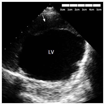
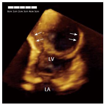
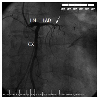
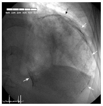
Left ventricular aneurysms are a frequent complication of acute extensive myocardial infarction and are most commonly located at the ventricular apex. A timely diagnosis is vital due to the serious complications that can occur, including heart failure, thromboembolism, or tachyarrhythmias. We report the case of a 78-year-old male with history of previous anterior myocardial infarction and currently under evaluation by chronic heart failure. Transthoracic echocardiogram revealed a huge thrombosed and calcified anteroapical left ventricular aneurysm. Coronary angiography demonstrated that the left anterior descending artery was chronically occluded, and revealed a big and spherical mass with calcified borders in the left hemithorax. Left ventriculogram confirmed that this spherical mass was a giant calcified left ventricular aneurysm, causing very severe left ventricular systolic dysfunction. The patient underwent cardioverter-defibrillator implantation for primary prevention.
Core tip: Early diagnosis of ventricular aneurysms following acute transmural myocardial infarction is vital due to the serious complications that can occur. We report the case of a 78-year-old male with history of previous anterior myocardial infarction and currently under evaluation by chronic decompensated heart failure. Subsequent investigation revealed a huge thrombosed and calcified anteroapical left ventricular aneurysm. The peculiar findings of echocardiography, fluoroscopy and left ventriculography are shown with demonstrative images.
- Citation: de Agustin JA, de Diego JJG, Marcos-Alberca P, Rodrigo JL, Almeria C, Mahia P, Luaces M, Garcia-Fernandez MA, Macaya C, de Isla LP. Giant and thrombosed left ventricular aneurysm. World J Cardiol 2015; 7(7): 431-433
- URL: https://www.wjgnet.com/1949-8462/full/v7/i7/431.htm
- DOI: https://dx.doi.org/10.4330/wjc.v7.i7.431
True left ventricular aneurysms are a frequent complication following acute extensive myocardial infarction. Early diagnosis is crucial due to the serious complications that can potentially occur, including heart failure, thromboembolism, or tachyarrhythmias.
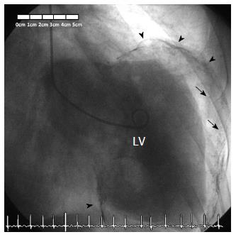
A 78-year-old male with history of previous anterior myocardial infarction and currently under evaluation by chronic decompensated heart failure (NYHA functional class III), underwent transthoracic echocardiogram revealing the presence of a huge and peripherally calcified anteroapical left ventricular aneurysm with a giant mural thrombus (Figures 1, 2, 3). Elective coronary angiography was performed which demonstrated that the left anterior descending artery was chronically occluded (Figure 4) and nonsignificant lesions in the other coronary arteries. Fluoroscopic imaging revealed a complete oval calcified image enclosed within an abnormal cardiac silhouette (Figure 5). Left ventriculogram confirmed that this image corresponded of a giant calcified and thrombosed left ventricular aneurysm, causing severe left ventricular systolic dysfunction (Figure 6). The calculated left ventricular ejection fraction was only 7%. The patient underwent cardioverter-defibrillator implantation for primary prevention.
Left ventricular aneurysms are a frequent complication of acute extensive myocardial infarction and are most commonly located at the ventricular apex[1,2]. A timely diagnosis is vital due to the serious complications that can occur, including heart failure, thromboembolism, or tachyarrhythmias. The benefits of surgical repair of left ventricular aneurysm have long been debated. Although a large amount of studies have showed that aneurysmectomy might improve the outcome[3], the results from the STICH trial have questioned the benefit of this treatment[4]. Therefore, indication for aneurysmectomy depends on the decision of individual surgeons, and should be based on the assessment of the left ventricular dimensions, mitral valve regurgitation severity, extent of myocardial scar tissue and viability of the other regions of the left ventricle, and surgery should be performed in centers with a high surgical experience.
A 78-year-old male with history of previous anterior myocardial infarction and currently under evaluation by chronic decompensated heart failure.
Giant thrombosed left ventricular aneurysm.
Intrathoracic mass.
Echocardiography and coronary angiography were used for the diagnosis of left ventricular aneurysm.
The patient received an implantable cardioverter-defibrillator for primary prevention and was referred for consideration of cardiac transplantation.
True left ventricular aneurysms are widely recognized as a common and serious complication following acute transmural myocardial infarction. However, this case is particular because of the huge size of the aneurysm.
The recognition of ventricular aneurysms is of great importance due to the numerous complications that can potentially occur. Echocardiography and catheterism are fundamental tests for diagnosis.
It is a interesting case and well described.
| 1. | Tikiz H, Atak R, Balbay Y, Genç Y, Kütük E. Left ventricular aneurysm formation after anterior myocardial infarction: clinical and angiographic determinants in 809 patients. Int J Cardiol. 2002;82:7-14; discussion 14-16. [RCA] [PubMed] [DOI] [Full Text] [Cited by in Crossref: 24] [Cited by in RCA: 25] [Article Influence: 1.0] [Reference Citation Analysis (0)] |
| 2. | Abrams DL, Edelist A, Luria MH, Miller AJ. Ventricular aneurysm. a reappraisal based on a study of sixty-five consecutive autopsied cases. Circulation. 1963;27:164-169. [RCA] [PubMed] [DOI] [Full Text] [Cited by in Crossref: 123] [Cited by in RCA: 122] [Article Influence: 4.1] [Reference Citation Analysis (0)] |
| 3. | Castelvecchio S, Menicanti L, Donato MD. Surgical ventricular restoration to reverse left ventricular remodeling. Curr Cardiol Rev. 2010;6:15-23. [RCA] [PubMed] [DOI] [Full Text] [Full Text (PDF)] [Cited by in Crossref: 24] [Cited by in RCA: 25] [Article Influence: 1.7] [Reference Citation Analysis (0)] |
| 4. | Jones RH, Velazquez EJ, Michler RE, Sopko G, Oh JK, O’Connor CM, Hill JA, Menicanti L, Sadowski Z, Desvigne-Nickens P. Coronary bypass surgery with or without surgical ventricular reconstruction. N Engl J Med. 2009;360:1705-1717. [RCA] [PubMed] [DOI] [Full Text] [Full Text (PDF)] [Cited by in Crossref: 572] [Cited by in RCA: 552] [Article Influence: 32.5] [Reference Citation Analysis (0)] |
Open-Access: This article is an open-access article which was selected by an in-house editor and fully peer-reviewed by external reviewers. It is distributed in accordance with the Creative Commons Attribution Non Commercial (CC BY-NC 4.0) license, which permits others to distribute, remix, adapt, build upon this work non-commercially, and license their derivative works on different terms, provided the original work is properly cited and the use is non-commercial. See: http://creativecommons.org/licenses/by-nc/4.0/
P- Reviewer: Bagur R, Cebi N, Lee TS S- Editor: Ji FF L- Editor: A E- Editor: Wang CH


