Published online Oct 26, 2015. doi: 10.4330/wjc.v7.i10.703
Peer-review started: July 12, 2015
First decision: August 14, 2015
Revised: August 24, 2015
Accepted: September 10, 2015
Article in press: September 16, 2015
Published online: October 26, 2015
Processing time: 131 Days and 5.4 Hours
Bacterial endocarditis following atrial septal defect closure using Amplatzer device in a child is extremely rare. We report a 10-year-old girl who developed late bacterial endocarditis, 6 years after placement of an Amplatzer atrial septal occluder device. Successful explantation of the device and repair of the resultant septal defect was carried out using a homograft patch. The rare occurrence of this entity prompted us to highlight the importance of long-term follow up, review the management and explore preventive strategies for similar patients who have multiple co-morbidities and a cardiac device. A high index of suspicion is warranted particularly in pediatric patients.
Core tip: Bacterial endocarditis following atrial septal defect closure using Amplatzer device in a child is extremely rare. We report a 10-year-old girl who developed late bacterial endocarditis, 6 years after placement of an Amplatzer atrial septal occluder device. This case report demonstrates the need for long-term follow up of patients with intracardiac device especially those with multiple co-morbidities or who have vulnerability to infection due to poor general condition or extremes of age such as our’s. A high index of suspicion for device complication is required if sepsis or embolic phenomenon found. Incomplete endothelization of the prosthetic devices may be linked to endocarditis and needs to be explored.
- Citation: Jha NK, Kiraly L, Murala JS, Tamas C, Talo H, Badaoui HE, Tofeig M, Mendonca M, Sajwani S, Thomas MA, Doory SAA, Khan MD. Late endocarditis of Amplatzer atrial septal occluder device in a child. World J Cardiol 2015; 7(10): 703-706
- URL: https://www.wjgnet.com/1949-8462/full/v7/i10/703.htm
- DOI: https://dx.doi.org/10.4330/wjc.v7.i10.703
Bacterial endocarditis of intracardiac devices including Amplatzer atrial septal occluder in pediatric population is extremely rare. In view of the limited experience, this is an opportunity to highlight such cases in order to review management and explore preventive strategies for a successful outcome in the future. We, therefore, presenting herewith a child with multiple co-morbidities who developed late bacterial endocarditis of an Amplatzer device following closure of secundum atrial septal defect and underwent successful management.
A 10-year-old girl was referred to us with a high-grade intermittent fever of 2 wk duration being treated for septic shock and altered sensorium. She also had a pyopericardium which was drained. She required ventilatory and minimal inotropic support. She had generalised anasarca, bilateral mild pleural effusions and generalised muscle spasticity. Additionally, there was cellulitis on the chest wall requiring surgical debridement. Blood and wound cultures were positive for Streptococcus Pyogenes, sputum for Pseudomonas and urine for Escherichia coli. She was managed with appropriate antibiotics. Other medications included diuretics, anti-convulsive therapy and ACE-inhibitors. She was known to have global developmental delay associated with cerebral atrophy and epilepsy with clonic seizures. In the past, at the age of 4 years, she underwent device closure of atrial septal defect in a hospital abroad. However, the details of the procedure were not available.
Routine blood tests revealed elevated markers of infection in addition to evidence of hepato-renal dysfunction. Computerized tomography of the chest revealed minor effusions in the pleural cavities and an Amplatzer device in the atrium.
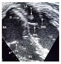
A two dimensional echocardiogram confirmed moderately depressed biventricular function, thickened pericardium and a prosthetic device in the inter-atrial septum with attached mobile vegetation (Figure 1). Other cardiac structures were normal.
It was suspected that the atrial septal occluder device was infected and possibly was a source of multiple systemic embolization and persistent bacteraemia. Therefore, we proceeded for surgical removal of the device.
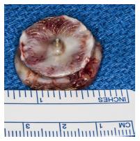
Surgery was performed through median sternotomy under systemic heparinization, standard cardiopulmonary bypass using aortic and bicaval cannulation, aortic cross-clamping and cardioplegic arrest at moderate systemic hypothermia via right atrial approach. The pericardium was thickened and adherent all around featuring constrictive pericarditis. The Amplatzer device’s surface was partially covered with the soft tissue (endothelized) with patchy bare areas. However, there was no active vegetation found (Figure 2). Other cardiac structures including valves were grossly normal. After explantation of the device, a resultant atrial septal defect was repaired using a patch obtained from a pulmonary homograft.
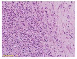
The patient had slow but steady recovery. The post-operative echocardiography showed improved cardiac function without residual defects. There was a constant decline in the levels of inflammatory markers. She recovered fully except that generalized spasticity persisted. The histopathological examination of the tissue attached to the device showed evidence of severe acute and chronic inflammation in the connective tissue (Figure 3). However, a stain for fungal organism and culture of the tissue within the device was negative. Further studies to investigate the tissue infection within the soft tissue such as biofilm study or electron microscopy was not available.
Transcatheter occlusion technique using Amplatzer device has become a preferred approach for atrial septal defects in selected patients. The common complications associated with occluding devices are mal-positioning or migration of device, thromboembolism, arrhythmias or endocarditis[1-5]. Bacterial endocarditis of an Amplatzer septal occluder device in the pediatric population is very rare. However, few reports have described early and late endocarditis associated with such device in adult population[2-4].
Early device infection could be due to inoculation of organisms during implantation. However, hematogenous infection is the primary source of late endocarditis. In our patient, the source of infection could have been cellulitis or respiratory infection. In addition, there was purulent pericarditis. This combination suggests a hematogenous spread of infection leading to prosthesis endocarditis. In the only published report in a 4-year-old child, authors have proposed incomplete endothelization of the device as a mechanism of late endocarditis[3]. Upon closer look of the explanted device, we also have noticed gross evidence of incomplete endothelization in the form of exposed metallic surface of the device in places without soft tissue coverage.
There are no established guidelines for the management of late endocarditis involving intra cardiac devices. We suggest that intensive management involving prolonged antibiotic therapy, monitoring of inflammatory markers and frequent blood cultures may be the first step. However, surgery is warranted if there is evidence of septal perforation, dehiscence, fistula formation, vegetation or embolization[6,7]. The relative indication may include persistent positive blood cultures in spite of maximal medical therapy[6,7]. The homograft patch may be the preferred choice for repair of resultant septal defect after explantation of the device in this situation presumably due to the resistant nature of the homograft tissue against infection and better antibiotic penetration as compared to synthetic materials. Bovine pericardium may be an alternative.
In the clinical and experimental studies, it has been demonstrated that it takes 3-6 mo for complete neo-endothelization of the device[3]. Therefore, appropriate length of bacterial endocarditis prophylaxis for patients with atrial septal device closure was arbitrarily determined and usually extends from 6 mo to 1 year after implantation[2-4]. We hope that in future, additional investigations, imaging techniques or biochemical markers will allow identification of patients with incomplete endothelization who warrant long-term endocarditis prophylaxis.
This case report demonstrates the need for long-term follow up of patients with intracardiac devices especially those with multiple co-morbidities or who have vulnerability to infection due to poor general condition or extreme of age. A high index of suspicion for device complication is required, if sepsis or embolic phenomenon is found. An Intensive medical or surgical management and prolonged follow up is warranted for successful outcome.
A 10-year-old girl presented with late bacterial endocarditis of a cardiac Amplatzer device in addition to multiple co-morbidities and pancarditis.
Late bacterial endocarditis of cardiac prosthesis with cellulitis and pancarditis.
Primary endocarditis of cardiac device, septicemia, pericarditis.
Leucocytosis and mildly elevated hepato-renal function markers.
CT scan of the chest and an echocardiograhy showed evidence of a cardiac prosthesis (Amplatzer atrial septal occluder device) with a large vegetation.
The histopathology of the soft tissue attached to the explanted cardiac device showed the presence of acute and chronic inflammatory infiltrates. In addition, gross examination of the explanted Amplatzer device confirmed the presence of bare metal exposed to the surface and the blood within the device.
The patient was treated with intravenous antibiotics according to the culture and sensitivity reports of the blood, urine and pericardial fluids. Additionally, explantation of the atrial septal occluder device was done on cardiopulmonary bypass on urgent basis in order to remove the source of persistent bacteraemia and to avid thromboembolism.
The biofilm and electron microscopic studies were not available which may have a precise-diagnostic value to prove the presence of specific infection within the device.
Cardiopulmonary bypass is a term used to commonly indicate open heart surgery using a cardiopulmonary bypass machine with an oxygenator.
This case report not only represents a very rare occurrence of the late endocarditis of the cardiac device in association with pancarditis and multiple co-morbidities in a child but also provides authors an opportunity to focus their attention on the mechanism and prevention of this pathology for a better outcome in future. They have substantiated the hypothesis of the role of exposed bare metal surface and deficient soft tissue coverage (incomplete endothelization) as a cause of prosthetic endocarditis. This fact not only guides us to explore the preventive measures while designing cardiac devices in future but also to be aware of a possibility of endocarditis in similar patients with multiple comorbidities and low resistance especially in pediatric population in order to have a cautious long-term follow-up and antibiotic prophylaxis.
The case report is very interesting and well described.
| 1. | Chessa M, Carminati M, Butera G, Bini RM, Drago M, Rosti L, Giamberti A, Pomè G, Bossone E, Frigiola A. Early and late complications associated with transcatheter occlusion of secundum atrial septal defect. J Am Coll Cardiol. 2002;39:1061-1065. [RCA] [PubMed] [DOI] [Full Text] [Cited by in Crossref: 396] [Cited by in RCA: 406] [Article Influence: 16.9] [Reference Citation Analysis (0)] |
| 2. | Aruni B, Sharifian A, Eryazici P, Herrera CJ. Late bacterial endocarditis of an Amplatzer atrial septal device. Indian Heart J. 2013;65:450-451. [RCA] [PubMed] [DOI] [Full Text] [Cited by in Crossref: 11] [Cited by in RCA: 15] [Article Influence: 1.2] [Reference Citation Analysis (0)] |
| 3. | Slesnick TC, Nugent AW, Fraser CD, Cannon BC. Images in cardiovascular medicine. Incomplete endothelialization and late development of acute bacterial endocarditis after implantation of an Amplatzer septal occluder device. Circulation. 2008;117:e326-e327. [RCA] [PubMed] [DOI] [Full Text] [Cited by in Crossref: 37] [Cited by in RCA: 44] [Article Influence: 2.4] [Reference Citation Analysis (0)] |
| 4. | Zahr F, Katz WE, Toyoda Y, Anderson WD. Late bacterial endocarditis of an amplatzer atrial septal defect occluder device. Am J Cardiol. 2010;105:279-280. [RCA] [PubMed] [DOI] [Full Text] [Cited by in Crossref: 25] [Cited by in RCA: 28] [Article Influence: 1.8] [Reference Citation Analysis (0)] |
| 5. | Balasundaram RP, Anandaraja S, Juneja R, Choudhary SK. Infective endocarditis following implantation of amplatzer atrial septal occluder. Indian Heart J. 2005;57:167-169. [PubMed] |
| 6. | Johnston LB, Conly JM. Intracardiac device and prosthetic infections: What do we know? Can J Infect Dis Med Microbiol. 2004;15:205-209. [PubMed] |
| 7. | Karchmer AW, Longworth DL. Infections of intracardiac devices. Cardiol Clin. 2003;21:253-271, vii. [PubMed] |
P- Reviewer: Polewczyk A S- Editor: Qiu S L- Editor: A E- Editor: Lu YJ
Open-Access: This article is an open-access article which was selected by an in-house editor and fully peer-reviewed by external reviewers. It is distributed in accordance with the Creative Commons Attribution Non Commercial (CC BY-NC 4.0) license, which permits others to distribute, remix, adapt, build upon this work non-commercially, and license their derivative works on different terms, provided the original work is properly cited and the use is non-commercial. See: http://creativecommons.org/licenses/by-nc/4.0/


