Peer-review started: August 20, 2014
First decision: November 19, 2014
Revised: December 3, 2014
Accepted: December 18, 2014
Article in press: January 4, 2015
Published online: January 26, 2015
Processing time: 157 Days and 15.5 Hours
Cor triatriatum dextrum is an extremely rare congenital heart abnormality in which the right atrium is separated into two chambers by a persistent fibrous membrane. A transvenous approach to place a dual-chamber pacemaker in such patients is technically challenging. We report the first case of a transvenous permanent pacemaker placement in a patient with cor triatriatum dextrum. An 87-year-old woman was diagnosed with paroxysmal atrial fibrillation. She was accidentally found to have cor triatriatum dextrum during the transesophageal echocardiography (TEE) prior to cardioversion. Later during her hospital stay, it was indicated to place a permanant pacemaker due to high grade atrioventricular block. After thorough reviewing TEE imagings, a transvenous catheter-based approach was decided feasible. Patient successfully received a dual chamber pacemaker through left subclavian venous approach. Furthermore in our case, using specially designed pacemaker leads and cautious intra-procedural maneuvering under fluoroscopic guidance ensured procedural success. In summary, a thorough pre-operative evaluation with transesophageal echocardiography is critical for the planning and eventual success of the transvenous placement of right-sided leads.
Core tip: Cor triatriatum dextrum is an extremely rare congenital heart abnormality in which the right atrium is separated by a persistent fibrous membrane. This membrane poses a technical challenge for dual-chamber pacemaker placement through the transvenous approach. Here we report the first transvenous pacemaker placement in a patient with cor triatriatum dextrum. A thorough pre-operative evaluation by transesophageal echocardiogram was critical for the planning of transvenous catheter-based right-sided leads placement. Using specially designed pacemaker leads and cautious intra-procedural maneuvering under fluoroscopic guidance ensured procedural success.
- Citation: Xiang K, Moukarbel GV, Grubb B. Permanent transvenous pacemaker implantation in a patient with Cor triatriatum dextrum. World J Cardiol 2015; 7(1): 43-46
- URL: https://www.wjgnet.com/1949-8462/full/v7/i1/43.htm
- DOI: https://dx.doi.org/10.4330/wjc.v7.i1.43
Cardiovascular disease is the leading cause of mortality and mobility according to recent statistics[1]. Adult congenital heart disease has become an important entity in the current cardiology practice due to the advances in pediatric cardiac care and improved survival[2]. Commonly seen congenital heart conditions include septal and valvular defects. Cor triatriatum dextrum is an extremely rare congenital heart anomaly in which the right atrium is separated into two chambers by the persistence of the right sinus venosus valve[3]. It is uncommon to encounter such patients requiring placement of a permanent pacemaker. We report the placement of a dual-chamber pacemaker through the transvenous approach in a patient with cor triatriatum dextrum. To our knowledge, there are no similar reports in the published literature.
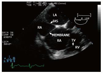
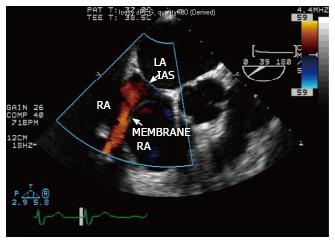
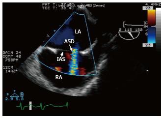
An 87-year-old woman presented to the hospital with complaints of dyspnea on exertion. She has no significant medical history other than prior cigarette smoking. On her arrival to the emergency room she was found to be in atrial fibrillation with rapid ventricular response. She was treated with heart rate control medications and anticoagulation. A transesophageal echocardiogram (TEE) was performed prior to cardioversion to normal sinus rhythm given poor rate control. This revealed the presence of a well-defined transverse membrane dividing the right atrium (Figure 1) consistent with the diagnosis of cor triatriatum dextrum. Color Doppler evaluation indicated separation of blood flow in the two divisions of the right atrium. However the division of the right atrium by this membrane was not complete. Color Doppler confirmed partial obstruction in the superior portion (Figure 2). The right atrium was enlarged without significant tricuspid regurgitation. An enlarged coronary sinus was noted with a measured diameter of 15.8 mm. Agitated saline injections via the left arm showed no saline contrast in the coronary sinus, indicating the absence of a persistent left superior vena cava. In addition, a small atrial septal defect (ASD) (measured diameter 3 mm) with a left to right shunt was demonstrated by color flow Doppler (Figure 3). No thrombus was noted in the left atrium and left atrial appendage. Subsequently the patient was cardioverted to normal sinus rhythm without complication.
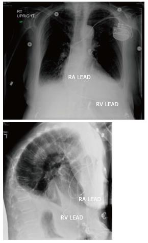
During her hospital course, she was found to have Morbitz type II AV block along with periods of complete heart block by remote cardiac monitoring station. Considering patient’s daily functional capacity predicting reasonable life expectancy, the decision was made to implant a permanent dual-chamber pacemaker. The presence of cor triatriatum dextrum with a membrane partially obstructing the cavity of the right atrium, presented a technical challenge in regards to adequate placement of the right ventricular and right atrial leads via the transvenous approach. After obtaining left subclavian venous access, a 6-French St. Jude Medical pacing electrode was inserted via a breakaway introducer sheath. The lead was gently guided to the area near the opening of the interatrial membrane. The area was gently probed with the pacing lead until it was seen to dip into the lower atrial area. Care was taken not to cross the ASD with the lead. Once the inner atrial membrane had been crossed, the lead was then advanced and fluoroscopically guided into the right ventricular apex. Once adequate positioning had been determined, the active fixation coil was deployed. Via a second breakaway introducer sheath, a Boston Scientific Dextrus active fixation lead was fluoroscopically guided into an area just below the membrane within the right atrial appendage, and the active fixation coil deployed (Figure 4). Each lead was then connected to a Boston Scientific Ingenio dual-chamber pulse generator. Via off field telemetry, adequate sensing and pacing levels of the leads were determined. The patient was discharged to home the next day.
The prevalence of adult congenital heart disease has increased in the past 10 years and its management has proposed new challenges to current cardiology practice[2]. We presented a case of successful pacemaker lead placement in a patient with non-obstructive cor triatriatum dextrum. Cor triatriatum dextrum is an extremely rare congenital heart abnormality in which the right atrium is separated into two chambers by the persistence of the right sinus venosus valve[3]. The superior chamber receives the venous blood from both vena cava and the inferior chamber is in contact with the tricuspid valve and the right atrial appendage. The size of the communicating orifice between the superior and inferior atrial chambers determines the natural course of cor triatriatum dextrum. If the communicating orifice is small, the patient shows symptoms of congestive heart failure during infancy or childhood and usually requires surgical intervention for survival. If the connection is large and non-obstructive, patient may remain asymptomatic for many years, as in our case. The clinical presentation therefore is somewhat variable. Patients with cor triatriatum dextrum may present with recurrent supraventricular tachycardia, right-side heart failure, or cyanosis in the presence of ASD with right-to-left shunt. In our case, patient presented initially with atrial fibrillation with rapid ventricular response. The disorder can be treated surgically in symptomatic patients by removing the membrane dividing the atrium.
During normal embryogenesis, the right atrium is formed by two different portions joining together: the right horn of the sinus venosus that forms the smooth posterior portion, and the original embryologic right atrium that forms the trabeculated anterior portion. The connection between these two portions is called sinoatrial orifice. The sinoatrial orifice is sided by two valvular folds that are called the right and left venous valves. During the development of right atrium, the right valve of the right horn of the sinus venosus forms a membranous valve that divides the right atrium in two parts. This valve directs oxygenated venous return from the inferior vena cava across the foramen ovale to the left side of the heart. This membranous valve normally regresses by the 12th week of gestation. Incomplete regression of the superior portion of right venosus valve forms membranes attached to the crista terminalis, while remnant of the inferior portion results in the Eustachian valve of the inferior vena cava, web-like remnant as Chiari network or the Thebesian valve of the coronary sinus. Failure of regression of this membrane causing persistent partition between the venous (smooth) and trabeculated portions of the right atrium leads to the formation of cor triatriatum dextrum[4].
Cor triatriatum dextrum has been associated with other congenital abnormalities, including ASD, patent foramen ovale, ventricular septal defect, hypoplastic right ventricle, hypoplastic tricuspid valve, and pulmonary atresia[3]. In our case, the presence of a small ASD posed the risk of crossing into the left atrium during manipulation of the lead across the atrial membrane. We also found an enlarged coronary sinus, measured diameter 15.8 mm (normal range 6.6 ± 1.5 mm)[5]. This is likely related to the elevated right atrial pressure. A rare but important congenital vascular anomaly associated with an enlarged coronary sinus is a persistent left superior vena cava draining into the coronary sinus[6]. To investigate this possibility, an agitated saline injection via the left arm was performed during TEE and showed no saline contrast in the coronary sinus, indicating the absence of a persistent left superior vena cava.
Considering the rarity of cor triatriatum dextrum, a patient with such a congenital abnormality who requires a permanent pacemaker is unique. To our knowledge, this is the first reported case of a permanent pacemaker placement through a transvenous approach in a patient with cor triatriatum dextrum. Although it is technically challenging, transvenous catheter-based approach is feasible if the membrane in the right atrium is non-obstructive and caution exercised during the procedure. A thorough pre-operative evaluation by transesophageal echocardiogram was critical for the planning of the transvenous catheter-based right-sided leads placement. Using specially designed pacemaker leads and cautious intra-procedural maneuvering under fluoroscopic guidance ensured procedural success.
A 87-year-old woman presented with paroxysmal atrial fibrillation.
Atrial fibrillation with raid ventricular response, later was also diagnosed with high grade AV block.
Any cause for atrial fibrillation and/or AV block such as hypertension, heart failure etc,.
Lab test result was unremarkable.
A transesophageal echocardiogram revealed a rare congenital condition called cor triatriatum dextrum.
A permanent pacemaker placement through a transvenous approach.
To the knowledge, this is the first reported case of a permanent pacemaker placement through a transvenous approach in a patient with cor triatriatum dextrum.
Cor triatriatum dextrum is an extremely rare congenital heart abnormality in which the right atrium is separated into two chambers by the persistence of the right sinus venosus valve.
Although it is technically challenging, transvenous catheter-based approach for a permanent pacemaker placement is feasible in a patient with cor triatriatum dextrum.
It is interesting.
| 1. | Santulli G. Epidemiology of cardiovascular disease in the 21st century: updated numbers and updated facts. J Cardiovasc Dis. 2013;1:1-2 Available from: http: //researchpub.org/journal/jcvd/number/vol1-no1/vol1-no1-1.pdf. |
| 2. | Marelli AJ, Mackie AS, Ionescu-Ittu R, Rahme E, Pilote L. Congenital heart disease in the general population: changing prevalence and age distribution. Circulation. 2007;115:163-172. [PubMed] |
| 3. | Hansing CE, Young WP, Rowe GG. Cor triatriatum dexter. Persistent right sinus venosus valve. Am J Cardiol. 1972;30:559-564. [PubMed] |
| 4. | Mahy IR, Anderson RH. Division of the right atrium. Circulation. 1998;98:2352-2353. [PubMed] |
| 5. | Cohen GI, White M, Sochowski RA, Klein AL, Bridge PD, Stewart WJ, Chan KL. Reference values for normal adult transesophageal echocardiographic measurements. J Am Soc Echocardiogr. 1995;8:221-230. [PubMed] |
P- Reviewer: Panduranga P, Santulli G S- Editor: Ji FF L- Editor: A E- Editor: Lu YJ
Open-Access: This article is an open-access article which was selected by an in-house editor and fully peer-reviewed by external reviewers. It is distributed in accordance with the Creative Commons Attribution Non Commercial (CC BY-NC 4.0) license, which permits others to distribute, remix, adapt, build upon this work non-commercially, and license their derivative works on different terms, provided the original work is properly cited and the use is non-commercial. See: http://creativecommons.org/licenses/by-nc/4.0/


