Published online Sep 26, 2014. doi: 10.4330/wjc.v6.i9.1045
Revised: April 14, 2014
Accepted: July 17, 2014
Published online: September 26, 2014
Processing time: 215 Days and 0.7 Hours
A 24-year-old healthy man consulted to our center because of typical on-and-off chest-pain and an electrocardiogram showing ST-segment elevation in inferior leads. An urgent coronary angiography showed angiographically normal coronary arteries. Cardiovascular magnetic resonance imaging confirmed acute myocarditis. Although acute myocarditis triggering coronary spasm is an uncommon association, it is important to recognize it, particularly for the management for those patients presenting with ST-segment elevation and suspect myocardial infarction and angiographically normal coronary arteries. The present report highlights the role of cardiovascular magnetic resonance imaging to identify acute myocarditis as the underlying cause.
Core tip: The present report highlights the role of cardiovascular magnetic resonance imaging to identify acute myocarditis as the underlying cause of coronary spasm presenting with ST-segment elevation myocardial infarction in a young healthy man.
- Citation: Kumar A, Bagur R, Béliveau P, Potvin JM, Levesque P, Fillion N, Tremblay B, Larose &, Gaudreault V. Acute myocarditis triggering coronary spasm and mimicking acute myocardial infarction. World J Cardiol 2014; 6(9): 1045-1048
- URL: https://www.wjgnet.com/1949-8462/full/v6/i9/1045.htm
- DOI: https://dx.doi.org/10.4330/wjc.v6.i9.1045
Myocarditis has been frequently associated in patients with acute chest pain syndrome and angiographically normal coronary arteries[1]. When the clinical presentation plus dynamic electrocardiographic (ECG) changes is quite suggestive of an acute coronary syndrome, coronary angiography is currently the first imaging diagnostic assessment in this setting. As a complementary imaging tool, cardiovascular magnetic resonance (CMR) imaging provides a strong evidence for tissue characterization while completing the differential diagnosis.
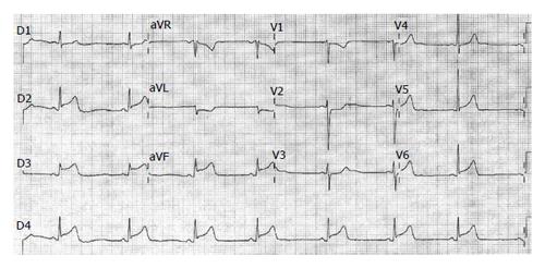
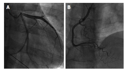
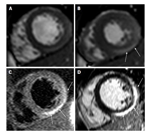
A 24-year-old male consulted our emergency room complaining of 24 h of typical, intense on-and-off chest-pain. He had no previous medical history and no risk factors for coronary disease. There was no history suggesting a recent virus infection or drug use. During a chest pain episode in the emergency room, the ECG showed ST-segment elevation in the inferior leads (Figure 1). Troponin was positive on admission. An urgent coronary angiogram was performed showing angiographically normal coronary arteries (Figure 2), and the ECG normalized spontaneously. On the coronary care unit, 8-10 h after cardiac catheterization, the patient experienced a new episode of chest-pain with recurrence of inferior ST-segment elevation. A treatment with intravenous nitroglycerin was started which led to resolution of chest-pain and ST-segment normalization. Two-dimensional Doppler echocardiography showed a very mild infero-lateral hypokinesis with preserved left ventricular ejection fraction. Of note, the creatine kinase and the Troponin-I peaked at 1600 IU/L (normal value < 150 IU/L) and 51.8 micrograms/mL (normal value < 0.02 micrograms/mL) respectively, within 24 h. In order to characterize the nature of this clinical scenario, the patient underwent CMR imaging confirming the mild infero-lateral hypokinesis (Figure 3A and B). In addition, tissue characterization showed myocardial edema localized in the epicardium of the lateral and infero-lateral walls (Figure 3C), the same area showed late gadolinium enhancement (Figure 3D). The subendocardial tissue appeared normal; therefore, highly compatible with acute myocarditis. The patient was discharged home seven days after admission on long acting Nifedipine and anti-inflammatory therapy.
Acute myocarditis triggering coronary vasospasm is a rare association. Especially myocarditis caused by Parvovirus B19, which affects endothelial cells, has been associated with coronary vasospasm[2]. While coronary vascular smooth muscle cell dysfunction leading to Prinzmetal angina is an important differential diagnosis as well as coronary spasm on atherosclerotic coronary disease, myocarditis is an important but probably less frequent diagnosis to consider. The present report highlights the role of CMR imaging to identify acute myocarditis as the underlying cause. The epicardial distribution of edema and necrosis is a hallmark of myocarditis, as opposed to ischemic injury caused by epicardial coronary artery disease which necessarily leads to injury including the subendocardium[3-6]. Myocarditis and epicardial coronary artery disease imply differences in medical treatment, therefore CMR enables the non-invasive assessment of changes in myocardial tissue composition (myocardial edema, hyperemia, and necrosis) and thus allowed for establishing the diagnosis of acute myocarditis[3-6].
A healthy young man presenting with typical chest pain and an electrocardiogram showing inferior wall ST-elevation.
Acute myocardial infarction was the most likely clinical diagnosis given the description of chest pain and the electrocardiographic findings.
Coronary vascular smooth muscle cell dysfunction leading to Prinzmetal angina as well as coronary spasm on atherosclerotic coronary disease are important differential diagnosis.
Serial troponin levels progressively increased.
Coronary angiography demonstrated angiographically normal coronary arteries and cardiovascular magnetic resonance imaging showed myocardial edema localized in the epicardium of the lateral and infero-lateral walls, the same area showed late gadolinium enhancement.
The patient was medically managed and discharged home seven days after admission on long acting Nifedipine and anti-inflammatory therapy.
Myocarditis caused by Parvovirus B19, which affects endothelial cells, has been associated with coronary vasospasm.
Although an uncommon association, it is important to recognize it, particularly for the management of those patients presenting with typical chest pain and electrocardiographic ST-segment elevation and therefore mimicking myocardial infarction.
The authors present a case that reports an uncommon association of an acute myocarditis triggering coronary spasm and presenting as ST-elevation myocardial infarction. The manuscript is clearly written, well organized, comprehensive, appropriate referenced and concise in its content.
| 1. | Assomull RG, Lyne JC, Keenan N, Gulati A, Bunce NH, Davies SW, Pennell DJ, Prasad SK. The role of cardiovascular magnetic resonance in patients presenting with chest pain, raised troponin, and unobstructed coronary arteries. Eur Heart J. 2007;28:1242-1249. [PubMed] |
| 2. | Yilmaz A, Mahrholdt H, Athanasiadis A, Vogelsberg H, Meinhardt G, Voehringer M, Kispert EM, Deluigi C, Baccouche H, Spodarev E. Coronary vasospasm as the underlying cause for chest pain in patients with PVB19 myocarditis. Heart. 2008;94:1456-1463. [RCA] [PubMed] [DOI] [Full Text] [Cited by in Crossref: 121] [Cited by in RCA: 115] [Article Influence: 6.4] [Reference Citation Analysis (0)] |
| 3. | Abdel-Aty H, Boyé P, Zagrosek A, Wassmuth R, Kumar A, Messroghli D, Bock P, Dietz R, Friedrich MG, Schulz-Menger J. Diagnostic performance of cardiovascular magnetic resonance in patients with suspected acute myocarditis: comparison of different approaches. J Am Coll Cardiol. 2005;45:1815-1822. [PubMed] |
| 4. | Friedrich MG, Sechtem U, Schulz-Menger J, Holmvang G, Alakija P, Cooper LT, White JA, Abdel-Aty H, Gutberlet M, Prasad S. Cardiovascular magnetic resonance in myocarditis: A JACC White Paper. J Am Coll Cardiol. 2009;53:1475-1487. [RCA] [PubMed] [DOI] [Full Text] [Full Text (PDF)] [Cited by in Crossref: 1951] [Cited by in RCA: 1787] [Article Influence: 105.1] [Reference Citation Analysis (2)] |
| 5. | Friedrich MG, Marcotte F. Cardiac magnetic resonance assessment of myocarditis. Circ Cardiovasc Imaging. 2013;6:833-839. [RCA] [PubMed] [DOI] [Full Text] [Cited by in Crossref: 103] [Cited by in RCA: 132] [Article Influence: 10.2] [Reference Citation Analysis (0)] |
| 6. | Kumar A, Friedrich MG. Acute chest pain syndrome: will MRI shake up cardiovascular care in the emergency room? Expert Rev Cardiovasc Ther. 2007;5:139-141. [PubMed] |
P- Reviewer: Carbucicchio C, Monti L, Patanè S, Robert KI S- Editor: Wen LL L- Editor: A E- Editor: Liu SQ


