©The Author(s) 2016.
World J Cardiol. Sep 26, 2016; 8(9): 496-503
Published online Sep 26, 2016. doi: 10.4330/wjc.v8.i9.496
Published online Sep 26, 2016. doi: 10.4330/wjc.v8.i9.496
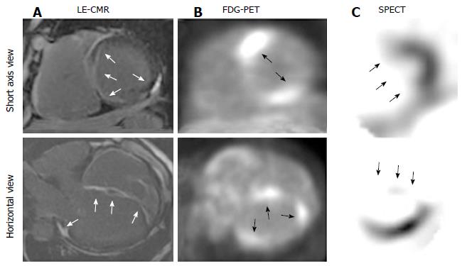
Figure 1 Non-invasive cardiac imaging in a 61-year-old male patient with cardiac involvement of systemic sarcoidosis.
LE-CMR (A) shows diffuse LE in the subepicardium (RV side) and subendocardium (LV side) of basal to apical ventricular septum and patchy LE in the midwall of posterior LV (white arrows); Corresponding FDG-PET (B) demonstrates focal uptake in basal and apical ventricular septum and posterior LV wall (black arrows); 99mTc-sestamibi SPECT (C) exhibits a defect only in ventricular septum (black arrows). CMR: Cardiac magnetic resonance; FDG-PET: 18F-fluorodeoxyglucose-positron emission computed tomography; LE: Late gadolinium enhancement; LV/RV: Left and right ventricles; SPECT: Single photon emission computed tomography.
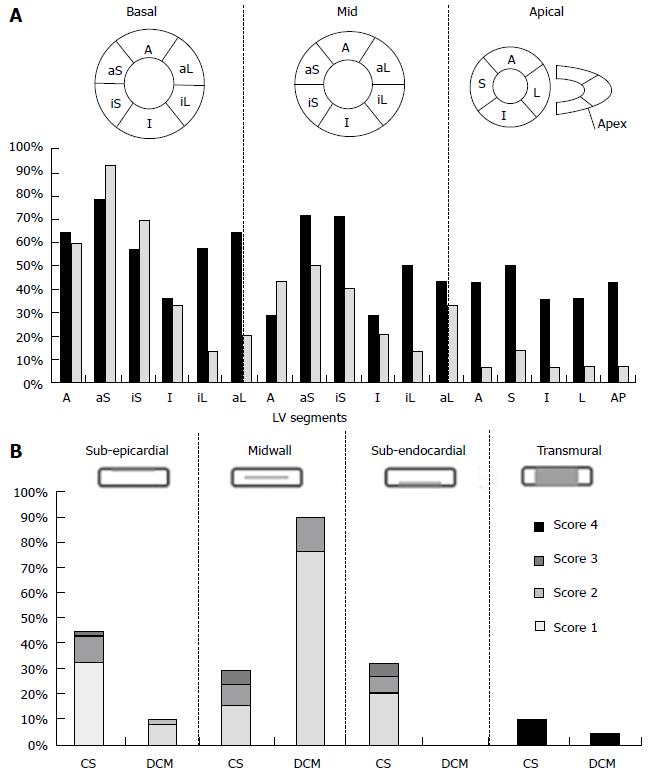
Figure 2 Intra-left ventricles (A) and intra-mural (B) late gadolinium enhancement distribution in patients with cardiac sarcoidosis and with dilated cardiomyopathy.
A: Columns indicate prevalence of LE at each LV segment in patients with CS (black) and with DCM (gray). A: Anterior; aL: Antero-lateral; aS: Anterior septal; I: Inferior; iL: Infero-lateral wall in basal, mid and apical LV; AP: LV apex; B: Columns consist of prevalence of LE with scores 1 to 3 at different intra-mural distribution in patients with CS and with DCM. Score 4 indicates the transmural distribution. CS: Cardiac sarcoidosis; DCM: Dilated cardiomyopathy; LV: Left ventricles.
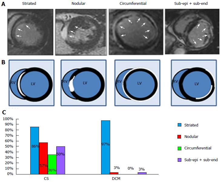
Figure 3 Typical late gadolinium enhancement distribution profiles.
Characteristic patterns of LE distribution in LE-MRI (A) and the cartoons (B). Striated: Striated LE distribution in midwall; Nodular: Nodular (transmural) LE distribution; Circumferential: Subepicardial LE distribution in > 50% circumferential LV wall; Sub-epi + sub-end: Subepicardial and subendocardial LE distribution with spared midwall (white arrows); C: The prevalence of characteristic patterns of LE distribution in patients with CS and with DCM. CS: Cardiac sarcoidosis; DCM: Dilated cardiomyopathy; LE: Late gadolinium enhancement; LV/RV: Left and right ventricles; MRI: Magnetic resonance imaging.
- Citation: Sano M, Satoh H, Suwa K, Saotome M, Urushida T, Katoh H, Hayashi H, Saitoh T. Intra-cardiac distribution of late gadolinium enhancement in cardiac sarcoidosis and dilated cardiomyopathy. World J Cardiol 2016; 8(9): 496-503
- URL: https://www.wjgnet.com/1949-8462/full/v8/i9/496.htm
- DOI: https://dx.doi.org/10.4330/wjc.v8.i9.496


