©The Author(s) 2015.
World J Cardiol. Sep 26, 2015; 7(9): 525-538
Published online Sep 26, 2015. doi: 10.4330/wjc.v7.i9.525
Published online Sep 26, 2015. doi: 10.4330/wjc.v7.i9.525
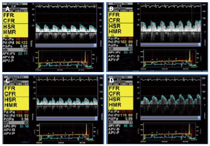
Figure 1 Microvascular function measured by intracoronary Doppler.
A shows an intracoronary Doppler tracing at baseline condition, with an APV of 22 cm/s; B shows Doppler at the same position during maximal hyperemia, induced with an adenosine intracoronary bolus of 200 μg, with an APV of 53 cm/s. CFR is therefore 2.4, indicating a normal endothelium-independent microvascular function; in C and D, increasing doses of intracoronary acetylcholine have been administered; the APV is compared with the baseline tracing to determine the degree of microvascular vasodilation induced by acetylcholine, which in this case is normal, suggesting a normal endothelium-dependent microvascular function. CFR: Coronary flow reserve; APV: Average peak velocity.
Figure 2 Coronary flow reserve measured by thermodilution.
A 60-year-old woman with episodes of atypical chest pain was referred to the catheterization laboratory for coronary angiography. A and B: Show angiographically normal epicardial coronary arteries. A microcirculation study was performed, using a thermodilution PressureWire, and intravenous adenosine for hyperemia; C: Shows the thermodilution curves, with a mean transit time of 0.70 s at baseline and 0.15 s during hyperemia, and a resulting coronary flow reserve of 4.5. Index of Microvascular Resistance was 15, within normal range. We concluded that microvascular function was preserved.
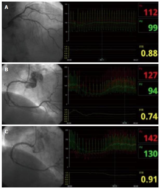
Figure 3 Functional assessment of epicardial stenosis.
A 77-year-old woman with exertional angina was referred for coronary angiography. Two intermediate lesions were found in the proximal and distal segments of the left anterior descending artery (LAD) (A), and in the proximal and mid segments of the right coronary artery (RCA) (B). Fractional Flow Reserve was non-significant on the LAD (FFR 0.84) and significant on the RCA (FFR 0.74). Accordingly, percutaneous coronary intervention was performed with a drug-eluting stent on the RCA (C). A post-intervention FFR confirmed that the good angiographic result was also associated with functional improvement (FFR 0.91). Six months after the procedure the patient remains asymptomatic. FFR: Fractional flow reserve.
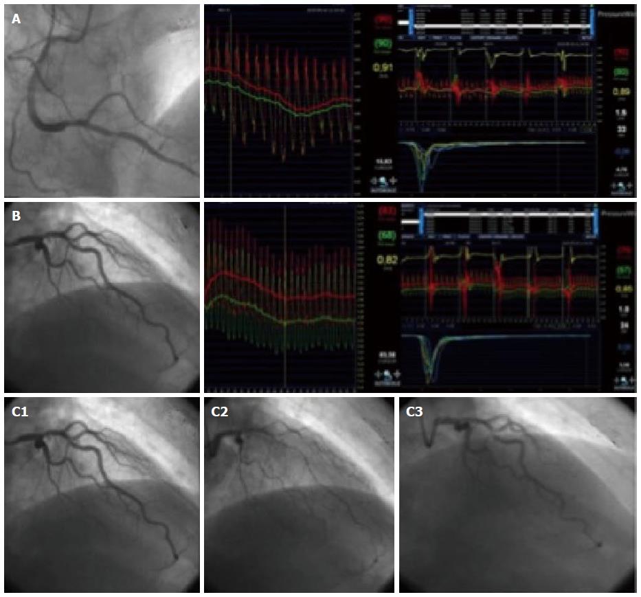
Figure 4 Thorough physiological assessment: Fractional flow reserve, microvascular and endothelial function.
A 66-year-old man with typical angina, both resting and exertional, was referred for coronary catheterization. The coronary angiogram showed moderate lesions on the posterior descending artery of the RCA (50%) and on the middle LAD (40%). Panel A depicts the coronary stenosis of the RCA on the left box; FFR of this lesion on the centre box, with a value of 0.91; and microvascular study on the right box, with a CFR of 1.5 (low) and IMR of 33 (elevated); Similarly, Panel B shows the angiogram of the LAD (left), the FFR of 0.82 (centre), and the microvascular study (right), with a CFR 1 (low) and IMR 24 (borderline); Panel C shows the successive angiograms at baseline (C1), after 20 μg of acetylcholine (C2), and after 200 μg of nitroglycerin (C3). As can be appreciated, a severe diffuse spasm of the left coronary artery was induced by acetylcholine. We concluded that both coronary stenosis were non-significant, and decided on optimal medical therapy. The study also revealed microvascular endothelium-independent dysfunction, and macrovascular vasospasm due to endothelial dysfunction. CFR: Coronary flow reserve; FFR: Fractional flow reserve; IMR: Index of microcirculatory resistance; LAD: Left anterior descending artery; RCA: Right coronary artery.
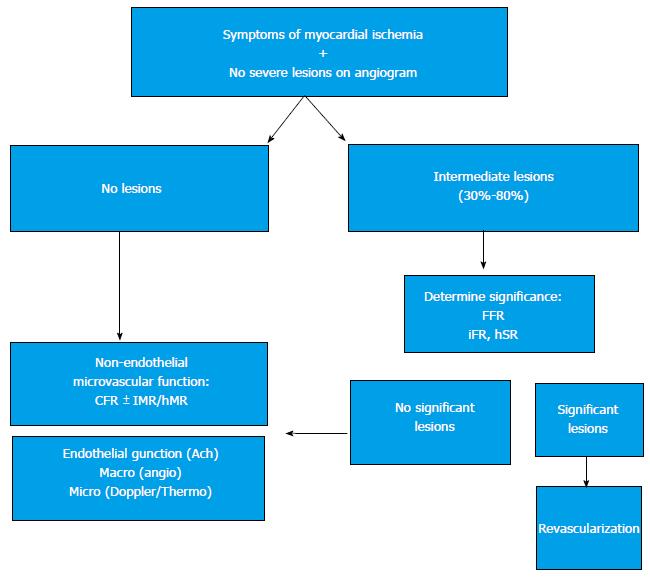
Figure 5 Summary of the proposed evaluation algorithm of patients with symptoms of ischemia without significant coronary lesions.
CFR: Coronary flow reserve; IMR: Index of microcirculatory resistance; FFR: Fractional flow reserve; hMR: Hyperemic microvascular resistance; hSR: Hyperemic Stenosis Resistance index.
- Citation: Díez-delhoyo F, Gutiérrez-Ibañes E, Loughlin G, Sanz-Ruiz R, Vázquez-Álvarez ME, Sarnago-Cebada F, Angulo-Llanos R, Casado-Plasencia A, Elízaga J, Diáz FFA. Coronary physiology assessment in the catheterization laboratory. World J Cardiol 2015; 7(9): 525-538
- URL: https://www.wjgnet.com/1949-8462/full/v7/i9/525.htm
- DOI: https://dx.doi.org/10.4330/wjc.v7.i9.525


