Copyright
©The Author(s) 2015.
Figure 1 Devices for percutaneous occlusion of the left atrial appendage.
A: PlAATO® device; B: Amplatzer® cardiac plug; C: Watchman® device.
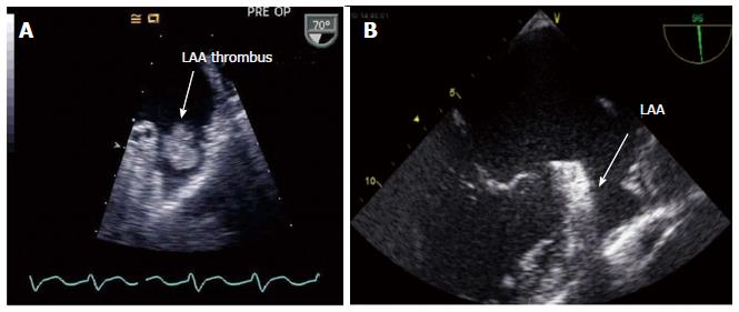
Figure 2 Left atrial appendage.
A: With thrombus; B: Without thrombus. LAA: Left atrial appendage.
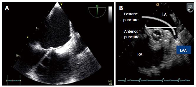
Figure 3 Echocardiography in transseptal puncture.
A: Tenting of transseptal needle at the middle part of the atrial septum; B: Access sheath passing through the atrial septum within left atrium. LAA: Left atrial appendage.
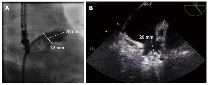
Figure 4 Measurement of the left atrial appendage during implantation procedure.
A: After angiography (RAO 25°, caudal 20°)-measurement of ostium size (22 mm) and depth (28 mm); B: Echocardiographic measurement at around 135° of ostium size (20 mm) and depth (25 mm).
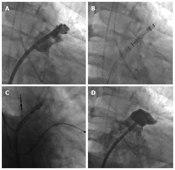
Figure 5 Deployment of a Watchman® device (fluoroscopic views).
A: Deployment sheath in correct position; B: Watchman device loaded within the sheath before deployment; C: Watchman device deployment; D: Watchman device completely deployed within the LAA. LAA: Left atrial appendage.
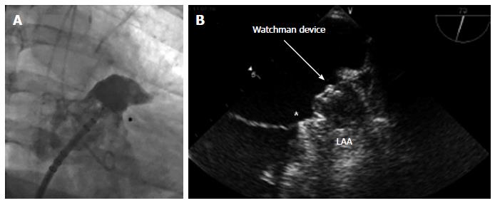
Figure 6 Optimal position of the Watchman device within the left atrial appendage.
A: Angiographic view (RAO 25° caudal 20°); B: Echocardiographic view. Indicates inferior transition from left atrial appendage to LA (star).
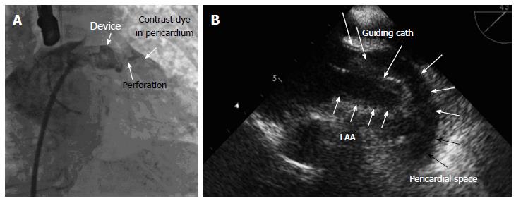
Figure 7 Pericardial effusion.
A: Angiographic view; B: Echo view. LAA: Left atrial appendage.
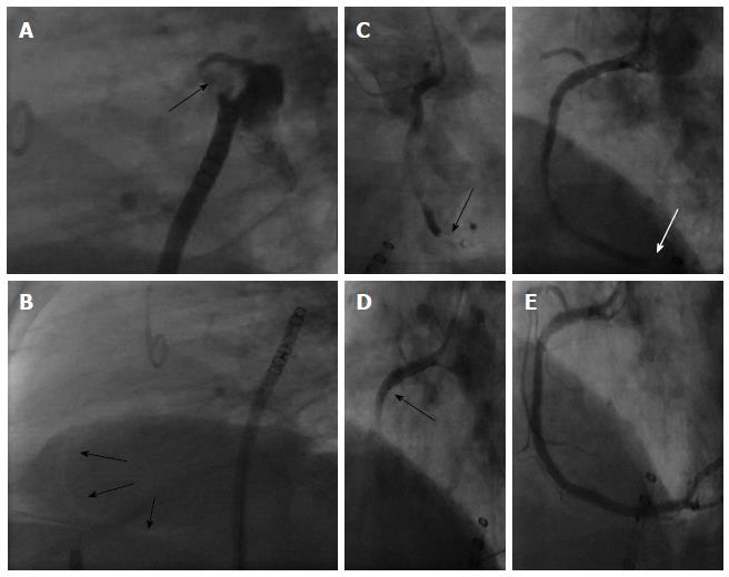
Figure 8 Air embolism.
A: Air bubble within the left atrial appendage; B: Right coronary artery (RCA) filled with air (bright shadows); C: RCA with large air bubble; D: Placement of a aspiration catheter (EXPORT, Medtronic) within the RCA; E: RCA after successful aspiration of air bubbles without filling defect. LAA: Left atrial appendage
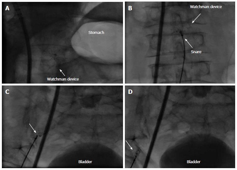
Figure 9 Device embolisation of watchman-device.
A: Within the left atrial appendage immediately after release (arrow indicates device); B: Embolisation from the left atrial appendage; C: Passage of the aortic valve; D: Device within the lower thoracic aorta after embolization.
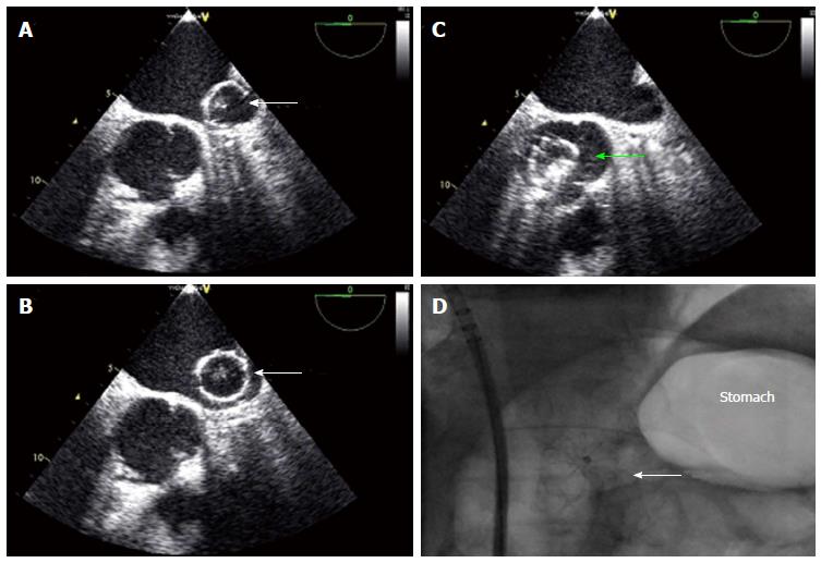
Figure 10 Device retrieval after embolization.
A: Watchman device within the lower thoracic aorta after embolization; B: Snaring of the Watchman device with a goose neck snare 20 mm; C: Retraction of the device into the right arteria iliaca; D: Device retracted into a large (14 Fr.) sheath.
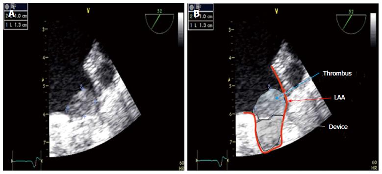
Figure 11 Thrombus formation (1.
0 cm x 1.3 cm) 45 d after implantation on a Watchman device (A) and schematic view: Thrombus, device and left atrial appendage are highlighted (B).
- Citation: Möbius-Winkler S, Majunke N, Sandri M, Mangner N, Linke A, Stone GW, Dähnert I, Schuler G, Sick PB. Percutaneous left atrial appendage closure: Technical aspects and prevention of periprocedural complications with the watchman device. World J Cardiol 2015; 7(2): 65-75
- URL: https://www.wjgnet.com/1949-8462/full/v7/i2/65.htm
- DOI: https://dx.doi.org/10.4330/wjc.v7.i2.65


