©2011 Baishideng Publishing Group Co.
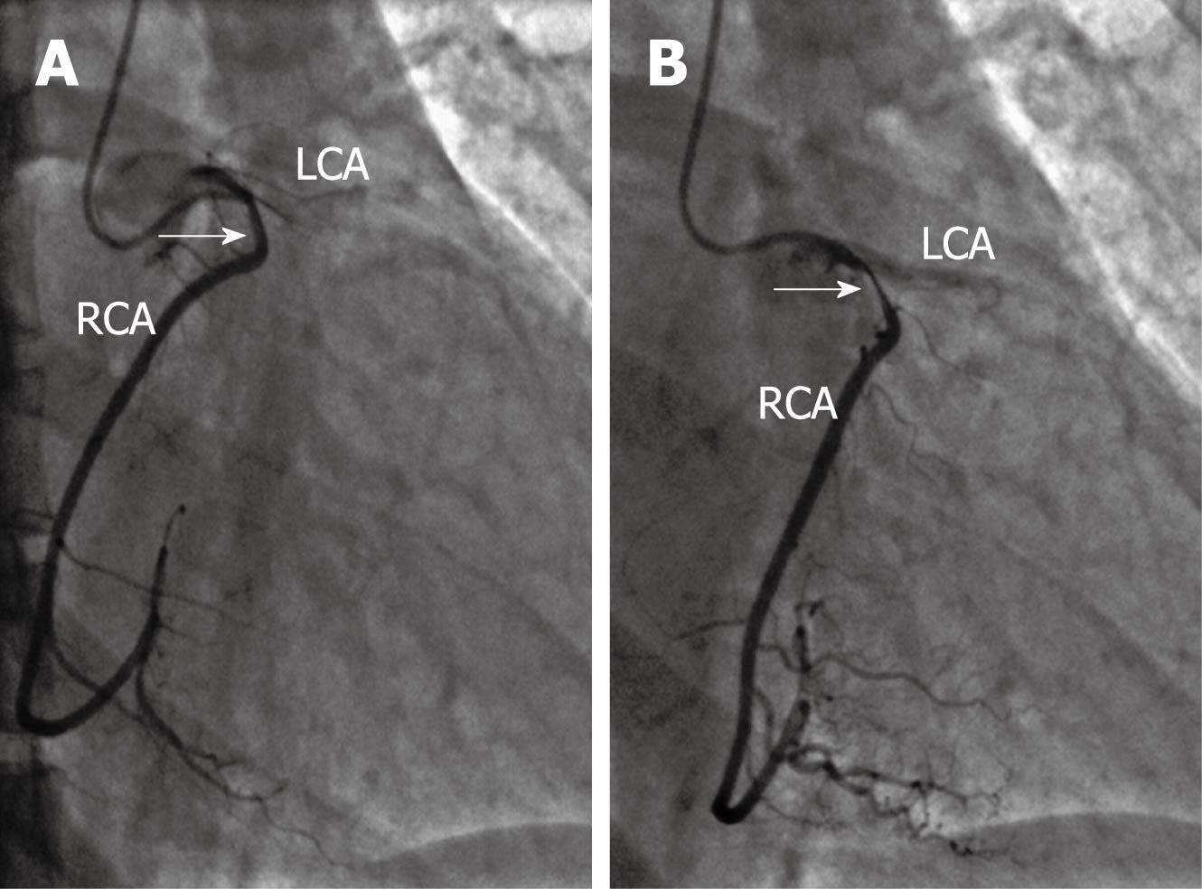
Figure 1 Coronary angiogram of the right coronary artery.
Coronary angiography showing a significant compression of the proximal right coronary artery (RCA) due to an inter-arterial trajectory between the pulmonary artery and the aorta during systole (arrows). A: Diastole; B: Systole. LCA: Left coronary artery
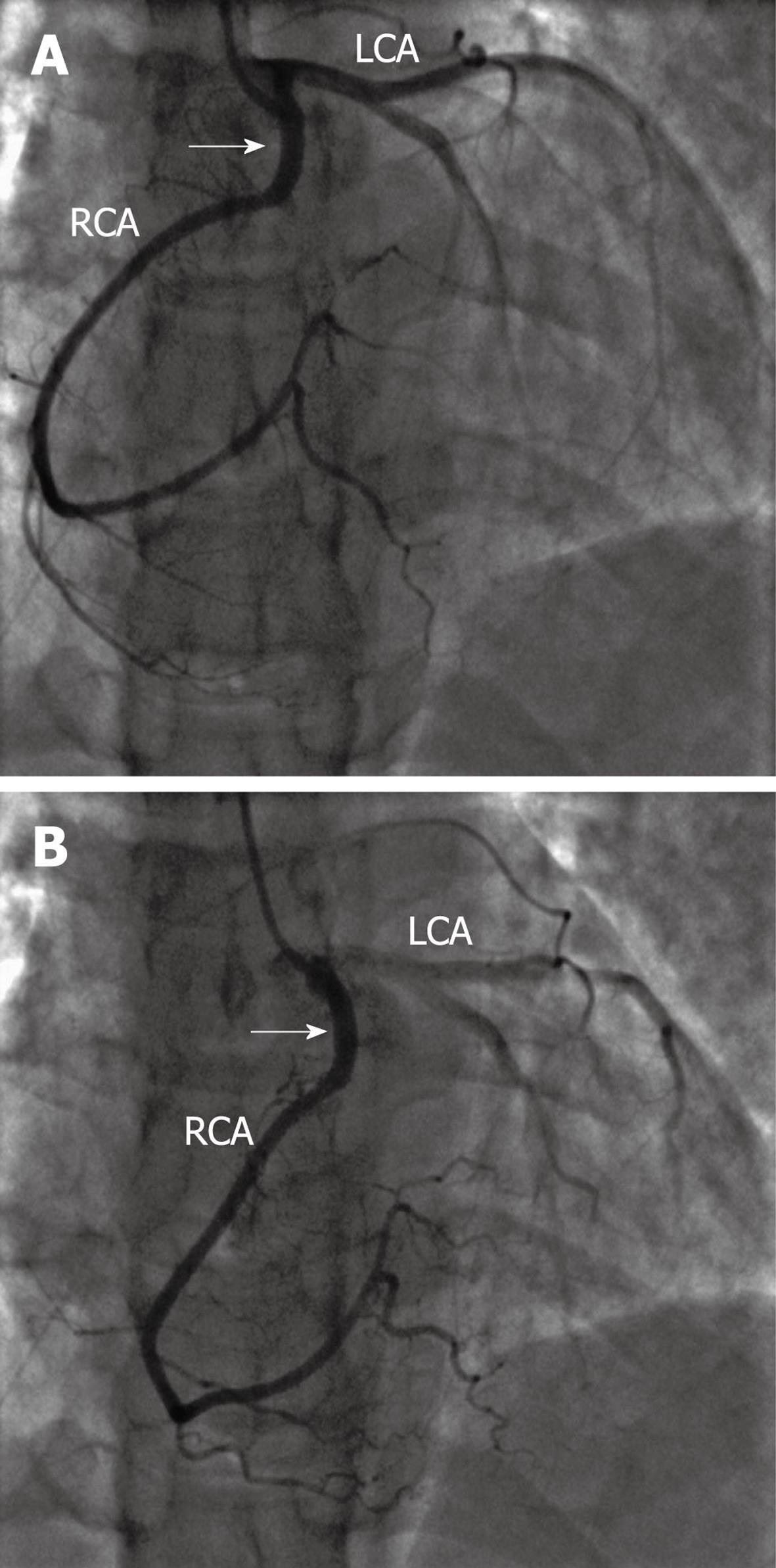
Figure 2 Coronary angiogram following successful transradial percutaneous coronary intervention.
Coronary angiography showing no compression effect (arrows) after successful stent implantation in the proximal right coronary artery (RCA). A: Diastole; B: Systole. LCA: Left coronary artery.
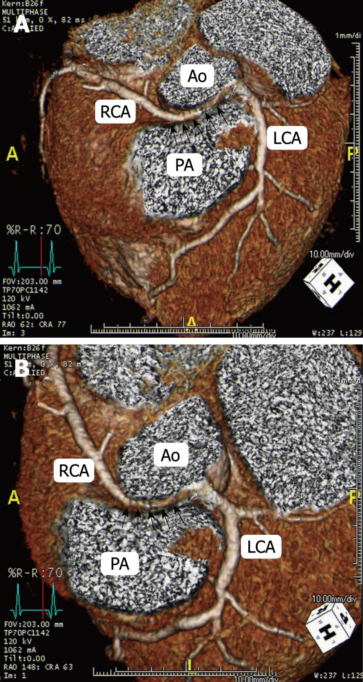
Figure 3 Three-dimensional volume rendered multislice computed tomography (A and B).
Computed tomography image showing the right coronary artery (RCA) arising from the left sinus of Valsalva, and an inter-arterial trajectory (black arrows) between the pulmonary artery (PA) and the aorta (Ao). LCA: Left coronary artery.
- Citation: Bagur R, Gleeton O, Bataille Y, Bilodeau S, Rodés-Cabau J, Bertrand OF. Right coronary artery from the left sinus of valsalva: Multislice CT and transradial PCI. World J Cardiol 2011; 3(2): 54-56
- URL: https://www.wjgnet.com/1949-8462/full/v3/i2/54.htm
- DOI: https://dx.doi.org/10.4330/wjc.v3.i2.54


