
















Copyright: ©Author(s) 2026.
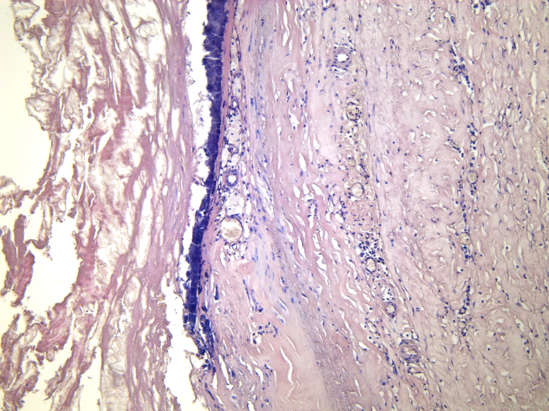
Figure 1 Aortic aneurysm.
Leukocyte infiltrates around the vasa vasorum and newly formed vessels in the adventitia and media (hematoxylin and eosin staining × 200).
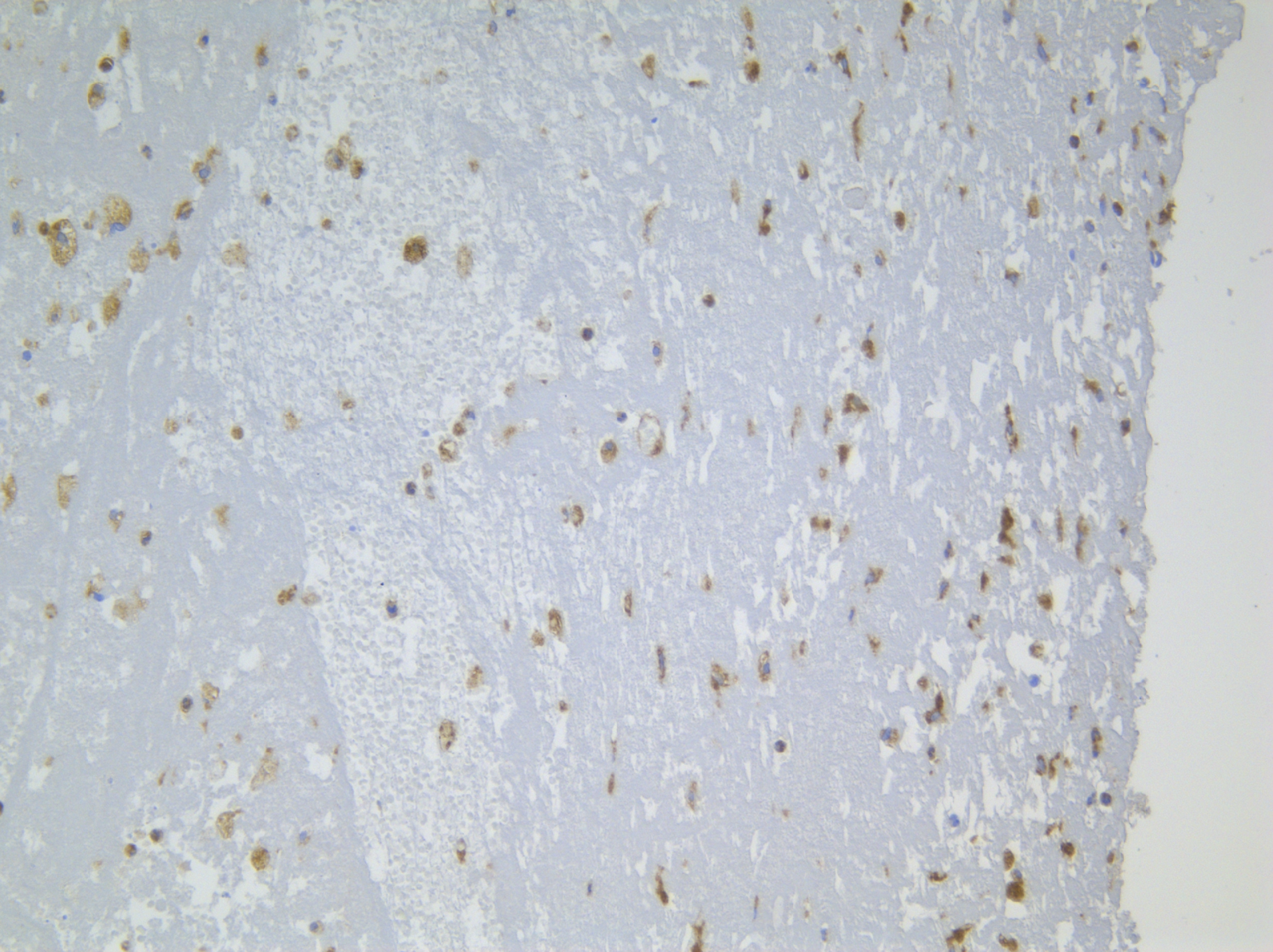
Figure 2 Immunohistochemical staining of an aortic aneurysm.
Large CD68positive macrophages containing numerous lysosomes actively resorb necrotic fibers (cell nuclei are stained with hematoxylin × 200).
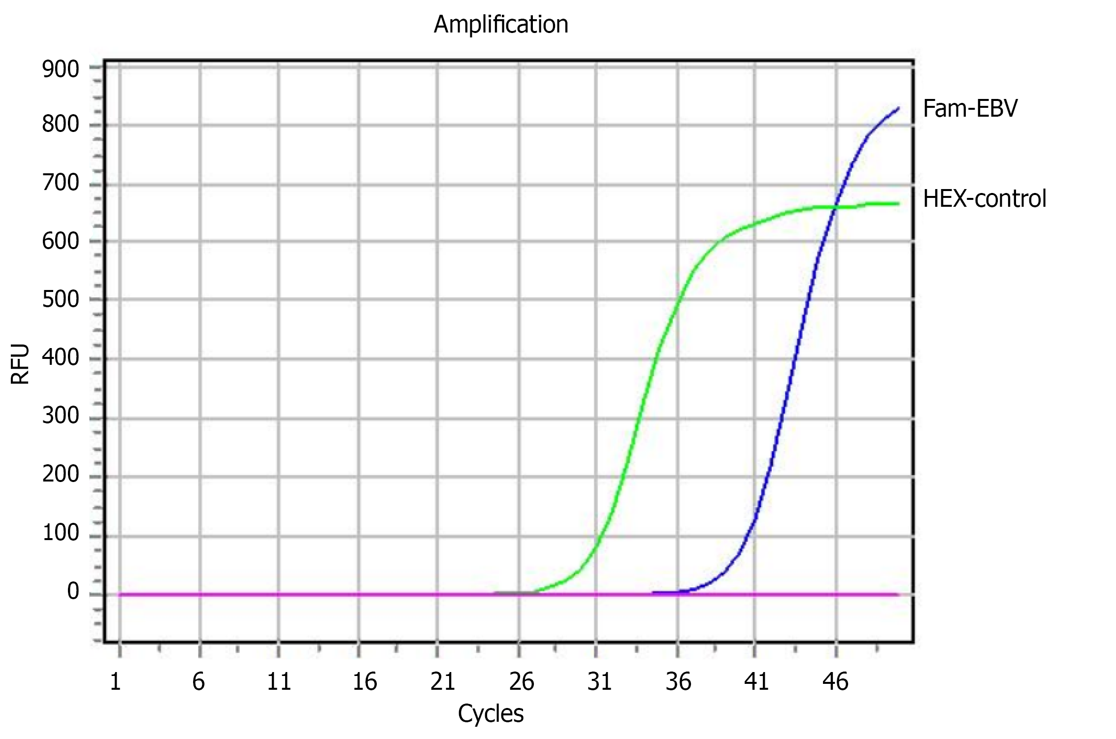
Figure 3 Fluorescence curves for the Fam channel (Epstein–Barr virus) and Hex channel (PCR internal control).
EBV: Epstein-Barr virus; RFU: Relative fluorescence unit.
- Citation: Suslov AV, Khasanova ZB, Buchaka AS, Doroschuk AD, Postnov AY. Herpesvirus infection and thoracic aortic aneurysm: Synthesis of evidence and contradictions. World J Cardiol 2026; 18(7): 122814
- URL: https://www.wjgnet.com/1949-8462/full/v18/i7/122814.htm
- DOI: https://dx.doi.org/10.4330/wjc.122814