
















Copyright: ©Author(s) 2026.
World J Cardiol. Oct 26, 2026; 18(10): 126812
Published online Oct 26, 2026. doi: 10.4330/wjc.126812
Published online Oct 26, 2026. doi: 10.4330/wjc.126812
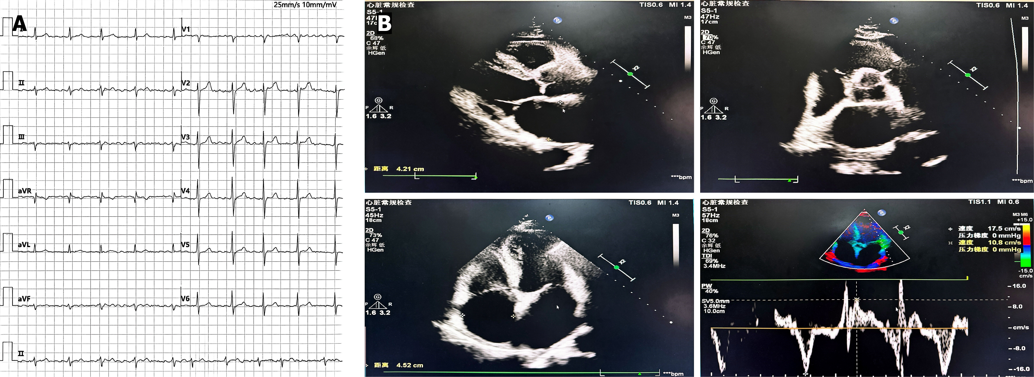
Figure 1 Electrocardiographic and echocardiographic findings in a patient with persistent atrial fibrillation.
A: Electrocardiogram test; B: Echocardiography.
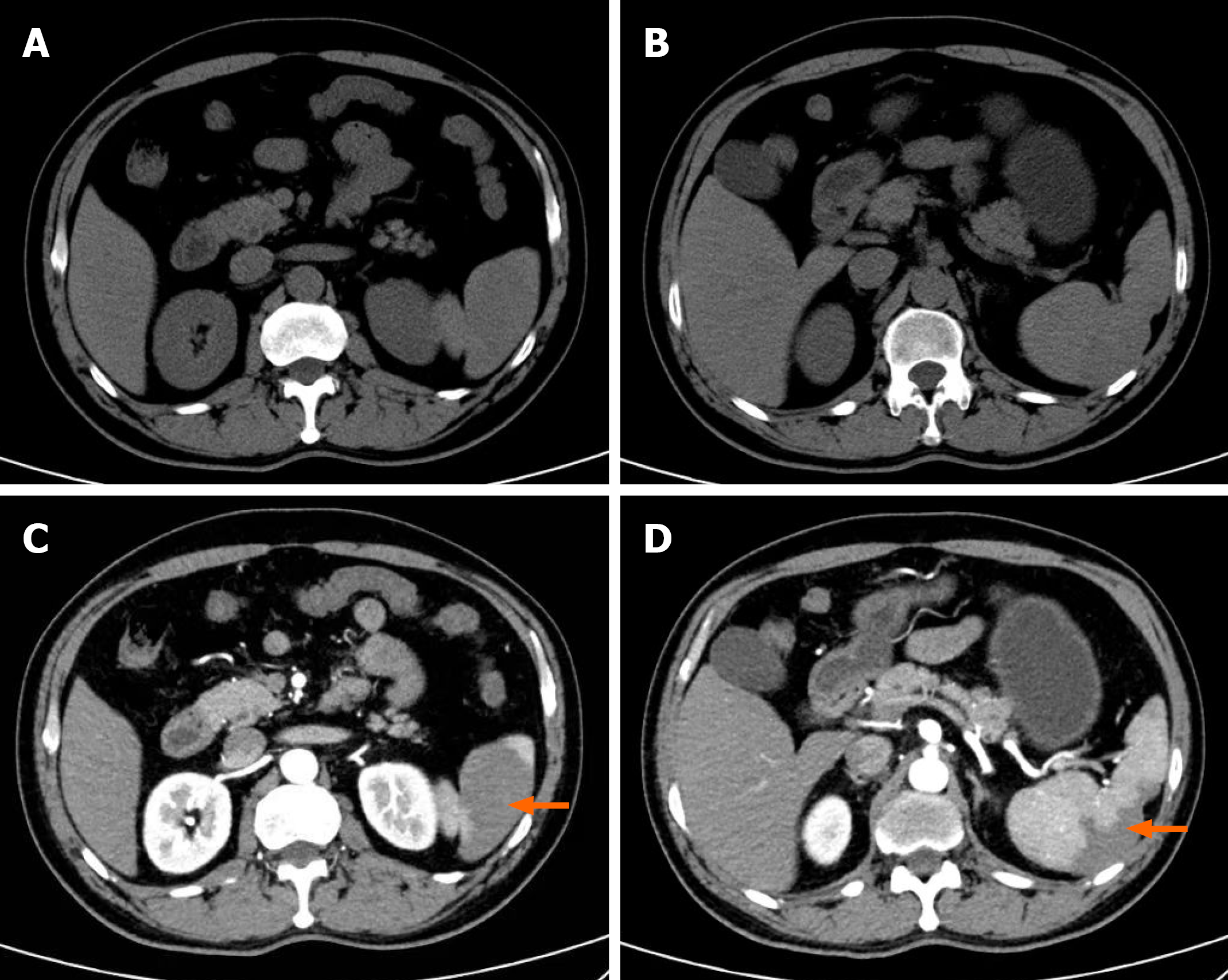
Figure 2 Imaging findings during the diagnostic evaluation ofcardioembolic splenic infarction associated with persistentatrial fibrillation.
A and B: Initial non-contrast computed tomography showedno definite abnormalities despite persistent abdominal pain; C and D: Follow-up contrast-enhanced computed tomography revealed a peripheral wedge-shaped low-attenuation lesion in the spleen, confirming the diagnosis of acute splenic infarction.
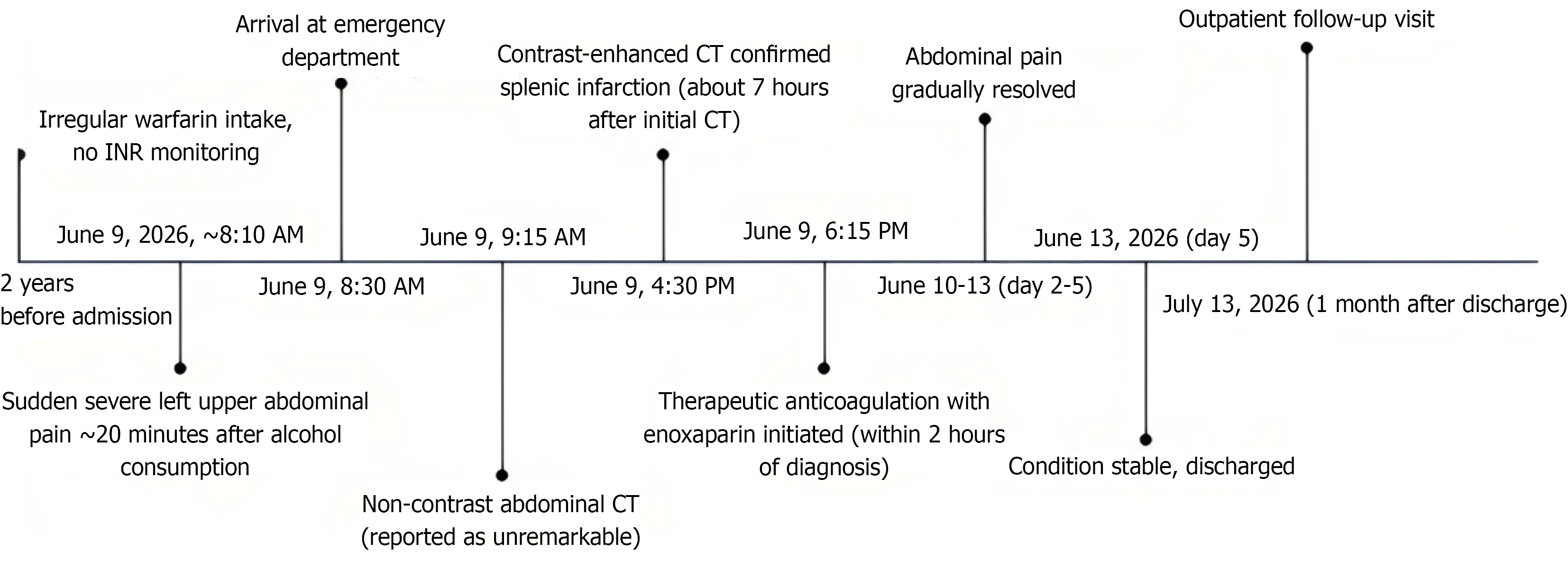
Figure 3 Timeline of clinical presentation, diagnosis, treatment, and follow-up of cardioembolic splenic infarction associated with persistent atrial fibrillation.
CT: Computed tomography; INR: International normalized ratio.
- Citation: Gao L, Lu P, Liu QL, Li ZL, Zhao PG, Zhou QY, Liu J, Feng YS, Li Z. Splenic infarction associated with persistent atrial fibrillation: A case report. World J Cardiol 2026; 18(10): 126812
- URL: https://www.wjgnet.com/1949-8462/full/v18/i10/126812.htm
- DOI: https://dx.doi.org/10.4330/wjc.126812