©The Author(s) 2025.
World J Cardiol. Jul 26, 2025; 17(7): 106828
Published online Jul 26, 2025. doi: 10.4330/wjc.v17.i7.106828
Published online Jul 26, 2025. doi: 10.4330/wjc.v17.i7.106828
Figure 1
Photographs of the snake.
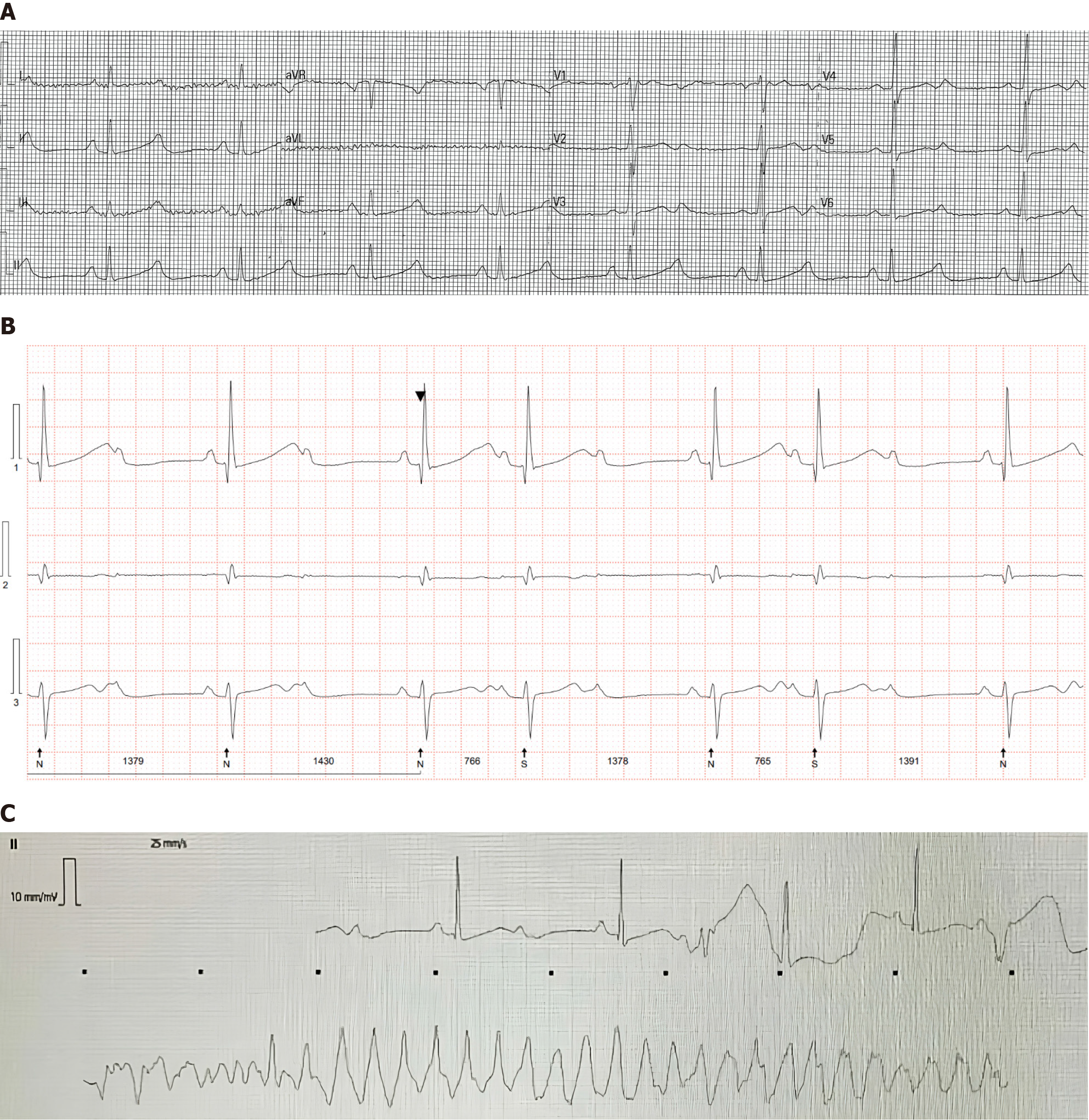
Figure 2 Imaging examinations.
A: Electrocardiogram showing sinus bradycardia and prolonged QT interval; B: 24-hour Holter monitoring showing advanced atrioventricular block; C: Patient monitor displaying wide complex tachycardia.
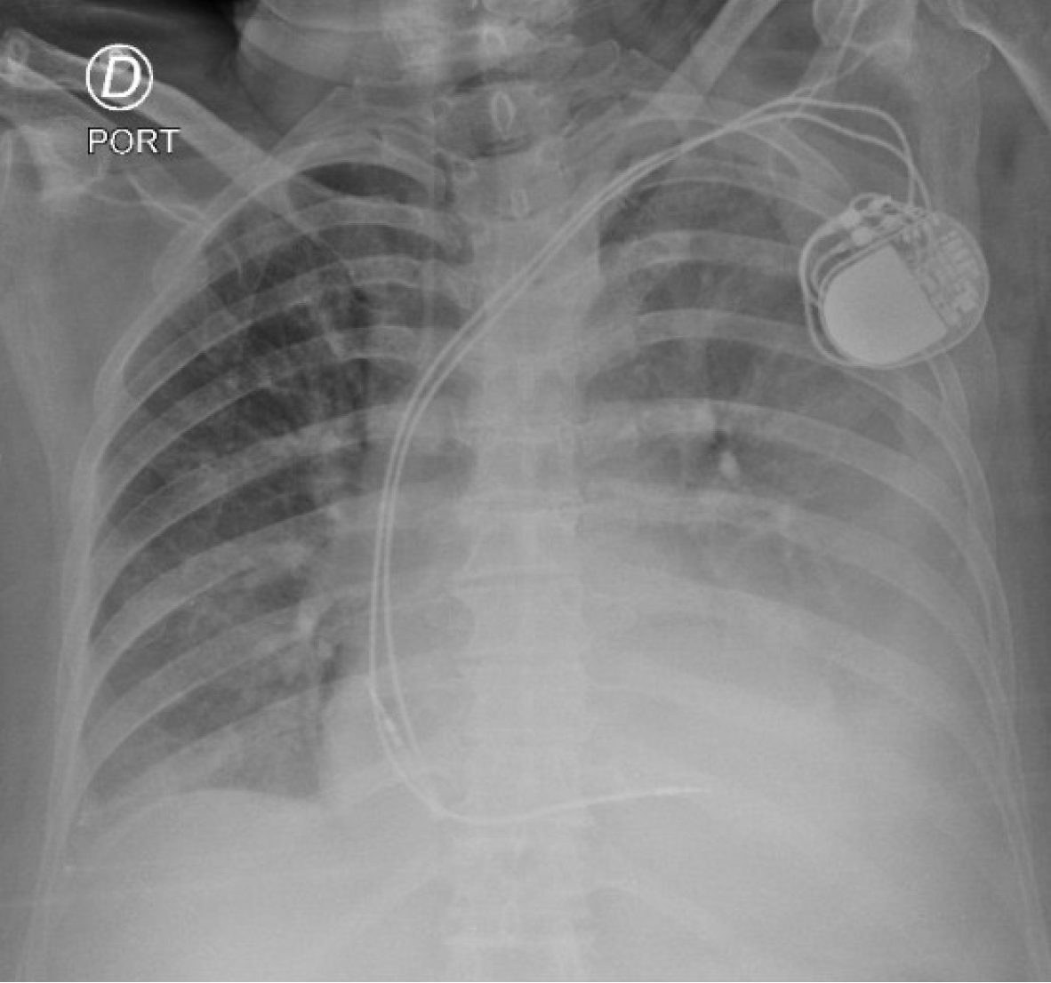
Figure 3
Chest X-ray showing permanent pacemaker.
- Citation: Acosta JS, Cifuentes Tarquino J, Arteaga JE. Bothrops bite and cardiac complications: A case report and review of literature. World J Cardiol 2025; 17(7): 106828
- URL: https://www.wjgnet.com/1949-8462/full/v17/i7/106828.htm
- DOI: https://dx.doi.org/10.4330/wjc.v17.i7.106828


