Copyright
©The Author(s) 2022.
World J Cardiol. Dec 26, 2022; 14(12): 657-664
Published online Dec 26, 2022. doi: 10.4330/wjc.v14.i12.657
Published online Dec 26, 2022. doi: 10.4330/wjc.v14.i12.657
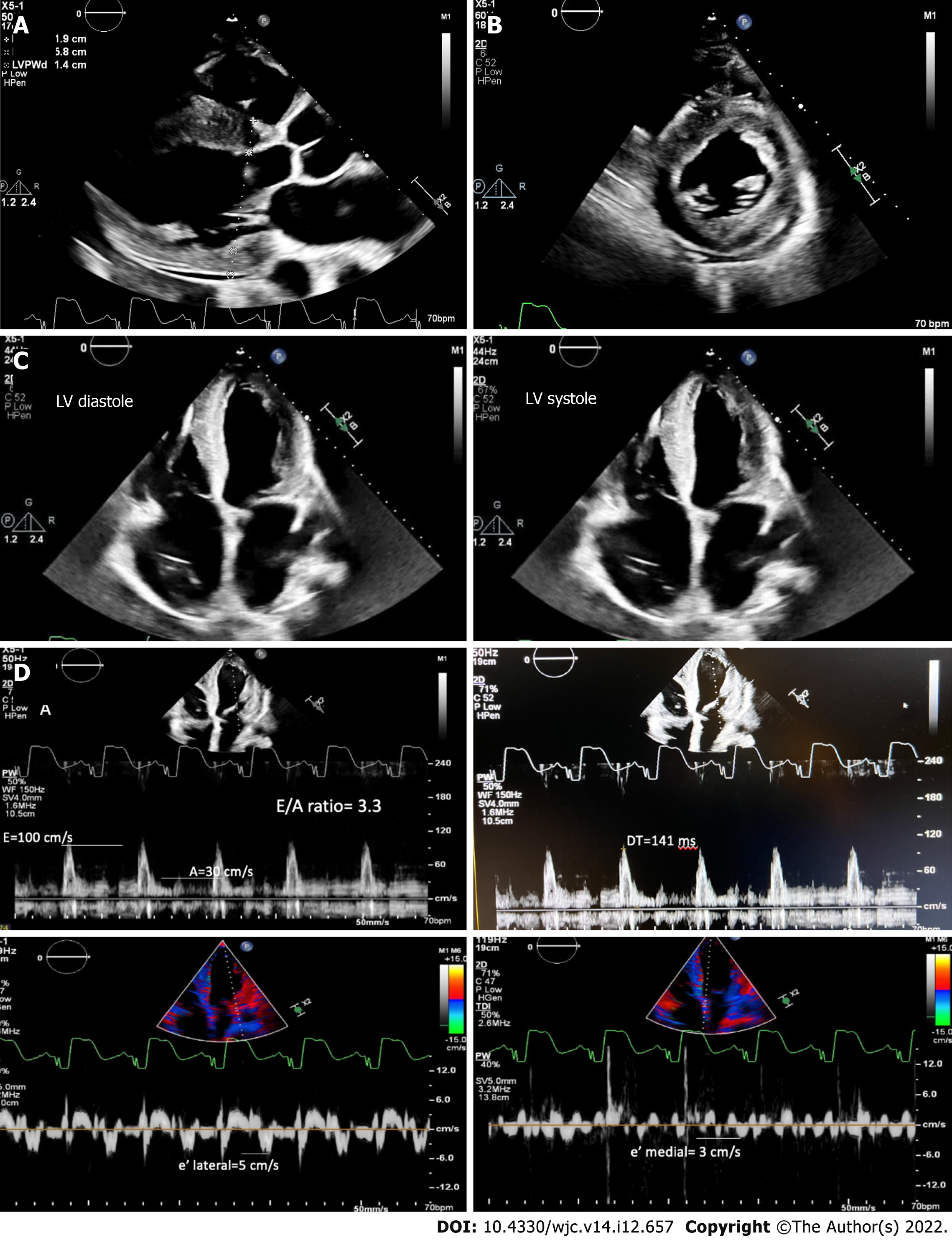
Figure 1 Transthoracic echocardiography.
A: Parasternal long axis of left ventricle shows concentric hypertrophy with an increased interventricular septum and posterior wall thickness and a trivial pericardial effusion; B: Parasternal short axis showing thickened walls of the left ventricular myocardium; C: Four-chamber view showing lehigh valley diastole and systole; D: E/A ratio of 3.3, severely reduced mitral annular tissue velocities (e' medial of 3 cm/s and e' lateral of 5 cm/s), E/e' of 25, and deceleration time time of 141 ms, consistent with restrictive pathology. LV: Lehigh valley.
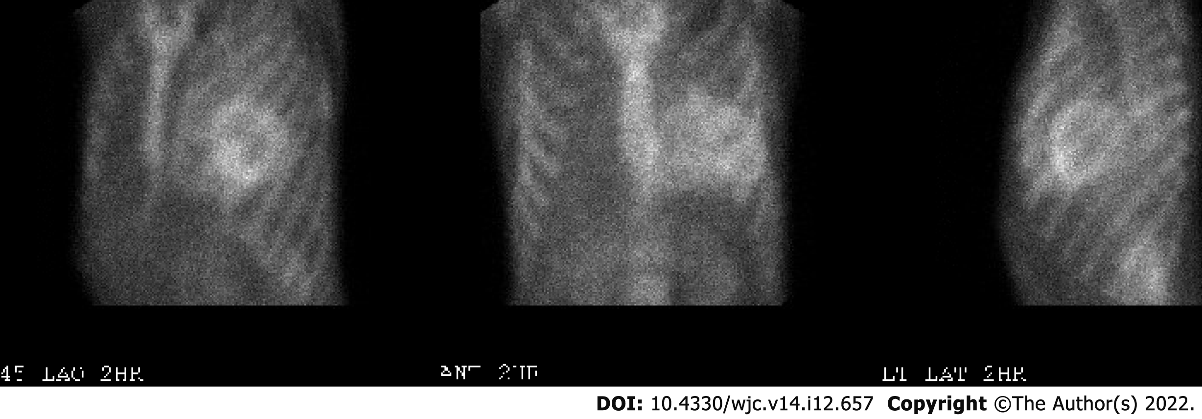
Figure 2 Technetium pyrophosphate scan showing increased myocardial uptake of tracer (visual grade 3) suggestive of transthyretin-mediated cardiac amyloidosis.
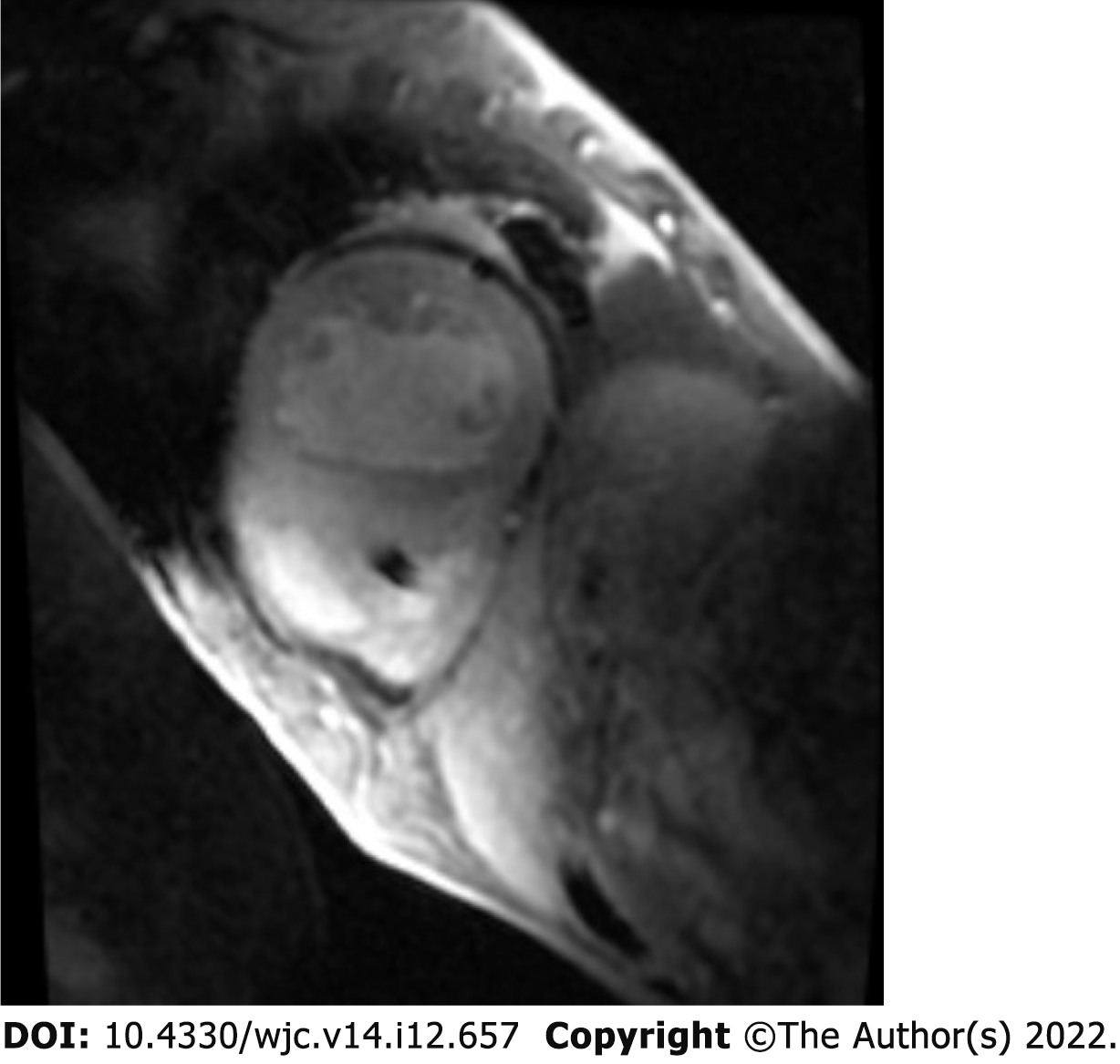
Figure 3 Cardiac magnetic resonance imaging showing delayed global hyperenhancement and failure to null likely due to cardiac amyloidosis.
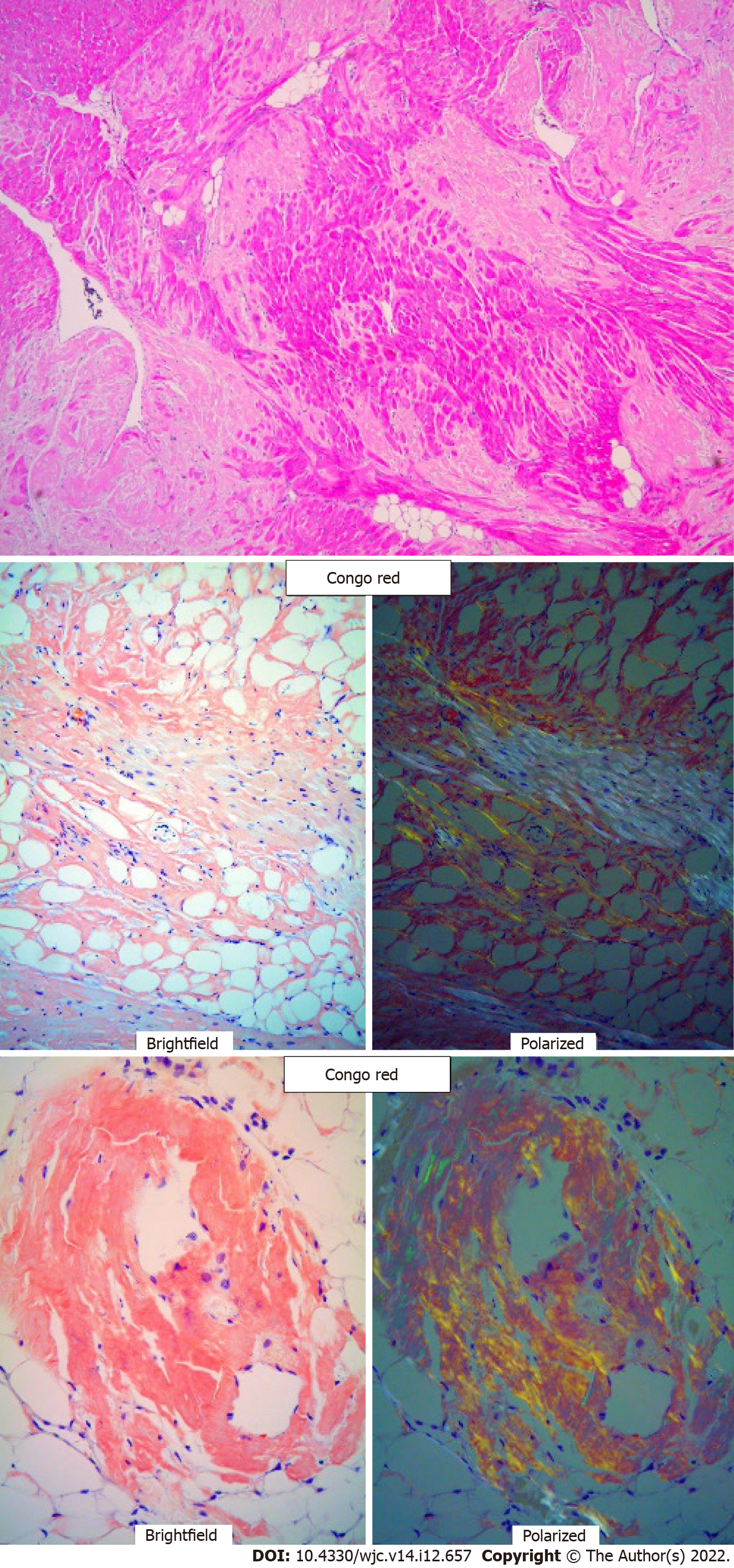
Figure 4 Native heart tissue pathology on H and E staining showing extracellular amyloid deposits, followed beneath by Congo red staining showing apple green refringence of amyloid deposits (100× magnification).
- Citation: Boda I, Farhoud H, Dalia T, Goyal A, Shah Z, Vidic A. Early and aggressive presentation of wild-type transthyretin amyloid cardiomyopathy: A case report. World J Cardiol 2022; 14(12): 657-664
- URL: https://www.wjgnet.com/1949-8462/full/v14/i12/657.htm
- DOI: https://dx.doi.org/10.4330/wjc.v14.i12.657


