Copyright
©The Author(s) 2021.
World J Cardiol. Sep 26, 2021; 13(9): 381-398
Published online Sep 26, 2021. doi: 10.4330/wjc.v13.i9.381
Published online Sep 26, 2021. doi: 10.4330/wjc.v13.i9.381
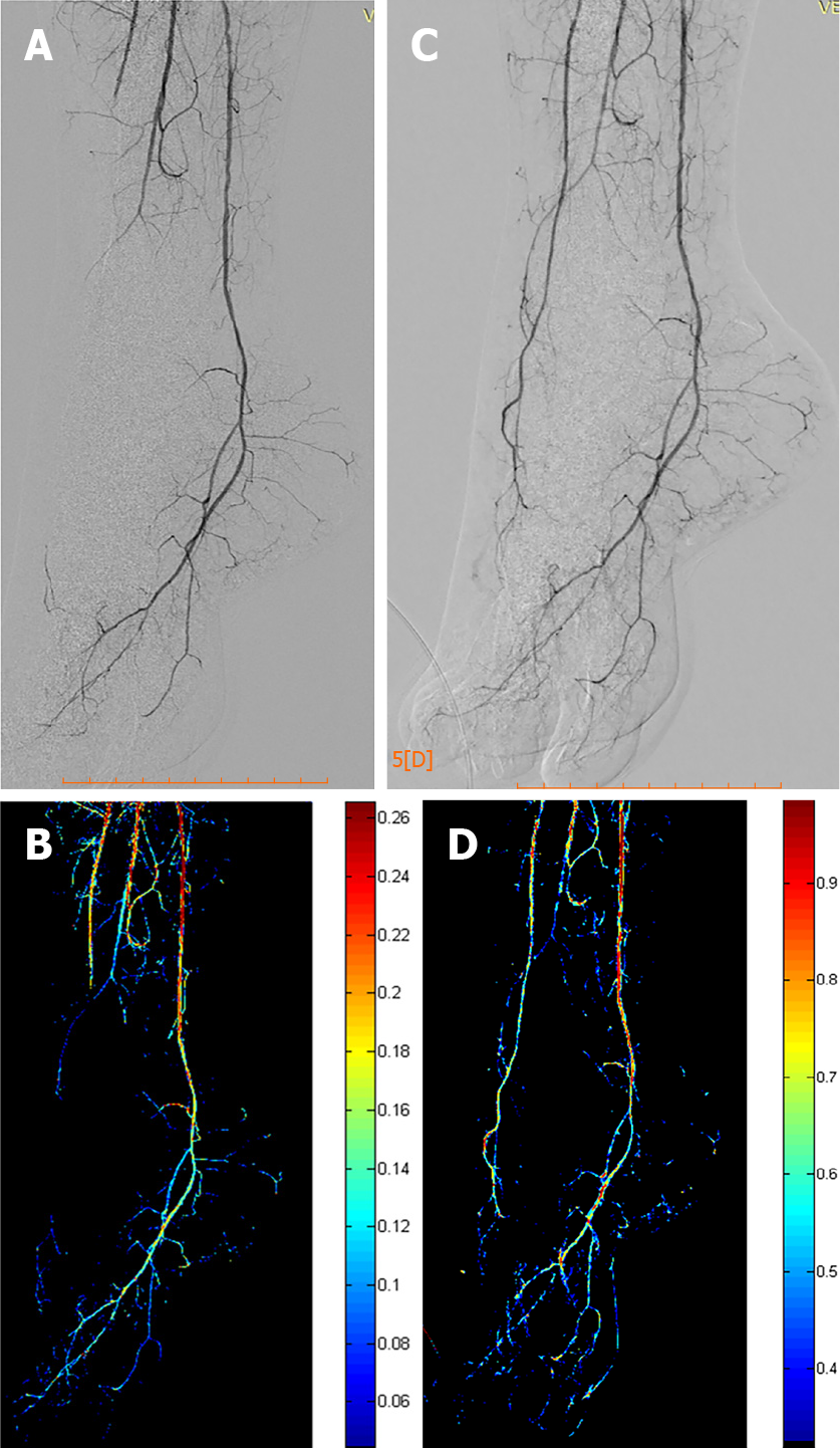
Figure 1 Two-dimensional perfusion digital subtraction angiography algorithm.
A and B: Pre-procedural digital subtraction angiography (DSA) depicting a chronic total occlusion of the distal anterior tibial and pedal arteries with the respective perfusion blood volume (PBV) map; C and D: Post-procedural DSA after balloon angioplasty of the occlusion and corresponding post-procedural PBV map.
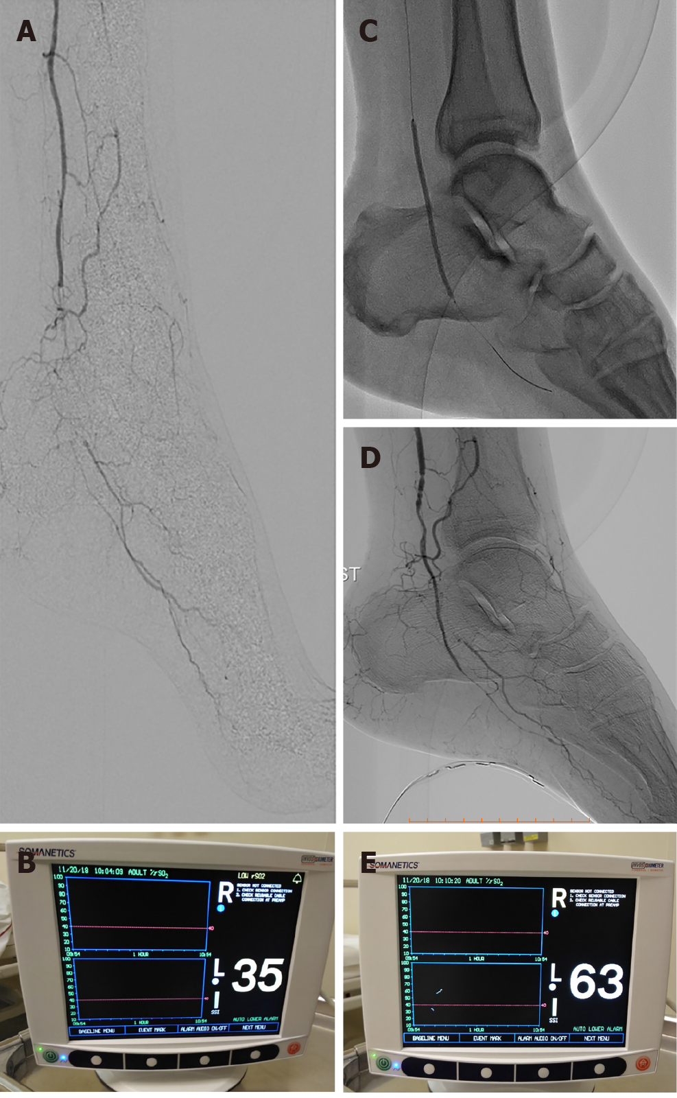
Figure 2 A case of 68-year-old male patient with insulin-dependent diabetes mellitus and advanced Rutherford-Becker class 6 gangrene of the left foot.
A and B: Digital subtraction angiography depicting a total occlusion of the distal posterior tibial artery and very low percentage value (35%) of regional hemoglobin oxygen saturation according to real-time near-infrared spectroscopy (NIRS) assessment of foot perfusion; C and D: Balloon angioplasty and final angiographic result demonstrating revascularization of the target occlusion. Note the NIRS electrode attached to the plantar surface of the treated foot; E: Immediately after revascularization, the percentage value of regional hemoglobin oxygen saturation was increased to 63%, demonstrating an 80% increase in foot tissue perfusion.
- Citation: Arkoudis NA, Katsanos K, Inchingolo R, Paraskevopoulos I, Mariappan M, Spiliopoulos S. Quantifying tissue perfusion after peripheral endovascular procedures: Novel tissue perfusion endpoints to improve outcomes. World J Cardiol 2021; 13(9): 381-398
- URL: https://www.wjgnet.com/1949-8462/full/v13/i9/381.htm
- DOI: https://dx.doi.org/10.4330/wjc.v13.i9.381


