Copyright
©The Author(s) 2021.
World J Cardiol. Nov 26, 2021; 13(11): 628-649
Published online Nov 26, 2021. doi: 10.4330/wjc.v13.i11.628
Published online Nov 26, 2021. doi: 10.4330/wjc.v13.i11.628
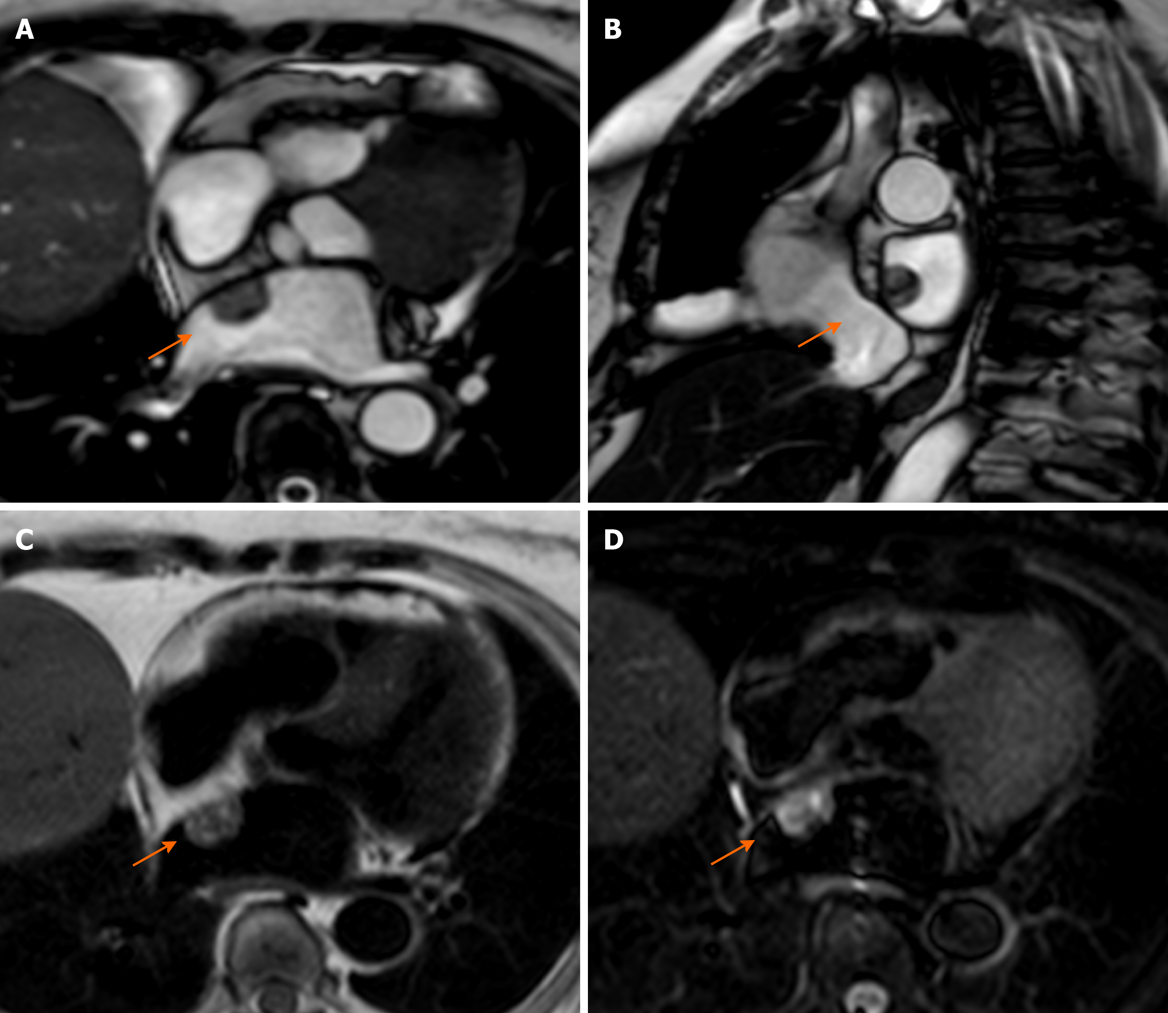
Figure 1 Cardiac mass in a 68-year-old male.
Transthoracic echocardiography shows (A) an apparently free mass in the right atrium mimicking a thrombus or a tumor (orange arrow). Cardiovascular magnetic resonance (B, C: 4-chamber cine steady state free precession diastolic and systolic frame respectively) reveals a pseudomass: A hypertrophied crista terminalis (orange arrow) with appearance of right atrial mass on transthoracic echocardiography.
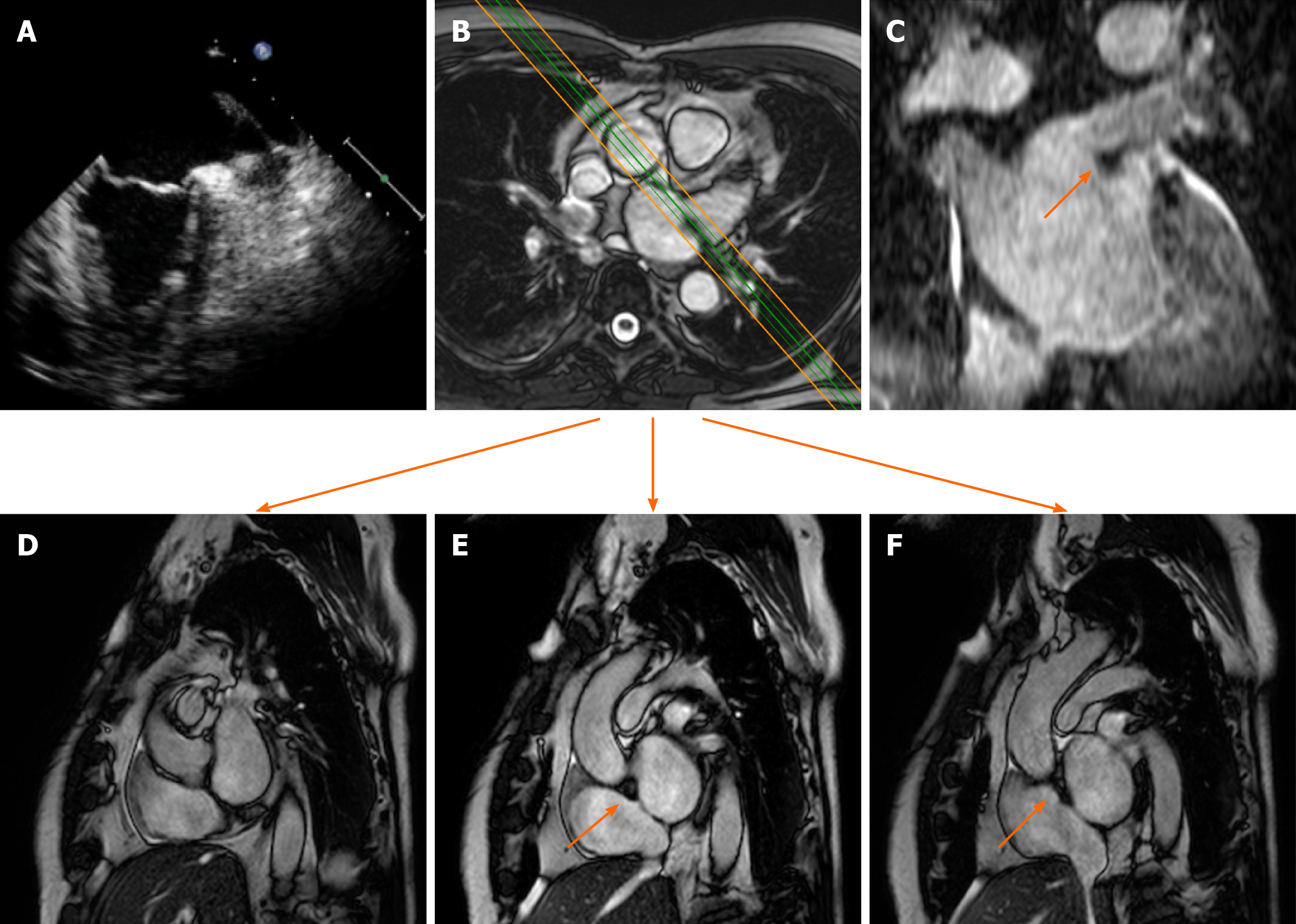
Figure 2 Cardiac mass in a 63-year-old male with atrial fibrillation.
Transthoracic echocardiography shows (A) a nodular mass that protrudes into the left atrium at the inlet of the left appendage. The patient underwent 2 mo of oral anticoagulant therapy, and the “mass” did not change. It was therefore requested a cardiovascular magnetic resonance (CMR) (B: axial cine- steady state free precession (SSFP) with the correspondent perpendicular plane oriented according to the green and orange lines and reported in D, E and F), moreover it was performed a three dimensional-steady state free precession SSFP acquisition with an oriented reconstruction (C). Overall the CMR shows the presence of pseudomass (orange arrow), a prominent coumadin ridge in the roof of the left atrium adjacent to the left upper pulmonary vein.
Figure 3 Sixty-five-year-old male patient with a dumbbell-shaped mass along the interatrial septum with negative Hounsfield Unit values on computed tomography scan (A), cardiovascular magnetic resonance cine-steady state free precession images (B and C, diastolic and systolic frame respectively) reveals the chemical shift artifact, also known as “India ink” artifact, typical of structures with adipose content.
These findings are in keeping with lipomatous hypertrophy of the interatrial septum (orange arrow).
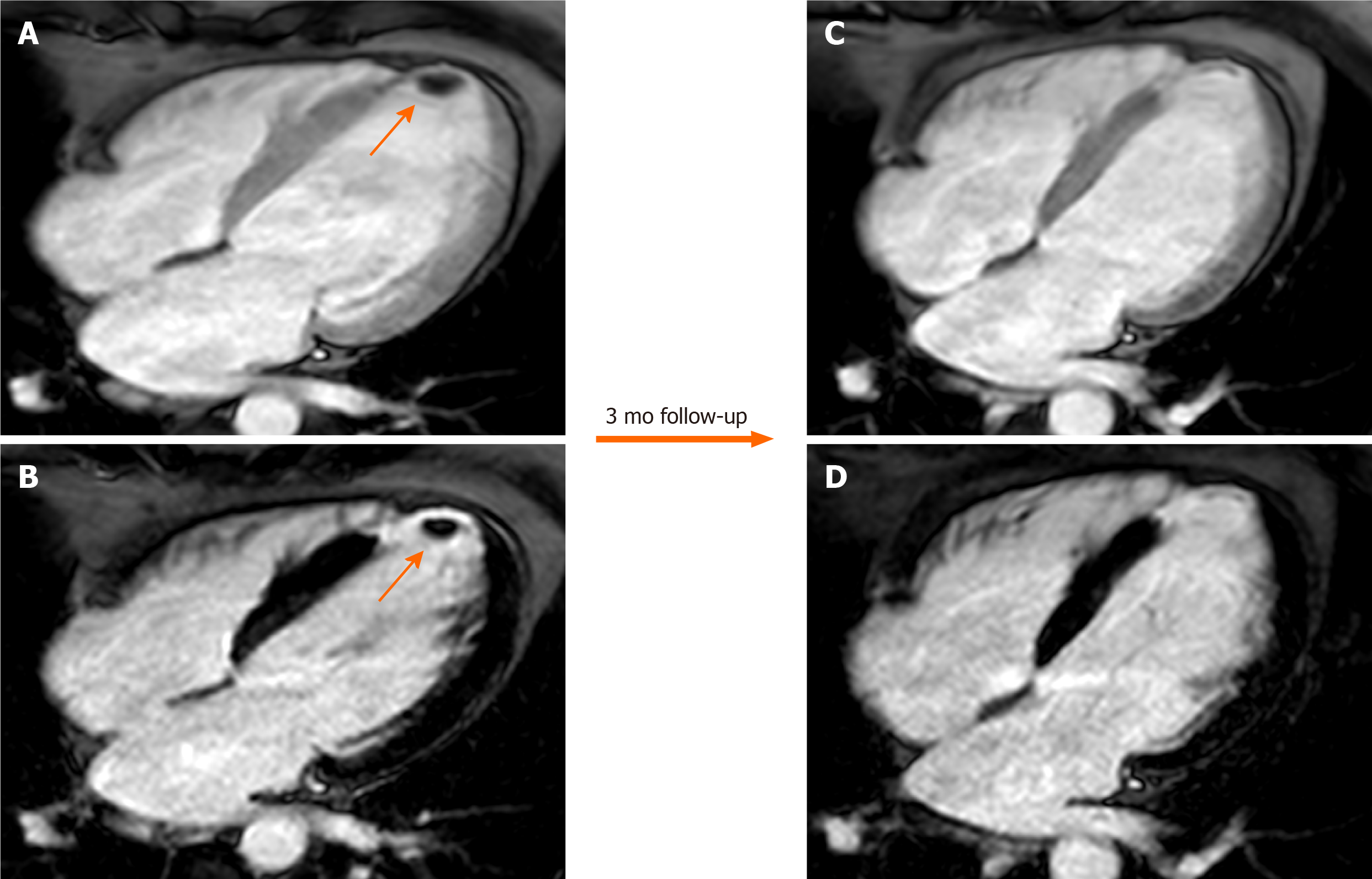
Figure 4 Sixty-six-year-old male patient with a history of myocardial infarction and suspected thrombotic formation at echocardiographic transthoracic examination.
Cardiovascular magnetic resonance shows a hypointense intraventricular “mass” at the left ventricular apex (orange arrow) in the early gadolinium enhancement (A) and late gadolinium enhancement (LGE) (B) sequences near the infarcted wall (i.e. with transmural LGE involvement). The patient underwent 3 mo of anticoagulant therapy with disappearance of the thrombus in the follow-up examination.
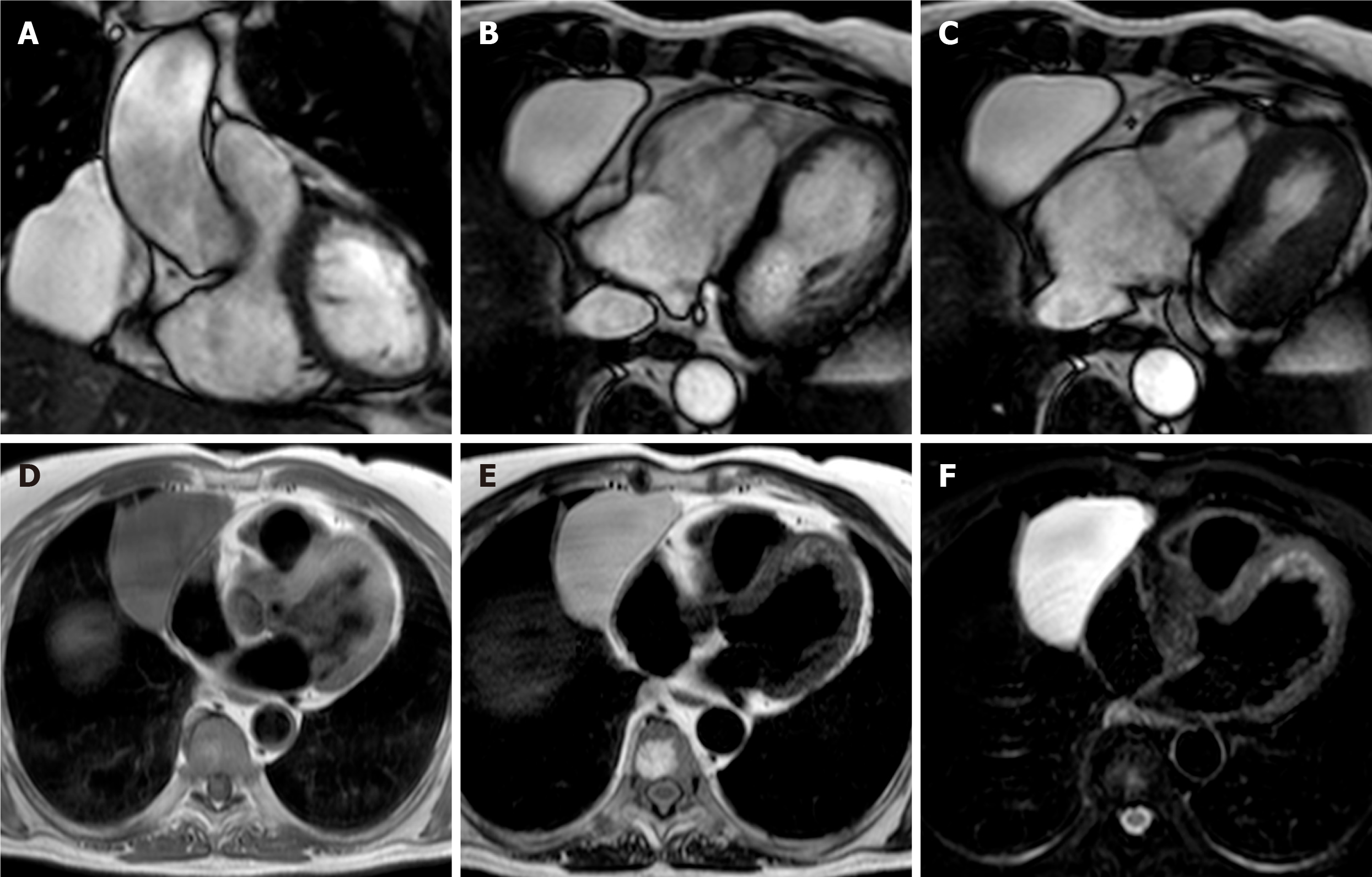
Figure 5 Fifty-seven-year-old female patient with parenchymal mass in the right cardiophrenic angle on chest X-ray.
This finding was first investigated by chest computed tomography and then by cardiovascular magnetic resonance (CMR) (A-C). The CMR confirm the presence of the mass on cine-steady state free precession images (A and B, two orthogonal diastolic frames of plane through the mass respectively), without sign of infiltration confirmed by the preserved movement of the heart chambers in relation to the mass itself. The mass shows low signal on T1-weighted sequences (D) and high signal on T2-weighted images (E and F, without and with fat suppression, respectively). These findings are in keeping with pericardial cyst.
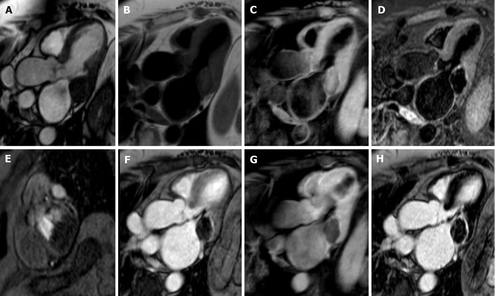
Figure 6 Eighty-year-old female with hyperechoic mass of uncertain significance in the left atrio-ventricular groove discovered at transthoracic echocardiography.
Cardiovascular magnetic resonance confirms the presence of the mass with the cine- steady state free precession images (A). The mass shows slight hyperintense signal on T1-weighted images without and with fat suppression (B and C, respectively), due to the presence of proteinaceous material, and hypointense signal on short tau inversion recovery images (D). The mass shows no contrast uptake during perfusion sequences (E) and no contrast enhancement both on early gadolinium enhancement images (F), T1- weighted images (G) repeated after the contrast medium injection, and late gadolinium enhancement images (H) with a peripheral rim of enhancement. These findings are in keeping with caseous calcification of the mitral valve.
Figure 7 Seventy-nine-year-old female patient with left atrial mass adherent to the interatrial septum discovered on cardiac computed tomography angiography.
The cardiovascular magnetic resonance confirms the presence of the mass using the cine- steady state free precession images (A and B, two orthogonal planes along the mass respectively). The site of lesion and its heterogeneous appearance on T2-weighet sequence (C and D) short tau inversion recovery, are in keeping with cardiac myxoma.
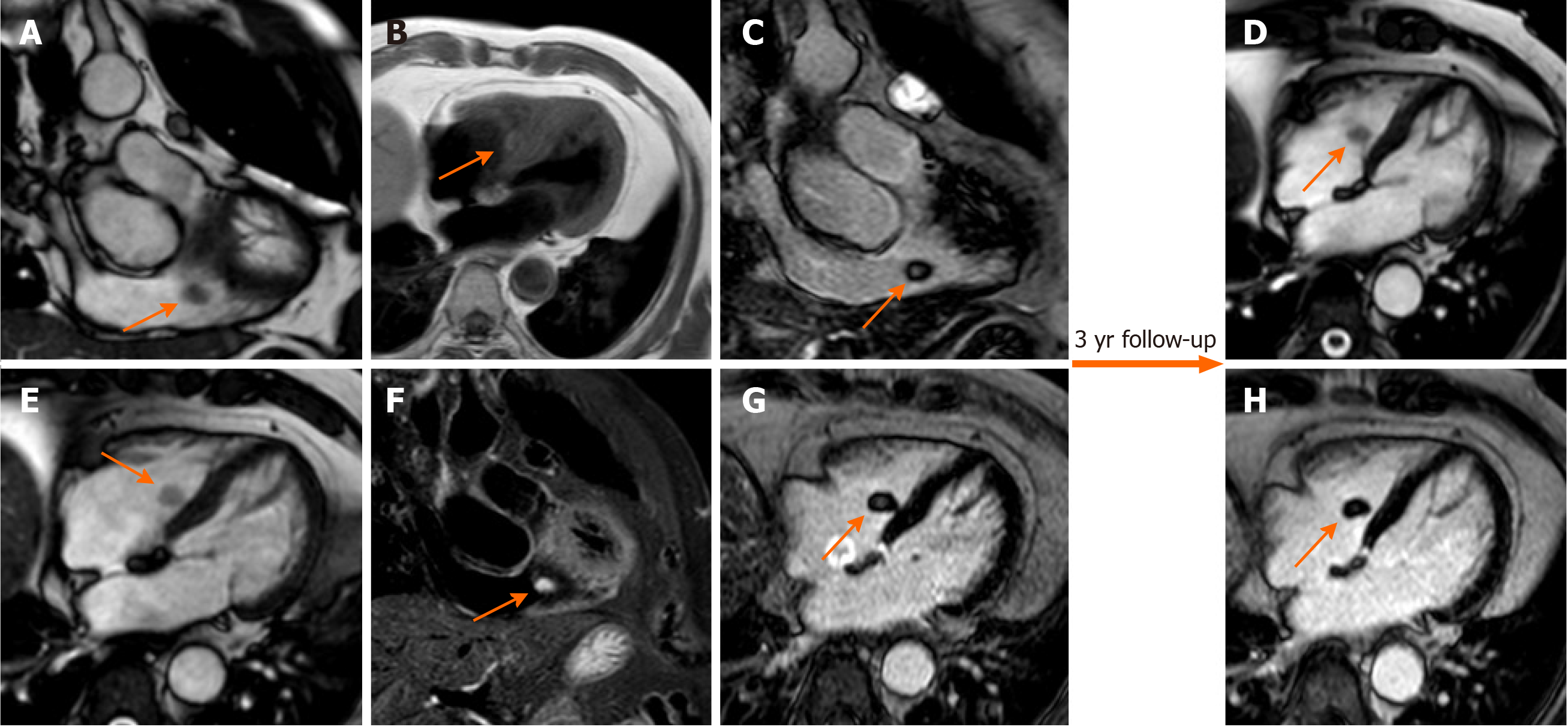
Figure 8 Seventy-one-year-old female with incidental finding of cardiac mass adherent to the septal leaflet of the tricuspid valve on cardiovascular magnetic resonance.
The examination was performed for suspected noncompact myocardium on a previous ultrasound examination in a patient with known metastatic thyroid cancer. Orange arrow shows a mobile “valvular” mass at cine-steady state free precession (SSFP) imaging (A and B), with intermediate signal on T1-weighted images (C), high signal on T2 short tau inversion recovery images (D) and with poor enhancement at late gadolinium enhancement (LGE) images (E and F). This location of the mass and its cardiovascular magnetic resonance (CMR) features are in keeping with fibroelastoma. The patient underwent a periodic CMR follow-up for 3 years (G: cine-SSFP and H: LGE sequences), and the mass shows no change.
Figure 9 Fifty-one-year-old male patient, with incidental finding of a suspicious intraseptal hyperechoic mass at transesophageal echocardiography.
Cine-steady state free precession magnetic resonance (A and B) confirms the presence of an intraseptal mass with a chemical shift artefact at the border. The mass shows high signal intensity on proton density-weighted images (C) and low signal intensity on short tau inversion recovery images (D). The mass shows no enhancement on perfusion images (E and F, two different frame). These findings are in keeping with cardiac lipoma.
Figure 10 Twenty-five-year-old male patient with a ventricular mass of uncertain significance on computed tomography scan performed after an episode of dyspnea and chest pain.
The cardiovascular magnetic resonance demonstrates a well-defined, solitary solid mass with intramural growth in the anterior wall of the left ventricle (A and B, cine steady state free precession diastolic and systolic frame, respectively). The mass shows homogeneous hypointense signal on short tau inversion recovery (C) and T1-weighted images (D). Homogeneous contrast uptake during perfusion sequences (E and F, two different frame of the perfusion sequence) and homogeneous hyperintensity in the early (G) and late gadolinium enhancement images (H). These findings are in keeping with cardiac fibroma. The patient finally underwent cardiac surgery, and the final histopathological diagnosis confirmed the radiological suspicion.
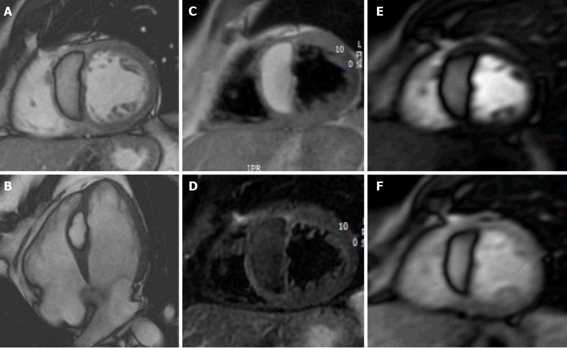
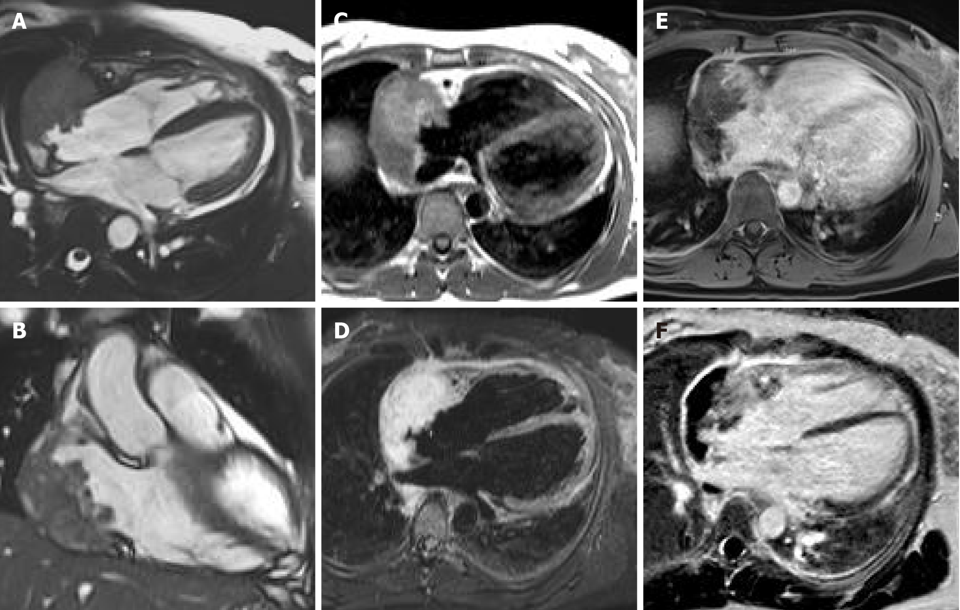
Figure 11 Sixty-five-year-old female patient with transthoracic echocardiogram finding of irregular mass originates from the right atrium wall with intracavitary expansion.
Cardiovascular magnetic resonance with cine-steady state free precession images (A and B) confirms the presence of an infiltrative atrial mass in the proximity of atrio-ventricular sulcus. T1-weighted and short tau inversion recovery images (C and D, respectively) show an heterogenous hyperintense signal intensity due to complex composition of the mass. The mass shows inhomogeneous enhancement at early gadolinium enhancement and late gadolinium enhancement sequences (E and F, respectively). These findings are in keeping with a malignant primary cardiac tumor. The patient underwent myocardial biopsy with diagnosis of angiosarcoma.
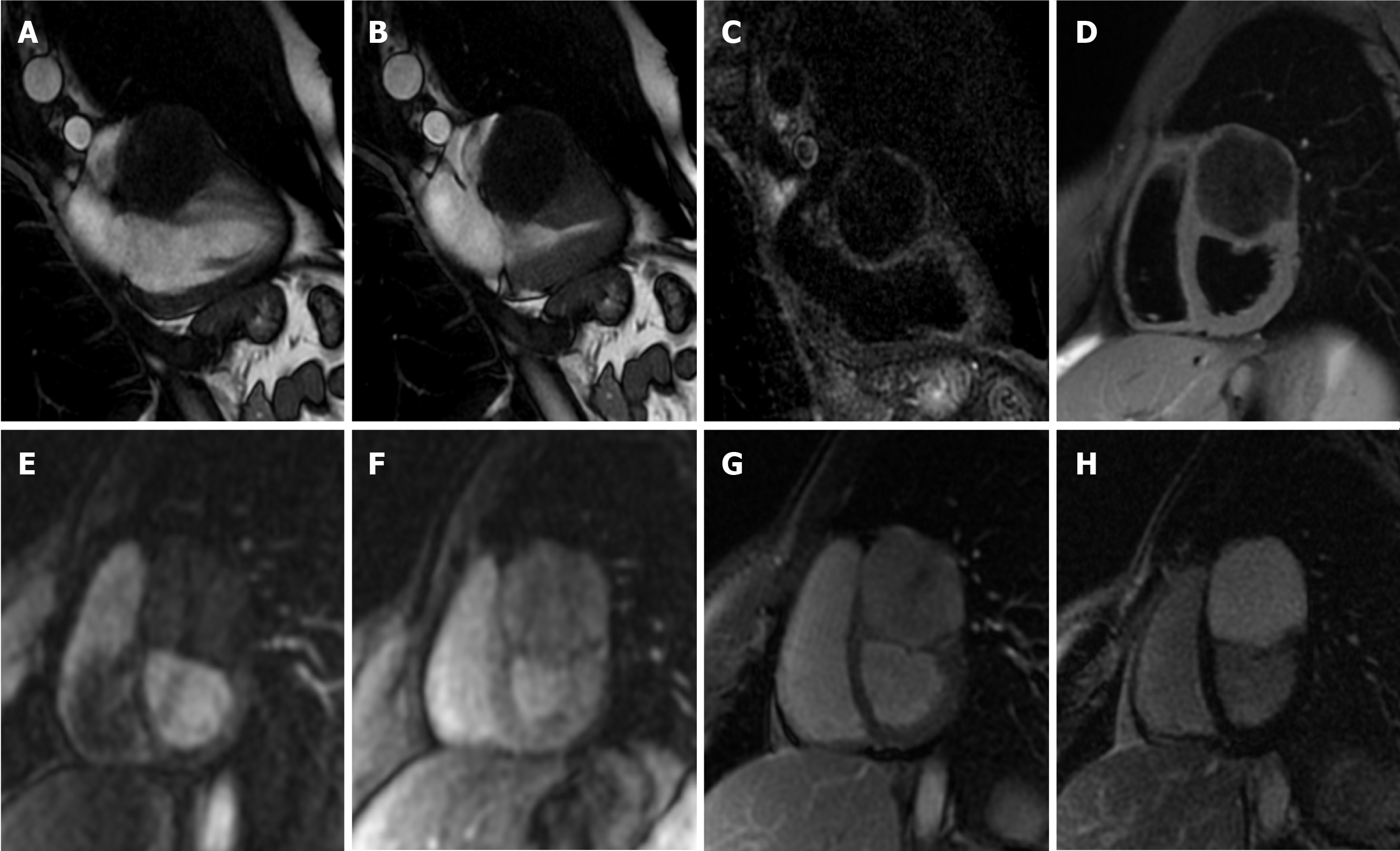
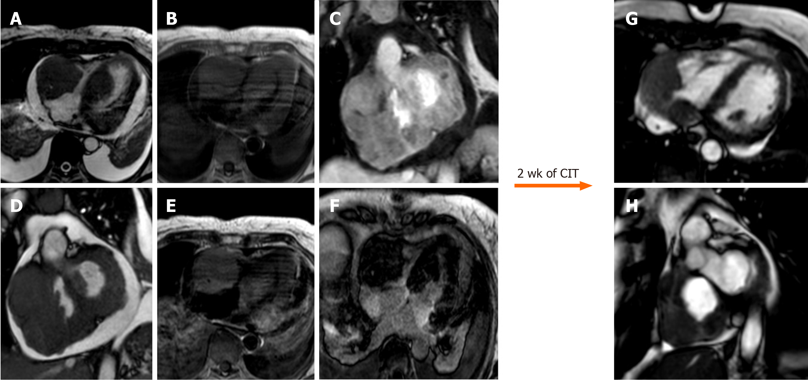
Figure 12 Sixty-five-year-old female with transthoracic echocardiogram finding of two masses in the left ventricle and right cavities following an episode of lipothymia.
Cardiovascular magnetic resonance (CMR) with cine-steady state free precession images (A and B) demonstrates the presence of four distinct masses, the most voluminous mass with infiltrative features and irregular margins, extending from the right atrio-ventricular groove to both atrial and the ventricular walls. These lesions are characterized by a substantially isointense signal to cardiac muscle in T1-weighted images (C), slightly hyperintense in T2-weighted images (D) with heterogeneous enhancement at early gadolinium enhancement and relatively homogeneous hypointensity at late gadolinium enhancement images (E, F). These findings are in keeping with cardiac lymphoma. The patient underwent biopsy with a diagnosis of cardiac large B-cell lymphoma and started a chemoimmunotherapy with an excellent response as shown on CMR (G and H, cine steady state free precession) after 2 wk. CIT: Chemo
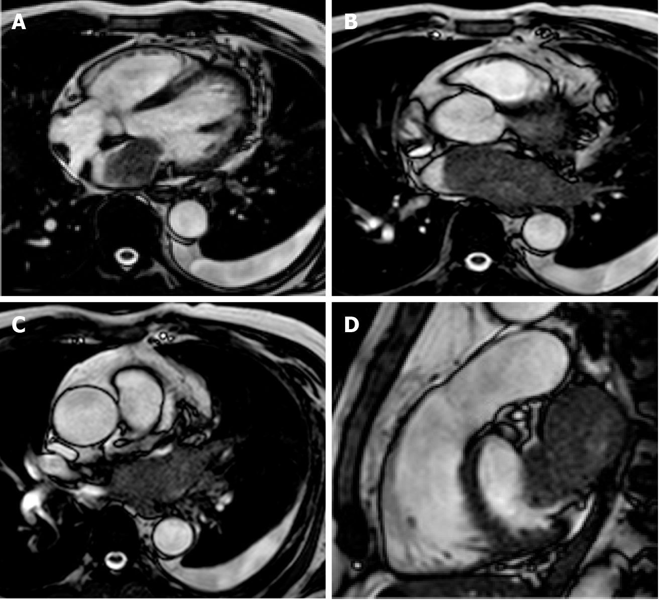
Figure 13 Sixty-five-year-old male with lung cancer.
Cardiovascular magnetic resonance examination with cine-steady state free precession sequences in planes of various orientations (A-D) clearly shows infiltration of the atrium through the pulmonary veins.
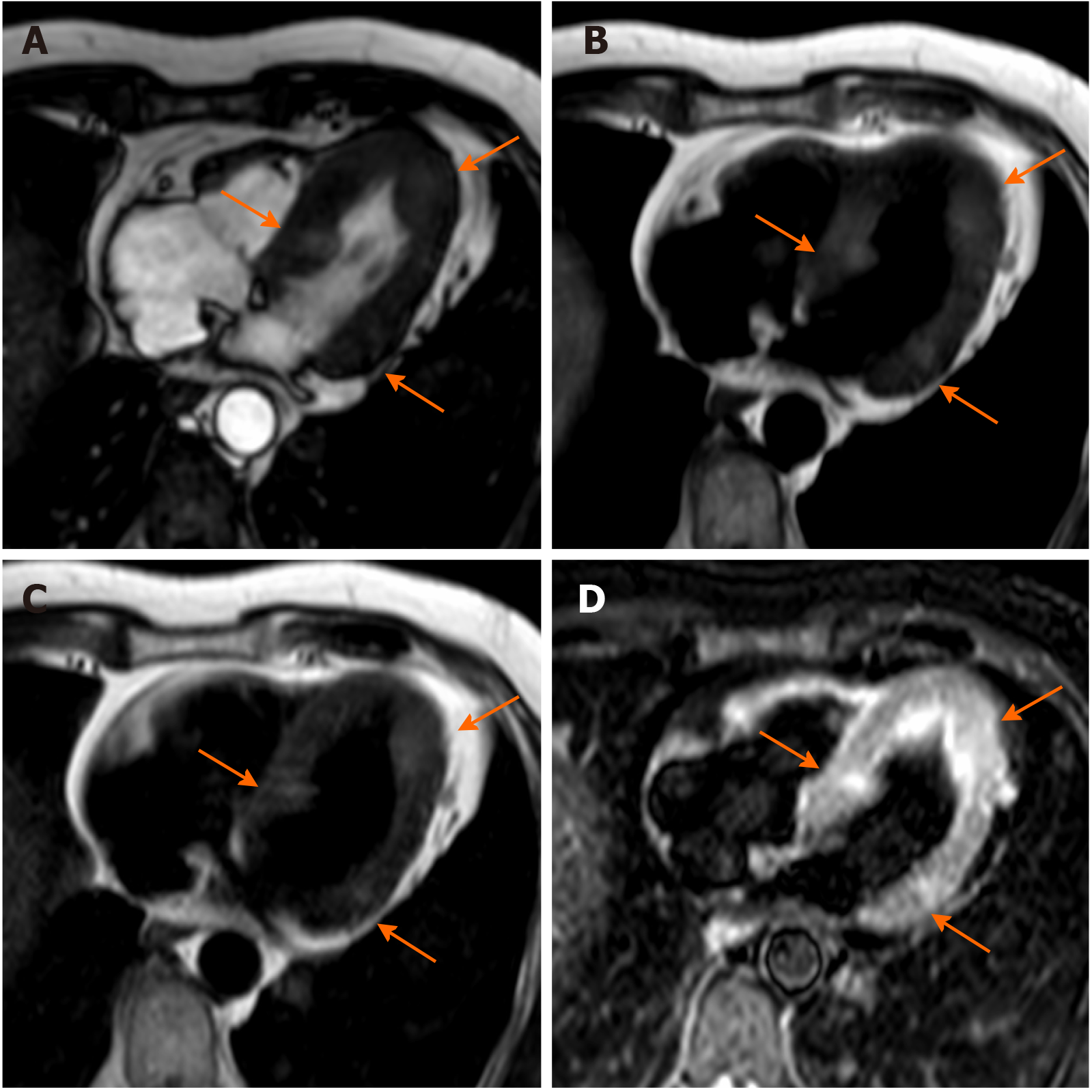
Figure 14 Sixty-four-year-old male with ocular melanoma with multiple metastases underwent cardiovascular magnetic resonance for characterization of a myocardial mass discovered at echocardiography.
Nine oval nodules (three are indicated by orange arrows) with diameters ranging from 5 mm to 19 mm, slightly hyperintense in cine-steady state free precession images, T1-weighted (due to the presence of melanin) and T2-weighted sequences. The findings are in keeping with cardiac melanoma localization.
- Citation: Gatti M, D’Angelo T, Muscogiuri G, Dell'aversana S, Andreis A, Carisio A, Darvizeh F, Tore D, Pontone G, Faletti R. Cardiovascular magnetic resonance of cardiac tumors and masses. World J Cardiol 2021; 13(11): 628-649
- URL: https://www.wjgnet.com/1949-8462/full/v13/i11/628.htm
- DOI: https://dx.doi.org/10.4330/wjc.v13.i11.628


