©The Author(s) 2020.
World J Cardiol. Jun 26, 2020; 12(6): 231-247
Published online Jun 26, 2020. doi: 10.4330/wjc.v12.i6.231
Published online Jun 26, 2020. doi: 10.4330/wjc.v12.i6.231
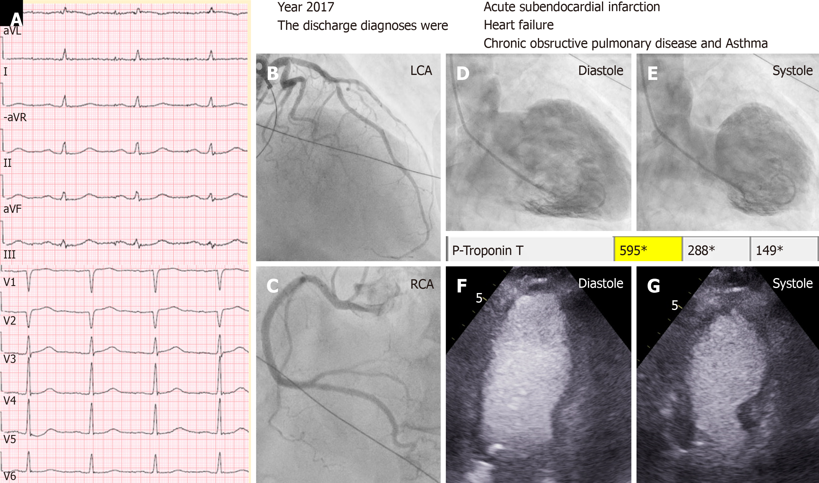
Figure 1 A case of midventricular takotsubo syndrome discharged under diagnosis of myocardial infarction.
A typical mid-ventricular takotsubo syndrome triggered by an emotional stress in a 79-years-old man with chronic co-morbidities in the form of chronic obstructive pulmonary disease and carcinoma of urinary bladder. The case was incorrectly diagnosed as type 2 myocardial infarction according to the universal definition of myocardial infarction as seen in the discharge diagnosis. A: Electrocardiogram during admission; B: Left coronary artery shows moderate stenosis in the left anterior descending artery, which cannot explain the circumferential mid-ventricular left ventricular wall motion abnormality; C: Normal right coronary artery (RCA); D and E: Contrast left ventriculography during diastole and systole showing mid-ventricular ballooning pattern. There was modest elevation of troponin T (595 ng/L); F and G: Echocardiography during diastole and systole confirming the left ventriculography finding of mid-ventricular ballooning.
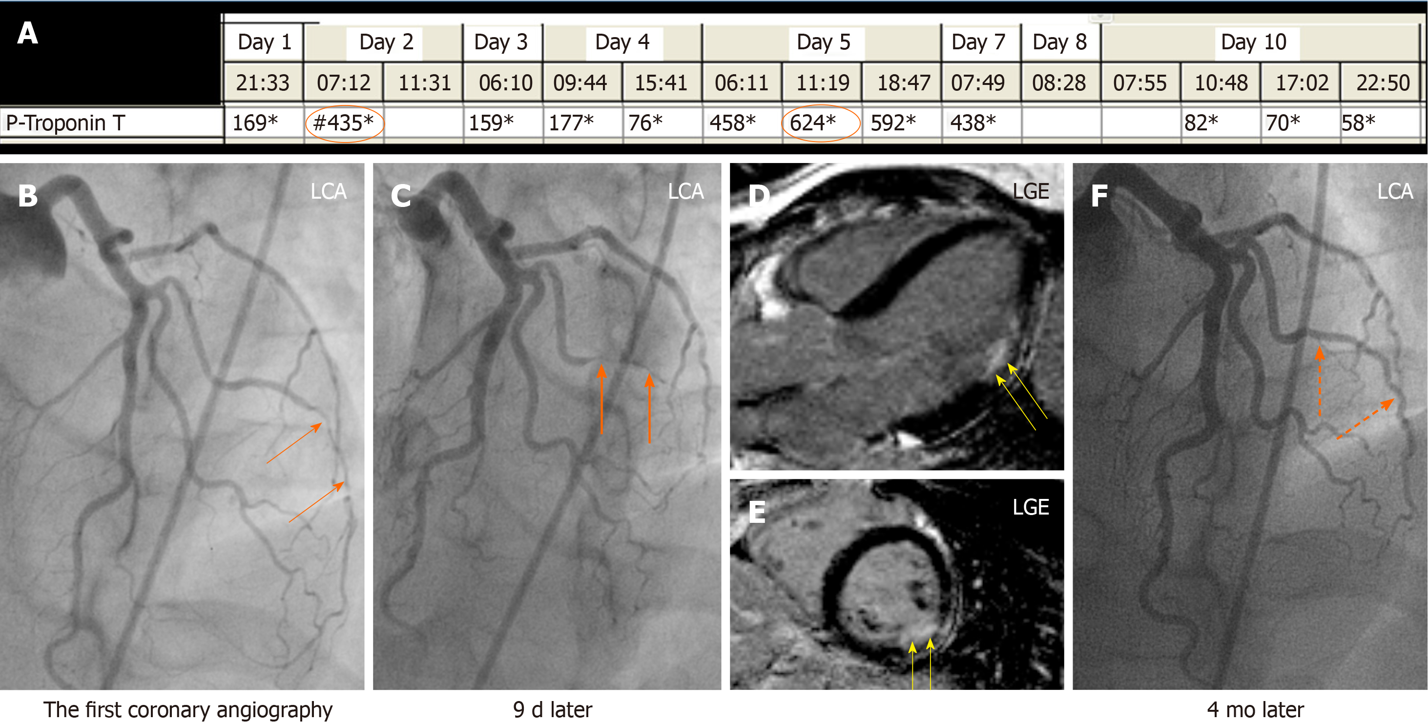
Figure 2 A case of obstructive coronary artery disease due spontaneous coronary artery dissection.
A 35-years-old female patient with a spontaneous coronary artery dissection (SCAD) of the diagonal artery with documented myocardial infarction (MI) by cardiac magnetic resonance imaging and obstructive coronary artery stenosis. A: Recurrent troponin elevation with a rise and/or fall pattern (orange circles); B: A diagonal artery with a peripheral stenosis (thin orange arrows); C: Proximal propagation of diagonal artery narrowing during repeated left coronary artery angiography 9 d later (thick orange arrows); D and E: Cardiac magnetic resonance imaging shows MI corresponding to the diagonal artery supply territory (yellow arrows); F: Follow up left coronary artery angiography 4 mo later reveals complete resolution of the diagonal artery lesion consistent with SCAD (broken orange arrows). Consequently, this patient has documented MI caused by obstructive coronary artery disease due to SCAD. LGE: Late gadolinium enhancement; LGA: Left coronary artery.
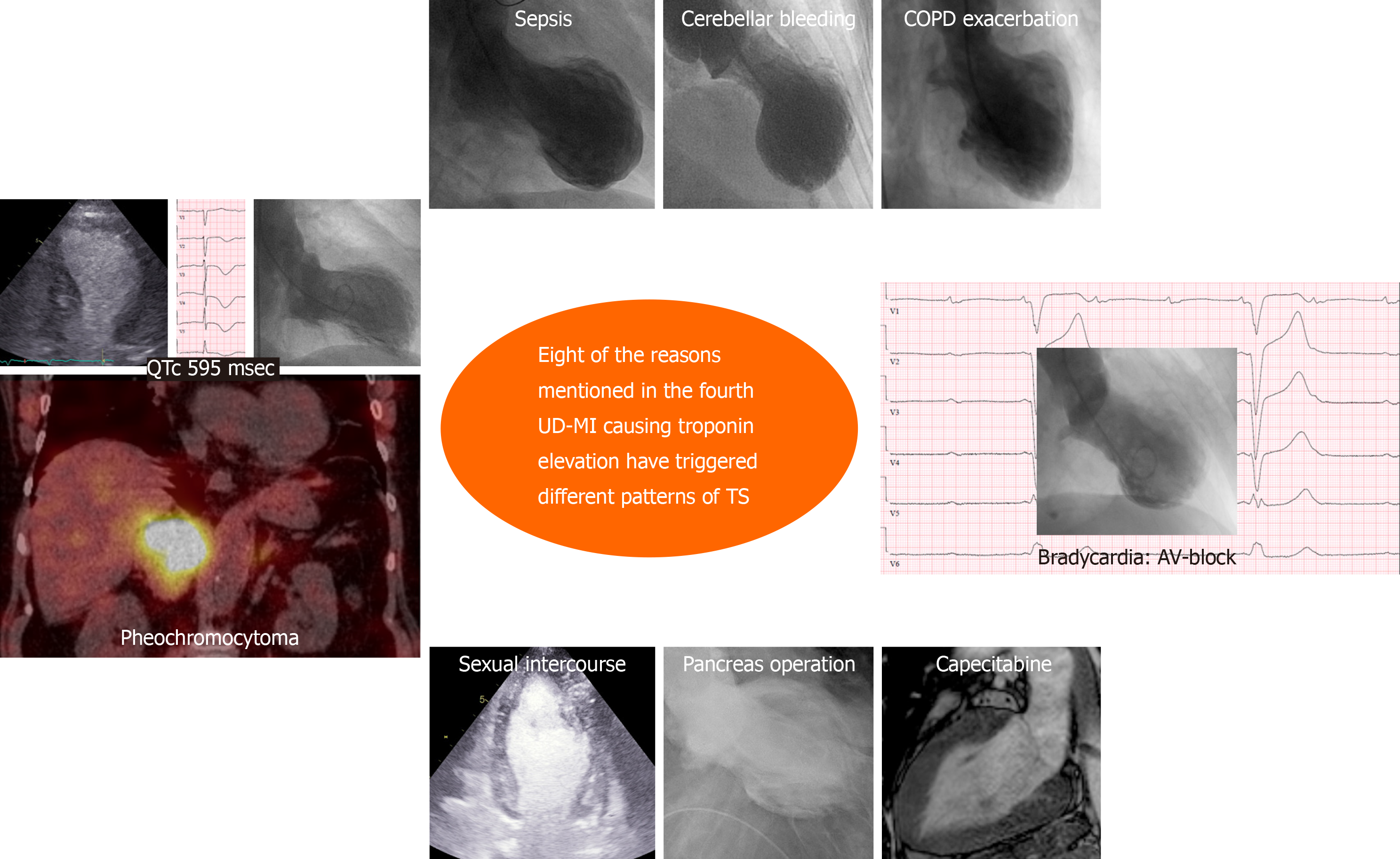
Figure 3 Eight clinical conditions causing troponin elevation have triggered different patterns of takotsubo syndrome.
Most of the reasons for troponin elevation mentioned in the fourth universal definition of myocardial infarction have been reported to trigger takotsubo syndrome. In this figure, 8 of the mentioned reasons (pheochromocytoma, sepsis, cerebellar bleeding, chronic obstructive pulmonary disease with acute exacerbation, heart block, capecitabine, pancreas operation, and strenuous physical exercise in the form of sexual intercourse) triggering different ballooning’s pattern of takotsubo syndrome are demonstrated.
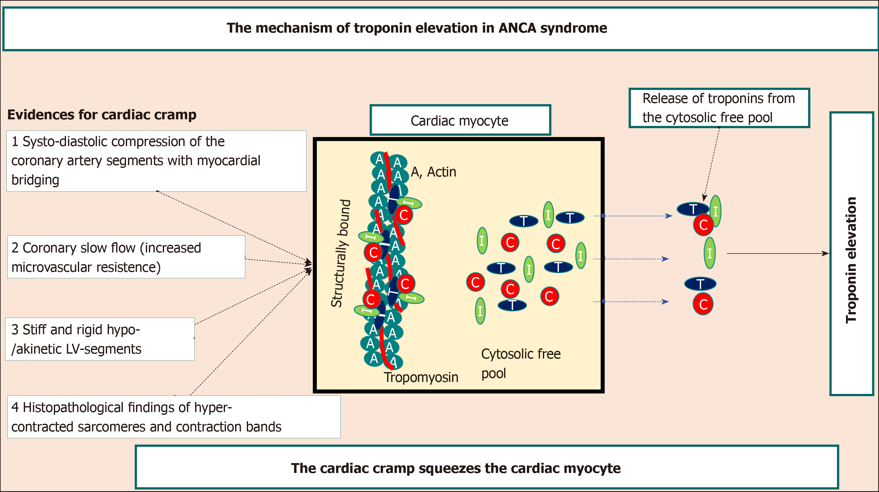
Figure 4 Mechanism of troponin elevation in autonomic neurocardiogenic syndrome.
Illustration of the hypothetical mechanism of troponin release in autonomic neurocardiogenic syndrome including takotsubo syndrome and takotsubo-related conditions. The systo-diastolic compression of a coronary artery segment specially left anterior descending artery with myocardial bridging, the coronary slow flow due to increased microvascular resistance, the stiff and rigid myocardial stunning, and the hyper-contracted sarcomeres indicate that the affected myocardium in autonomic neurocardiogenic syndrome including takotsubo syndrome is in a state of cardiac cramp. This cardiac cramp may squeeze the cardiac myocyte causing release of troponins from the cytosolic free pool resulting in mild-moderate troponin elevation as demonstrated in the figure. C: Troponin C; I: Troponin I; T: Troponin T; ANCA: Autonomic neurocardiogenic.
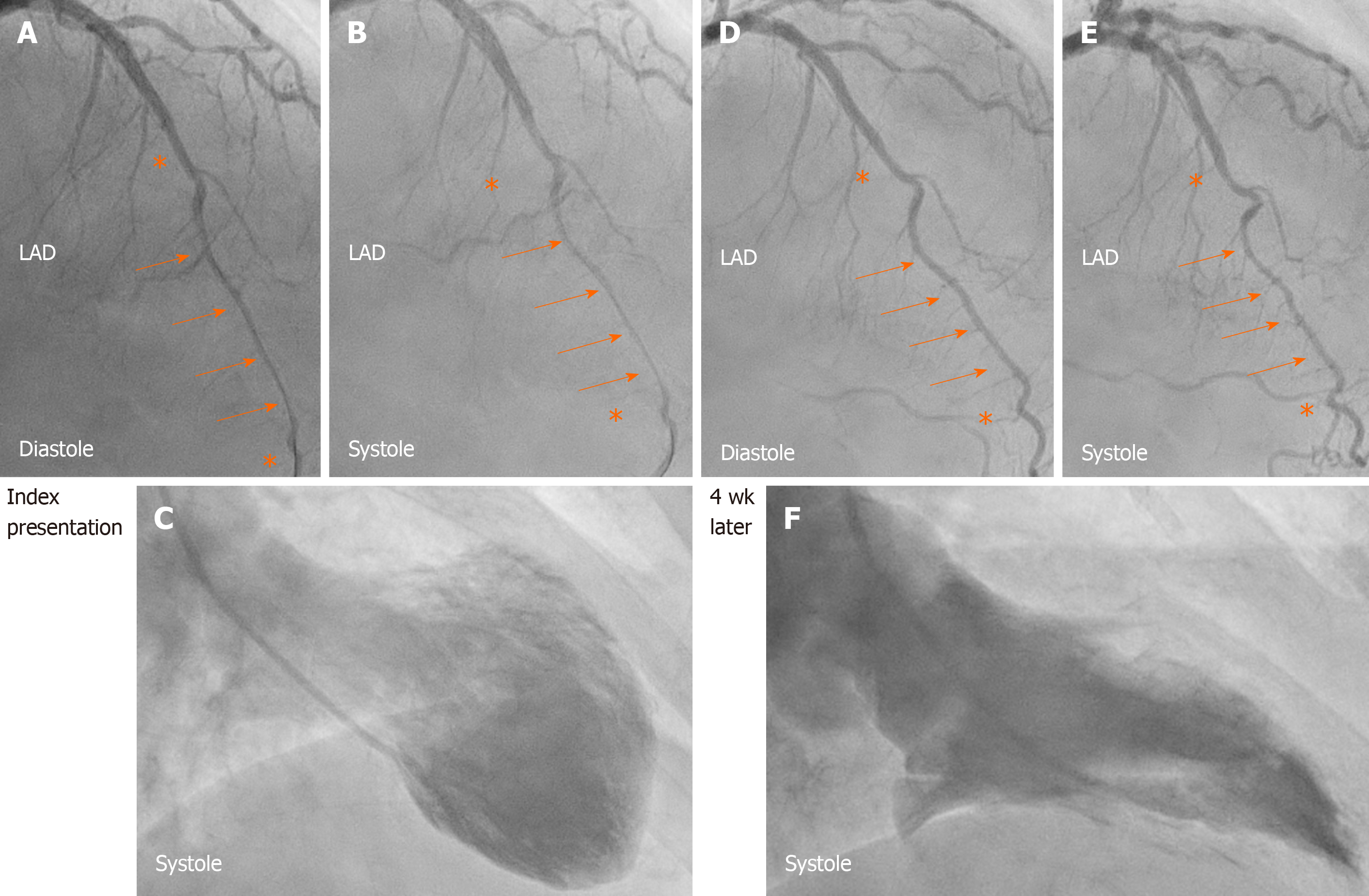
Figure 5 Systo-diastolic compression of a coronary artery segment with myocardial bridging.
A, B, and C: Left anterior descending artery (LAD) during diastole (A) and systole (B) in the acute stage of takotsubo syndrome (TS) in a patient where contrast left ventriculography revealed mid-apical pattern of TS (C). During the acute stage of the disease: Note the diastolic and systolic compression of a segment of LAD with myocardial bridging (orange arrows) bordered by normal segments (Asterix) before and after the segment with myocardial bridging. D, E, and F: Four weeks later, there was complete relief of LAD compression during diastole (D, orange arrows) and only mild systolic compression (E, orange arrows) after normalization of left ventricular function (F). This indicates that the stunned myocardium was in a cramp state during the acute-subacute stage of TS. LAD: Left anterior descending artery.
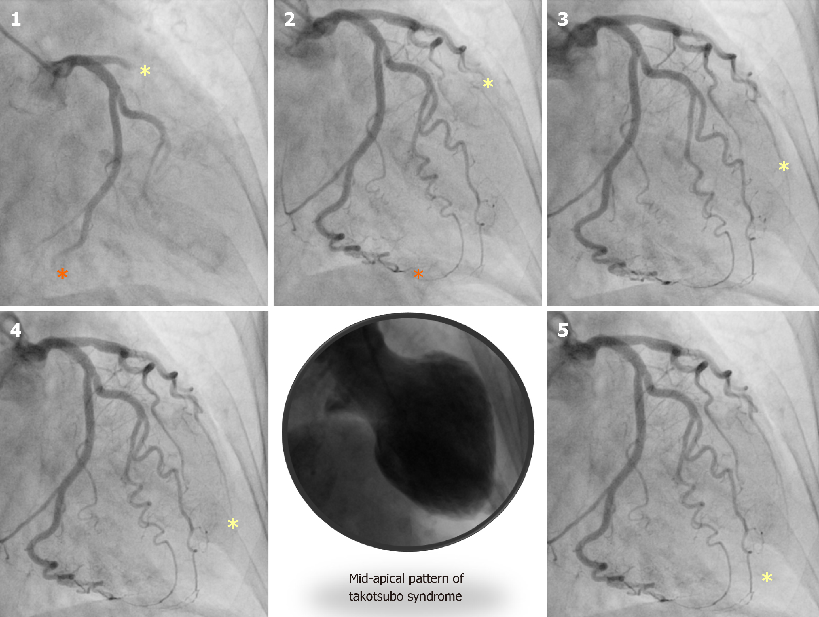
Figure 6 Slow flow in the left anterior descending artery in a patient with takotsubo syndrome.
A case of mid-apical ballooning pattern of takotsubo syndrome (middle, lower panel) showing the marked slow flow in the left anterior descending artery (yellow Asterix) compared to left circumflex artery (orange Asterix) in panel 1 to 5.
- Citation: Y-Hassan S. Autonomic neurocardiogenic syndrome is stonewalled by the universal definition of myocardial infarction. World J Cardiol 2020; 12(6): 231-247
- URL: https://www.wjgnet.com/1949-8462/full/v12/i6/231.htm
- DOI: https://dx.doi.org/10.4330/wjc.v12.i6.231


