Published online Feb 26, 2014. doi: 10.4331/wjbc.v5.i1.58
Revised: November 9, 2013
Accepted: December 17, 2013
Published online: February 26, 2014
Processing time: 201 Days and 9.8 Hours
Mobilization of intracellular Ca2+ stores is involved in many diverse cell functions, including: cell proliferation; differentiation; fertilization; muscle contraction; secretion of neurotransmitters, hormones and enzymes; and lymphocyte activation and proliferation. Cyclic adenosine diphosphate ribose (cADPR) is an endogenous Ca2+ mobilizing nucleotide present in many cell types and species, from plants to animals. cADPR is formed by ADP-ribosyl cyclases from nicotinamide adenine dinucleotide. The main ADP-ribosyl cyclase in mammals is CD38, a multi-functional enzyme and a type II membrane protein. It has been shown that many extracellular stimuli can induce cADPR production that leads to calcium release or influx, establishing cADPR as a second messenger. cADPR has been linked to a wide variety of cellular processes, but the molecular mechanisms regarding cADPR signaling remain elusive. The aim of this review is to summarize the CD38/cADPR/Ca2+ signaling pathway, focusing on the recent advances involving the mechanism and physiological functions of cADPR-mediated Ca2+ mobilization.
Core tip: This is a comprehensive review regarding the role and mechanism of the CD38/Cyclic adenosine diphosphate ribose (cADPR)/Ca2+ signaling pathway in various cellular processes. We introduce the structure and function of cADPR, together with its production and degradation pathways. We also describe CD38, the main enzyme that is responsible for synthesis of cADPR, through its structure and topology. Finally, we summarize the functions of the CD38/cADPR/Ca2+ signaling pathway under both physiological and pathological conditions.
- Citation: Wei W, Graeff R, Yue J. Roles and mechanisms of the CD38/cyclic adenosine diphosphate ribose/Ca2+ signaling pathway. World J Biol Chem 2014; 5(1): 58-67
- URL: https://www.wjgnet.com/1949-8454/full/v5/i1/58.htm
- DOI: https://dx.doi.org/10.4331/wjbc.v5.i1.58
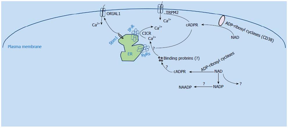
Discovered more than two decades ago, cyclic adenosine diphosphate ribose (cADPR) has been established as a second messenger, according to criteria first proposed by Sutherland and co-workers[1]. Together with inositol 1,4,5-trisphosphate (IP3) and nicotinic acid adenine dinucleotide phosphate (NAADP), cADPR has been recognized as a principal second messenger involved in cellular Ca2+ mobilization. Extracellular stimuli can induce cADPR production, which leads to Ca2+ mobilization from intracellular stores as well as Ca2+ entry from the extracellular compartment to initiate diverse cellular responses. cADPR is synthesized by ADP-ribosyl cyclases and the major ADP-ribosyl cyclase in mammals is CD38 (Figure 1). In this review, we will first introduce the structure and function of cADPR. Next, the structure and topology of CD38 will be reviewed. Finally, the physiological functions of CD38/cADPR/Ca2+ signaling and their involvement in pathological processes will be summarized.
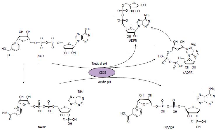
A suitable model system is the foundation of any novel finding and this concept is also true for the discovery of cADPR. Sea urchin eggs are large and amenable for microinjection studies so that Ca2+ mobilizing activities during fertilization can be readily observed, and it is easy to isolate endoplasmic reticulum (ER) from sea urchin eggs, making them the perfect system to investigate mechanisms of intracellular Ca2+ mobilization[2]. Taking advantage of the sea urchin homogenate preparation and use of the fluorescent Ca2+ indicator Fura 2, Lee et al[3] and Clapper et al[4] found that the pyridine nucleotide nicotinamide adenine dinucleotide (NAD) can invoke a delayed Ca2+ release from ER independent of IP3. They then determined that this delay was due to enzymatic conversion of NAD to cADPR by the homogenate. Later, Lee et al[5] solved the structure of cADPR by x-ray crystallography and showed that it is a novel cyclic nucleotide formed by the covalent linkage of the N1 nitrogen of the adenine ring to the anomeric carbon of the terminal ribose to become a closed cyclic structure (Figure 2). Benefiting from the identified structure, multiple cADPR analogs have been synthesized, which greatly promoted research on the role and mechanism of cADPR-mediated Ca2+ signaling[6-9].
From the very beginning of research on cADPR, several pharmacological studies have clearly shown that the mechanism of cADPR-induced Ca2+ release is different from that of IP3. For example, desensitization experiments demonstrated that the sea urchin homogenates which were desensitized to IP3 would still respond to cADPR[4], and the IP3 inhibitor heparin had no effect on the cADPR-induced Ca2+ release[10]. Using the sea urchin homogenate as the model, Galione et al[11] proposed that calcium-induced calcium release (CICR) may be modulated by cADPR, since concentrations of cADPR in the nanomolar range could greatly increase the sensitivity to Ca2+ during the CICR process. Thus, ryanodine receptors (RyRs) were proposed to be the cADPR receptors through which the CICR functions, and this idea was supported by several subsequent studies. For example, cADPR was shown to directly activate RyR2 that was incorporated into lipid bilayers[12]. In HEK293 cells transfected with an islet type RyR, which is a splice variant of the RyR2 gene by alternative splicing of exons 4 and 75, Ca2+ release was enhanced in the presence of 100 μmol/L cADPR, and the effect could be reversed by preincubating with a cADPR antagonist, 8-bromo-cADPR (8-Br-cADPR)[13]. Similarly, cADPR triggered a marked Ca2+ transient in HEK293 cells that stably expressed RyR1 and RyR3, and this Ca2+ transient was abolished by dantrolene, an RyR antagonist[14]. In summary, all these results suggested that RyRs might serve as cADPR receptors (Figure 1).
However, further experiments argued that the action of cADPR on ryanodine receptors might require the assistance of additional protein factors (Figure 1). For example, both calmodulin and FK506 binding protein (FKBP) have been shown to be required for cADPR action[15-20]. These data suggested that cADPR does not directly bind to the ryanodine receptors, but acts through some intermediate proteins, whose definitive identities remain to be established. Zheng et al[21] demonstrated in mouse bladder smooth muscle that Ca2+ release induced by cADPR is actually mediated by FKBP12.6 proteins. Nevertheless, additional research such as genome-wide RNAi screening is needed to elucidate the direct receptor of cADPR.
In addition, growing evidence has shown that cADPR also evokes Ca2+ influx (Figure 1)[22]. It has been shown that cADPR can significantly potentiate the transient receptor potential cation channel M2 (TRPM2) channel activity in a temperature dependent manner[23]. Similarly, we recently synthesized a novel fluorescent caged cADPR analogue, coumarin caged isopropylidene-protected cIDPRE (Co-i-cIDPRE), and found that it is a potent and controllable cell permeant cADPR agonist. Moreover, we demonstrated that uncaging of Co-i-cIDPRE activates RyRs for Ca2+ mobilization and triggers Ca2+ influx via TRPM2[24]. Yet, another experiment showed that TRPM2 is not involved in the effect of another membrane-permeant cADPR agonist, 8-bromo-cyclic IDP-ribose (8-Br-N1-cIDPR), which induced Ca2+ entry in T cells[25]. Thus, the channel that mediates the cADPR induced Ca2+ influx still needs to be elucidated.
As mentioned above, the effect of NAD to induce Ca2+ release in sea urchin eggs was shown to result from its enzymatic conversion to cADPR. Subsequently, a similar enzymatic activity was shown to exist in a wide variety of mammalian tissues[26]. The first purified enzyme shown to produce cADPR from NAD was identified in Aplysia and was later named ADP-ribosyl cyclase[27]. Surprisingly, the amino acid sequence of Aplysia ADP-ribosyl cyclase, a soluble 30 kDa protein, showed overall about 68% homology with human CD38, a lymphocyte antigen[28,29]. CD38 was indeed able to catalyze the cyclization of NAD to cADPR in pancreatic beta-cells[30]. Moreover, purified murine CD38 was able to convert NAD to cADPR in an in vitro assay[28]. Later, CD157, a GPI-anchored antigen that shared 30% homology with CD38, was found to have ADP-ribosyl cyclase activity as well[31].
Overall, these ADP-ribosyl cyclases share about 25%-30% sequence identity[32], and this family is likely to grow since researchers have continued to find ADP-ribosyl cyclase activity that is undefined. In addition, it appears that these unknown cyclases function differently in different tissues. For example, an unidentified cardiac ADPR cyclase can be inhibited by micromolar concentrations of Zn2+, which is different from the effects of this cation on CD38 and CD157[33,34]. A similar ADP-ribosyl cyclase that can be inhibited by the divalent cations Zn2+ and Cu2+ has also been found in the disks of bovine retinal rod outer segments[35]. Specific inhibitor based analysis confirmed the existence of a distinct ADP-ribosyl cyclase in the kidney since it responded differently to the inhibitor 4,4’-dihydroxy azobenzene (DHAB) treatment than CD38[36].
So far, CD38 is still considered to be the main mammalian ADP-ribosyl cyclase, as shown by the fact that extracts of tissues from CD38 knockout mice have little if any ADP-ribosyl activity compared to those from wild type mice. When incubated with NAD in vitro, CD38 only produced a small portion of cADPR, while the majority of the product is ADP-ribose; thus CD38 possesses both cyclase and NADase activities. In addition, CD38 can hydrolyze cADPR to ADP-ribose and, other than CD157, it remains the only ADP-ribosyl cyclase that has been identified in mammals[28]. Moreover, CD38 shows another bifunctional character in that it catalyzes the synthesis and hydrolysis of another secondary messenger, NAADP. In this reaction, CD38 catalyzes the exchange of the nicotinamide group of NADP with nicotinic acid under acidic conditions to generate NAADP; furthermore, NAADP can also be hydrolyzed by CD38 to ADPRP (Figure 2)[37,38]. Understanding the structure and function of CD38 is a crucial part of cADPR/Ca2+ signaling research.
CD38 is a transmembrane protein, containing a short 21 amino acid residue N-terminal cytoplasmic tail, a 23 amino acid residue hydrophobic transmembrane domain, and a large 256 amino acid residue carboxyl-terminal extracellular domain with four putative glycosylation sites[39]. The extracellular domain of human CD38 with the glycosylation sites removed has been expressed in yeast and purified. Structural analysis of the recombinant CD38 by X-ray crystallography showed that the secondary structure of CD38 is similar to that of the Aplysia cyclase. Overall, both CD38 and the cyclase have similar topology although the cyclase forms dimers in the crystals whereas CD38 does not. The middle cleft of both proteins forms a deep pocket as the active site, with a TLEDTL conserved sequence sitting in the bottom of the pocket[40,41]. Site-directed mutagenesis studies identified Glu226 as the catalytic residue of CD38[42]. Two other residues, Glu146 and Thr221, were found to be essential for the cyclization and hydrolysis activity of CD38, respectively[43]. Upon binding of NAD to the active site, the nicotinamide ring interacts with Trp189 by hydrophobic ring stacking, the 2’ and 3’ hydroxyls of the northern ribose form hydrogen bonds with Glu226, and the ribose diphosphate moiety interacts with amino acids Trp125, Ser126, Arg127, Thr221 and Phe222. Upon cleavage of the nicotinamide ring, the N1 nitrogen of the adenine ring gains access to the anomeric carbon to form a covalent bond and produce cADPR. Alternatively, a water molecule, rather than the adenine ring, attacks the intermediate to form ADP-ribose[44]. In contrast to the formation of cyclic ADP-ribose from NAD, CD38 also catalyzes the formation of NAADP from NADP. Under acidic pH and in the presence of nicotinic acid, the acidic residues in the active site of CD38 are protonated, thereby facilitating the nucleophilic attack of the intermediate of NADP by nicotinic acid to generate NAADP[44].
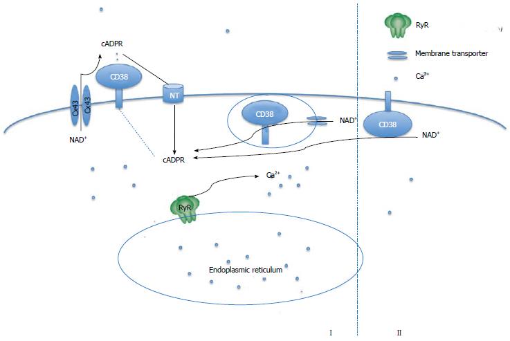
Structurally, CD38 is predicted to be a type-II transmembrane protein with its catalytic C-terminal domain located outside of the cell[39]. This circumstance presents a dilemma because the NAD substrate is located intracellularly whereas the enzyme is positioned extracellularly. If so, cytosolic NAD must be transported out of cells first and then cyclized by CD38 to produce cADPR in the extracellular space. Subsequently, the cADPR product must be transported back into the cytosol to induce Ca2+ release from the ER. This scenario obviously presents a “topological paradox” for the cADPR/Ca2+ signaling cascade. Two general hypotheses have been proposed to solve this puzzle (Figure 3). The first proposal is based on the presence of transporters, such as connexin 43 hemichannels, which allow intracellular NAD to move to the extracellular space so that it is available for access to the catalytic domain of CD38 to be converted to cADPR[45]. The cADPR product is then transferred back to cells via either CD38 or nucleoside transporters[46]. Besides this direct transport model via transporters, Zocchi et al[47] also suggested that CD38 undergoes an extensive internalization through invaginations of the plasma membrane to form endocytotic vesicles, which makes the active site of CD38 intravesicular and able to convert cytosolic NAD into cADPR. CD38 itself is a unidirectional transmembrane transporter of cADPR that mediates the cADPR efflux into the cytoplasm to reach the Ca2+ store, while influx of the cytosolic NAD+ substrate into the endocytotic CD38-containing vesicles is mediated by other transmembrane transporters, such as connexin 43 hemichannels[48]. The internalization of CD38 has been supported by several studies. For example, the internalization of CD38 can be induced by NADP in Chinese hamster ovary (CHO) cells[49] and hemin treatment can induce internalization of CD38 in K562 cells[50]. Rah et al[51] have also demonstrated that association of phospho-nonmuscle myosin heavy chain IIA with tyrosine kinase Lck and CD38 is critical for the internalization and activation of CD38. However, mechanisms regarding the transporter mediated CD38 activation process remain elusive. For example, connexin 43 hemichannels are opened for NAD export only when the cellular Ca2+ is 100 nmol/L; thus this system is unlikely to operate when Ca2+ is elevated above basal levels[45].
The second proposal offered to explain the topological paradox involves a consideration of the orientation of CD38. Bruzzone and coworkers have shown that treatment of granulocytes with 8-Br-cyclic adenosine monophosphate (cAMP), a cell-permeant analog of cAMP, induced serine phosphorylation of CD38, correlating with a cAMP-dependent intracellular cADPR synthesis[52]. Although the exact location of the phosphorylation sites is unknown, it was predicted to be in the catalytic C-terminal domain that contains multiple serine residues. However, if the catalytic domain of CD38 is phosphorylated by protein kinase A (PKA), this domain should be in the cytosol to directly cyclize NAD, thereby synthesizing cADPR intracellularly. This suggests that although CD38 is believed to be a type-II protein, at least a portion of the total CD38 is expressed as a type-III membrane protein with its C-terminal catalytic domain sitting in the cytosol[53]. Since the number of positive charges that determine the polarity of membrane protein is equal on each side of the CD38 transmembrane segment, studies from protease digestion[54] and electron microscopy[55] showed that the nuclear CD38 might be a type-III membrane protein. Most recently, Zhao et al[56] reported that expression of a cytosolic CD38 protein with deletion of both the N-terminal tail and transmembrane domain results in intact disulfides as well as active enzyme in spite of the cytosolic reductive environment; this result appears to solve the fundamental need of the six disulfides for CD38 enzymatic activity. Based on this finding, they consequently proved the co-expression of type II and type III CD38 on the surface of leukemia HL-60 cells during retinoic acid-induced differentiation and on interferon Υ-activated natural human monocytes and U937 cells[57]. They proposed that the type-III structure may take part in fast cellular responses, while the type-IIstructure may be more suitable for slower and long term responses (Figure 3)[58].
In addition to its role in cADPR production, another function of CD38 is to regulate the NAD level inside cells. It has been well established that NAD plays an essential role in energy metabolism and is involved in diverse signal transduction pathways. A rather surprising finding is that CD38 has a dramatic role in intracellular NAD metabolism. NAD levels in CD38 knockout mice are 10 to 20-fold higher than that in wild-type animals. These results suggest that CD38 is a major regulator of NAD levels in mammalian cells[59].
CD38 was originally identified as a lymphocyte antigen; thus it is not surprising that the CD38/cADPR/Ca2+ pathway plays an important role in inflammatory processes. In an ischemic stroke study, CD38-/- mice produced less monocyte chemoattractant protein-1 (MCP-1) after temporary middle cerebral artery occlusion and had fewer infiltrating macrophages and lymphocytes in the ischemic hemisphere than the wild type mice, whereas the amount of resident microglia was unaltered. The same study also demonstrated that CD38 affected immune cell migration as well as activation, two crucial postischemic inflammatory responses in secondary brain damage, suggesting that CD38 might be a therapeutic target to modulate the inflammatory mechanisms after cerebral ischemia[60]. Recently, Ng et al[61] used intravital multi-photon microscopy to observe the neutrophil granulocyte traffic into the injury site in the dermis of mice and found that the amplification phase, which is the attraction of more neutrophils toward the damage focus after the initial phase of migration by scouting neutrophils, was mediated by cADPR. cADPR and CD38 were also involved in the regulation of leukocyte adhesion and chemotaxis and were required for the deletion of T regulatory cells during inflammation as well[62]. In addition, 8-Br-cADPR, a cADPR antagonist, inhibited the MCP-1 induced Ca2+ increase, reactive oxygen species (ROS) production and apoptosis in human retinal pigment epithelium, suggesting that cADPR is also involved in the inflammatory responses of age-related macular degeneration (AMD)[63].
Recently, we demonstrated that cADPR is important for regulating cell proliferation and neuronal differentiation in PC12 cells. We found that acetylcholine (Ach) activates the CD38/cADPR pathway to induce Ca2+ release and the CD38/cADPR/Ca2+ signaling pathway is required for Ach-stimulated cell proliferation in PC12 cells. Interestingly, inhibition of the cADPR pathway accelerated nerve growth factor (NGF)-induced neuronal differentiation in PC12 cells. On the other hand, CD38 overexpression increased cell proliferation but delayed NGF-induced differentiation. Taken together, we demonstrated that cADPR plays a dichotomic role in regulating proliferation and neuronal differentiation of PC12 cells[64].
Abscisic acid (ABA) is an endogenous stimulator of insulin secretion in human and murine pancreatic beta cells. ABA triggered activation of CD38 and production of cADPR before insulin release, suggesting that CD38 is a regulator of insulin release[65]. Also, CD38 expression and cADPR production induced by ABA were required for ABA-induced upregulation of COX-2 and prostaglandin E2 in human mesenchymal stem cells (MSC) and for chemokinesis of MSC[66].
Since cADPR can activate RyRs for Ca2+ release from ER and can modulate the CICR process, the CD38/cADPR/Ca2+ pathway is predicted to participate in the regulation of cardiac activities, including cardiogenesis and the function of adult cardiac tissue. In fact, ever since the discovery of cADPR, researchers have vigorously explored its role in cardiac tissues. Galione et al[67] showed that application of cADPR through a patch electrode resulted in an increase in Ca2+ transients with a concomitant increase of the magnitude of contraction in guinea-pig cardiac ventricular myocytes, whereas application of the inhibitor 8-amino-cADPR resulted in a significant reduction in contractions and Ca2+ release from the SR. Similarly, in rat cardiac ventricular myocytes, cADPR increased the frequency of Ca2+“sparks”, which may contribute to the increase in subsequent whole-cell Ca2+ transients[68]. In addition, Prakash et al[69] found that microinjection of cADPR into adult rat ventricular myocytes not only induced sustained Ca2+ responses in a concentration dependent manner but also increased the frequency and amplitude of spontaneous Ca2+ waves, which were completely blocked by 8-amino-cADPR, a cADPR antagonist.
Interestingly, cardiac hypertrophy developed only in CD38 knockout male mice. The expression of RyR protein was increased only in female CD38 knockout mice compared with wild type, suggesting that the CD38/cADPR signaling plays an important role in intracellular Ca2+ homeostasis in cardiac myocytes in vivo, although its deficiency was compensated differentially according to gender[70].
cADPR was also shown to be involved in angiotensin II-induced cardiac hypertrophy[71]. In rat cardiomyocytes, angiotensin II evoked a Ca2+ increase via IP3R to activate PKC, which then activated the NAD(P)H oxidase to initiate ROS generation. The ROS together with Ca2+ then activated the ADP-ribose cyclase to synthesize cADPR, which induced a sustained increase of both Ca2+ and ROS and finally led to cardiac hypertrophy[72]. Most recently, Xu et al[73] demonstrated that CD38/cADPR was involved in the regulation of superoxide (O2•-) production in mouse coronary arterial myocytes (CAMs). NAD(P)H oxidase is responsible for O2•- production. Since CD38 can use NAD, an NAD(P)H oxidase product, to produce cADPR and cADPR production can result in an increase in NAD(P)H oxidase activity, the system contains a positive feedback loop. Xu et al[73] found that oxotremorine, a muscarinic type 1 receptor agonist, stimulated intracellular O2•- production in CAMs that was inhibited in CD38 knockout, CD38 knockdown, or nicotinamide-treated (a CD38 inhibitor) cells. On the other hand, direct application of cADPR into CAMs increased intracellular Ca2+ and O2•- production in CD38-/- CAMs. Moreover, CD38 knockout, Nox1 knockdown or Nox4 knockdown blocked oxotremorine-induced contraction in the isolated perfused coronary arteries in mice. Taken together, these data indicate that the CD38/cADPR pathway is an important regulator of Nox-mediated intracellular O2•- production.
The CD38/cADPR/Ca2+ pathway has also been shown to regulate the cardiogenesis process. We recently studied the role of CD38/cADPR/Ca2+ in the cardiomyogenesis of mouse embryonic stem (ES) cells. We found that beating cells appeared earlier and were more abundant in CD38 knockdown embryoid bodies (EBs) than control EBs, and the expression of several cardiac markers was increased significantly in CD38 knockdown EBs than control EBs. Similarly, more cardiomyocytes (CMs) existed in CD38 knockdown or cADPR antagonist-treated EBs compared to control EBs. Conversely, CD38 overexpression in mouse ES cells markedly inhibited CM differentiation. Surprisingly, CD38 knockdown ES cell derived CMs possess the functional properties characteristic of normal ES cell derived CMs. In addition, we found that the CD38/cADPR pathway inhibited the Erk1/2 cascade during CM differentiation of ES cells, and transient inhibition of Erk1/2 blocked the enhancive effects of CD38 knockdown on the differentiation of CM from ES cells. Taken together, we demonstrated that the CD38/cADPR/Ca2+ signaling pathway inhibits the CM differentiation of mouse ES cells[74].
The mechanism underlying cADPR regulation of Ca2+ sparks in cardiomyocyte remains elusive. Zhang et al[19]. showed that cADPR markedly increased the Ca2+ spark frequency in cardiomyocytes isolated from wild type mice, whereas cADPR failed to initiate Ca2+ sparks in cardiomyocytes isolated from FK506 binding protein 12.6 (FKBP12.6) knockout mice. They further demonstrated that cADPR induced FKBP12.6 dissociation from RyRs in a phosphorylation-dependent manner. Yet, another study showed that cAMP signaling is required for the role of cADPR in the beta-adrenergic receptor induced Ca2+increase in rat cardiomyocytes. They found that the isoproterenol-mediated increase of Ca2+ was blocked by pretreatment with 8-Br-cADPR, PKA inhibitor H89 or a high concentration of ryanodine. Moreover, incubation of ventricular lysates with isoproterenol, forskolin or cAMP resulted in activation of ADP-ribosyl cyclase of the ventricular lysates[34]. Interestingly, for comparison, estrogen increased CD38 expression and its cyclase activity, but did not affect its hydrolase activity, while progesterone eliminated the effects of estrogen on CD38 in the rat myometrium[75]. Nevertheless, the mechanism of how the CD38/cADPR is involved in the regulation of cardiac function is still unclear.
The CD38/cADPR/Ca2+ pathway has been suggested to be involved in various pathological processes. For example, CD38 deficiency accelerated diabetes in a non-obese diabetic (NOD) mice model[76]. It has also been shown that both the specific kidney ADP-ribosyl cyclase activity and cADPR production were increased in the kidneys of diabetic mice, suggesting that cADPR plays a role in the renal pathogenesis of diabetes[77]. Down-regulation of CD38 has also been shown to mediate the intermittent hypoxia induced impairment of glucose-induced insulin secretion, suggesting that CD38 plays a role in type 2 diabetes progression[78]. Numerous studies have been attempted to dissect the molecular mechanism of the role of CD38/cADPR/Ca2+ pathway in mediating diabetes in order to identify an alternative therapeutic tool. Tian et al[79] found that the content of cADPR was elevated with concomitant enhanced activity of RyR2 in ventricular myocytes isolated from a type 1 diabetic rat model, suggesting that cADPR mediates type 1 diabetes through regulating the function of RyR2. Chen et al[80] demonstrated that the ATP-gated ion channel P2X7 was required for the acceleration of type 1 diabetes induced by CD38 deficiency. Taken together, knowledge about the role of the CD38/cADPR/Ca2+ pathway in diabetes is accumulating rapidly and there is hope that understanding this pathway will facilitate the development of novel therapeutics for the disease.
The CD38/cADPR/Ca2+ pathway has been associated with inflammatory airway disorders. In human airway smooth muscle (ASM) cells, increased ASM contractility in inflammatory diseases such as asthma was due to enhanced Ca2+ sensitivity to cytokines, which was correlated with the increase of CD38 expression and cADPR level[81]. This increase of CD38 was induced by TNFαvia NFκB and could be inhibited by glucocorticoids[82]. In addition, the CD38/cADPR/Ca2+ pathway also mediated the 2-arachidonoylglycerol induced rapid actin rearrangement during differentiation of HL-60 cells into macrophage-like cells[83], and extracellular NAD+ induced stimulation and recruitment of human granulocytes during the inflammation process[84]. In addition, CD38 was involved in a neuroinflammatory disorder where CD38 expression level was markedly increased in IL-1beta- or HIV-1-activated human astrocytes, whereas CD38 knockdown significantly reduced proinflammatory cytokine and chemokine production in astrocytes[85]. Considering these results, the CD38/cADPR/Ca2+ pathway plays important roles in multiple inflammatory processes.
The CD38/cADPR/Ca2+ pathway modulates various processes of cells, including inflammation, insulin secretion, cardiogenesis, cardiac regulation etc. With further investigation, it is likely that other physiological roles of the CD38/cADPR/Ca2+ pathway will be revealed. For example, Yue et al[64] have shown that the CD38/cADPR/Ca2+ pathway delayed the nerve growth factor induced differentiation of PC12 cells; thus it is reasonable to predict that this pathway might also be involved in the regulation of neurogenesis. Using the mouse embryonic stem cell in vitro differentiation model, our preliminary results showed that the CD38/cADPR/Ca2+ pathway does play a role in neural differentiation of mES (unpublished data); however, further research is needed to decipher the underlying mechanism. A comprehensive understanding of the physiological and pathological roles of the CD38/cADPR/Ca2+ pathway in various cellular processes will undoubtedly be helpful for exploiting new molecular therapy targets. In addition, it still remains to be determined whether cADPR binds directly to RyRs or through some unknown proteins. Recently, the long-sought-after store-operated Ca2+ entry proteins were identified using a genome-wide RNAi screen by several groups[86-88]. A similar strategy could be applied to identify novel cADPR-interacting proteins or regulators.
We thank members of the Yue lab for advice on the manuscript.
P- Reviewers: Chiang HL, Schmitz ML S- Editor: Ma YJ L- Editor: Roemmele A E- Editor: Wu HL
1. | Sutherland EW, Robison GA, Butcher RW. Some Aspects of the Biological Role of Adenosine 3’,5’-monophosphate (Cyclic AMP). Circulation. 1968;37:279-306. [DOI] [Cited in This Article: ] [Cited by in Crossref: 761] [Cited by in F6Publishing: 759] [Article Influence: 13.3] [Reference Citation Analysis (0)] |
2. | Clapper DL, Lee HC. Inositol trisphosphate induces calcium release from nonmitochondrial stores i sea urchin egg homogenates. J Biol Chem. 1985;260:13947-13954. [PubMed] [Cited in This Article: ] |
3. | Lee HC, Walseth TF, Bratt GT, Hayes RN, Clapper DL. Structural determination of a cyclic metabolite of NAD+ with intracellular Ca2+-mobilizing activity. J Biol Chem. 1989;264:1608-1615. [PubMed] [Cited in This Article: ] |
4. | Clapper DL, Walseth TF, Dargie PJ, Lee HC. Pyridine nucleotide metabolites stimulate calcium release from sea urchin egg microsomes desensitized to inositol trisphosphate. J Biol Chem. 1987;262:9561-9568. [PubMed] [Cited in This Article: ] |
5. | Lee HC, Aarhus R, Levitt D. The crystal structure of cyclic ADP-ribose. Nat Struct Biol. 1994;1:143-144. [PubMed] [DOI] [Cited in This Article: ] [Cited by in Crossref: 119] [Cited by in F6Publishing: 123] [Article Influence: 4.0] [Reference Citation Analysis (0)] |
6. | Shuto S, Fukuoka M, Manikowsky A, Ueno Y, Nakano T, Kuroda R, Kuroda H, Matsuda A. Total synthesis of cyclic ADP-carbocyclic-ribose, a stable mimic of Ca2+-mobilizing second messenger cyclic ADP-ribose. J Am Chem Soc. 2001;123:8750-8759. [PubMed] [DOI] [Cited in This Article: ] [Cited by in Crossref: 60] [Cited by in F6Publishing: 64] [Article Influence: 2.7] [Reference Citation Analysis (0)] |
7. | Dong M, Si YQ, Sun SY, Pu XP, Yang ZJ, Zhang LR, Zhang LH, Leung FP, Lam CM, Kwong AK. Design, synthesis and biological characterization of novel inhibitors of CD38. Org Biomol Chem. 2011;9:3246-3257. [PubMed] [DOI] [Cited in This Article: ] [Cited by in Crossref: 27] [Cited by in F6Publishing: 29] [Article Influence: 2.1] [Reference Citation Analysis (0)] |
8. | Zhou Y, Yu P, Jin H, Yang Z, Yue J, Zhang L, Zhang L. Synthesis and calcium mobilization activity of cADPR analogues which integrate nucleobase, northern and southern ribose modifications. Molecules. 2012;17:4343-4356. [PubMed] [DOI] [Cited in This Article: ] [Cited by in Crossref: 5] [Cited by in F6Publishing: 7] [Article Influence: 0.5] [Reference Citation Analysis (0)] |
9. | Rosen D, Bloor-Young D, Squires J, Parkesh R, Waters G, Vasudevan SR, Lewis AM, Churchill GC. Synthesis and use of cell-permeant cyclic ADP-ribose. Biochem Biophys Res Commun. 2012;418:353-358. [PubMed] [DOI] [Cited in This Article: ] [Cited by in Crossref: 6] [Cited by in F6Publishing: 7] [Article Influence: 0.5] [Reference Citation Analysis (0)] |
10. | Dargie PJ, Agre MC, Lee HC. Comparison of Ca2+ mobilizing activities of cyclic ADP-ribose and inositol trisphosphate. Cell Regul. 1990;1:279-290. [PubMed] [Cited in This Article: ] |
11. | Galione A, Lee HC, Busa WB. Ca(2+)-induced Ca2+ release in sea urchin egg homogenates: modulation by cyclic ADP-ribose. Science. 1991;253:1143-1146. [PubMed] [DOI] [Cited in This Article: ] [Cited by in Crossref: 490] [Cited by in F6Publishing: 478] [Article Influence: 14.1] [Reference Citation Analysis (0)] |
12. | Mészáros LG, Bak J, Chu A. Cyclic ADP-ribose as an endogenous regulator of the non-skeletal type ryanodine receptor Ca2+ channel. Nature. 1993;364:76-79. [PubMed] [DOI] [Cited in This Article: ] [Cited by in Crossref: 272] [Cited by in F6Publishing: 273] [Article Influence: 8.5] [Reference Citation Analysis (0)] |
13. | Takasawa S, Kuroki M, Nata K, Noguchi N, Ikeda T, Yamauchi A, Ota H, Itaya-Hironaka A, Sakuramoto-Tsuchida S, Takahashi I. A novel ryanodine receptor expressed in pancreatic islets by alternative splicing from type 2 ryanodine receptor gene. Biochem Biophys Res Commun. 2010;397:140-145. [PubMed] [DOI] [Cited in This Article: ] [Cited by in Crossref: 33] [Cited by in F6Publishing: 35] [Article Influence: 2.3] [Reference Citation Analysis (0)] |
14. | Ogunbayo OA, Zhu Y, Rossi D, Sorrentino V, Ma J, Zhu MX, Evans AM. Cyclic adenosine diphosphate ribose activates ryanodine receptors, whereas NAADP activates two-pore domain channels. J Biol Chem. 2011;286:9136-9140. [PubMed] [DOI] [Cited in This Article: ] [Cited by in F6Publishing: 1] [Reference Citation Analysis (0)] |
15. | Lee HC. Physiological functions of cyclic ADP-ribose and NAADP as calcium messengers. Annu Rev Pharmacol Toxicol. 2001;41:317-345. [PubMed] [DOI] [Cited in This Article: ] [Cited by in F6Publishing: 1] [Reference Citation Analysis (0)] |
16. | Guse AH. Biochemistry, biology, and pharmacology of cyclic adenosine diphosphoribose (cADPR). Curr Med Chem. 2004;11:847-855. [PubMed] [DOI] [Cited in This Article: ] [Cited by in Crossref: 66] [Cited by in F6Publishing: 70] [Article Influence: 3.3] [Reference Citation Analysis (0)] |
17. | Galione A, Churchill GC. Cyclic ADP ribose as a calcium-mobilizing messenger. Sci STKE. 2000;2000:pe1. [PubMed] [DOI] [Cited in This Article: ] [Cited by in Crossref: 18] [Cited by in F6Publishing: 26] [Article Influence: 1.0] [Reference Citation Analysis (0)] |
18. | Noguchi N, Takasawa S, Nata K, Tohgo A, Kato I, Ikehata F, Yonekura H, Okamoto H. Cyclic ADP-ribose binds to FK506-binding protein 12.6 to release Ca2+ from islet microsomes. J Biol Chem. 1997;272:3133-3136. [PubMed] [Cited in This Article: ] |
19. | Zhang X, Tallini YN, Chen Z, Gan L, Wei B, Doran R, Miao L, Xin HB, Kotlikoff MI, Ji G. Dissociation of FKBP12.6 from ryanodine receptor type 2 is regulated by cyclic ADP-ribose but not beta-adrenergic stimulation in mouse cardiomyocytes. Cardiovasc Res. 2009;84:253-262. [PubMed] [DOI] [Cited in This Article: ] [Cited by in Crossref: 36] [Cited by in F6Publishing: 41] [Article Influence: 2.6] [Reference Citation Analysis (0)] |
20. | Thomas JM, Summerhill RJ, Fruen BR, Churchill GC, Galione A. Calmodulin dissociation mediates desensitization of the cADPR-induced Ca2+ release mechanism. Curr Biol. 2002;12:2018-2022. [PubMed] [DOI] [Cited in This Article: ] [Cited by in Crossref: 20] [Cited by in F6Publishing: 22] [Article Influence: 1.0] [Reference Citation Analysis (0)] |
21. | Zheng J, Wenzhi B, Miao L, Hao Y, Zhang X, Yin W, Pan J, Yuan Z, Song B, Ji G. Ca(2+) release induced by cADP-ribose is mediated by FKBP12.6 proteins in mouse bladder smooth muscle. Cell Calcium. 2010;47:449-457. [PubMed] [DOI] [Cited in This Article: ] [Cited by in Crossref: 23] [Cited by in F6Publishing: 26] [Article Influence: 1.7] [Reference Citation Analysis (0)] |
22. | Guse AH, Berg I, da Silva CP, Potter BV, Mayr GW. Ca2+ entry induced by cyclic ADP-ribose in intact T-lymphocytes. J Biol Chem. 1997;272:8546-8550. [PubMed] [DOI] [Cited in This Article: ] [Cited by in Crossref: 76] [Cited by in F6Publishing: 82] [Article Influence: 2.9] [Reference Citation Analysis (0)] |
23. | Togashi K, Hara Y, Tominaga T, Higashi T, Konishi Y, Mori Y, Tominaga M. TRPM2 activation by cyclic ADP-ribose at body temperature is involved in insulin secretion. EMBO J. 2006;25:1804-1815. [PubMed] [DOI] [Cited in This Article: ] [Cited by in F6Publishing: 1] [Reference Citation Analysis (0)] |
24. | Yu PL, Zhang ZH, Hao BX, Zhao YJ, Zhang LH, Lee HC, Zhang L, Yue J. A novel fluorescent cell membrane-permeable caged cyclic ADP-ribose analogue. J Biol Chem. 2012;287:24774-24783. [PubMed] [Cited in This Article: ] |
25. | Kirchberger T, Moreau C, Wagner GK, Fliegert R, Siebrands CC, Nebel M, Schmid F, Harneit A, Odoardi F, Flügel A. 8-Bromo-cyclic inosine diphosphoribose: towards a selective cyclic ADP-ribose agonist. Biochem J. 2009;422:139-149. [PubMed] [DOI] [Cited in This Article: ] [Cited by in Crossref: 17] [Cited by in F6Publishing: 20] [Article Influence: 1.3] [Reference Citation Analysis (0)] |
26. | Rusinko N, Lee HC. Widespread occurrence in animal tissues of an enzyme catalyzing the conversion of NAD+ into a cyclic metabolite with intracellular Ca2+-mobilizing activity. J Biol Chem. 1989;264:11725-11731. [PubMed] [Cited in This Article: ] |
27. | Lee HC, Aarhus R. ADP-ribosyl cyclase: an enzyme that cyclizes NAD+ into a calcium-mobilizing metabolite. Cell Regul. 1991;2:203-209. [PubMed] [Cited in This Article: ] |
28. | Howard M, Grimaldi JC, Bazan JF, Lund FE, Santos-Argumedo L, Parkhouse RM, Walseth TF, Lee HC. Formation and hydrolysis of cyclic ADP-ribose catalyzed by lymphocyte antigen CD38. Science. 1993;262:1056-1059. [PubMed] [DOI] [Cited in This Article: ] [Cited by in Crossref: 577] [Cited by in F6Publishing: 595] [Article Influence: 18.6] [Reference Citation Analysis (0)] |
29. | States DJ, Walseth TF, Lee HC. Similarities in amino acid sequences of Aplysia ADP-ribosyl cyclase and human lymphocyte antigen CD38. Trends Biochem Sci. 1992;17:495. [PubMed] [DOI] [Cited in This Article: ] [Cited by in Crossref: 158] [Cited by in F6Publishing: 172] [Article Influence: 5.2] [Reference Citation Analysis (0)] |
30. | Takasawa S, Tohgo A, Noguchi N, Koguma T, Nata K, Sugimoto T, Yonekura H, Okamoto H. Synthesis and hydrolysis of cyclic ADP-ribose by human leukocyte antigen CD38 and inhibition of the hydrolysis by ATP. J Biol Chem. 1993;268:26052-26054. [PubMed] [Cited in This Article: ] |
31. | Itoh M, Ishihara K, Tomizawa H, Tanaka H, Kobune Y, Ishikawa J, Kaisho T, Hirano T. Molecular cloning of murine BST-1 having homology with CD38 and Aplysia ADP-ribosyl cyclase. Biochem Biophys Res Commun. 1994;203:1309-1317. [PubMed] [DOI] [Cited in This Article: ] [Cited by in Crossref: 69] [Cited by in F6Publishing: 78] [Article Influence: 2.5] [Reference Citation Analysis (0)] |
32. | Lee HC. Structure and enzymatic functions of human CD38. Mol Med. 2006;12:317-323. [PubMed] [Cited in This Article: ] |
33. | Kannt A, Sicka K, Kroll K, Kadereit D, Gögelein H. Selective inhibitors of cardiac ADPR cyclase as novel anti-arrhythmic compounds. Naunyn Schmiedebergs Arch Pharmacol. 2012;385:717-727. [PubMed] [DOI] [Cited in This Article: ] [Cited by in Crossref: 17] [Cited by in F6Publishing: 20] [Article Influence: 1.5] [Reference Citation Analysis (0)] |
34. | Xie GH, Rah SY, Kim SJ, Nam TS, Ha KC, Chae SW, Im MJ, Kim UH. ADP-ribosyl cyclase couples to cyclic AMP signaling in the cardiomyocytes. Biochem Biophys Res Commun. 2005;330:1290-1298. [PubMed] [Cited in This Article: ] |
35. | Fabiano A, Panfoli I, Calzia D, Bruschi M, Ravera S, Bachi A, Cattaneo A, Morelli A, Candiano G. Catalytic properties of the retinal rod outer segment disk ADP-ribosyl cyclase. Vis Neurosci. 2011;28:121-128. [PubMed] [Cited in This Article: ] |
36. | Nam TS, Choi SH, Rah SY, Kim SY, Jang W, Im MJ, Kwon HJ, Kim UH. Discovery of a small-molecule inhibitor for kidney ADP-ribosyl cyclase: Implication for intracellular calcium signal mediated by cyclic ADP-ribose. Exp Mol Med. 2006;38:718-726. [PubMed] [Cited in This Article: ] |
37. | Aarhus R, Graeff RM, Dickey DM, Walseth TF, Lee HC. ADP-ribosyl cyclase and CD38 catalyze the synthesis of a calcium-mobilizing metabolite from NADP. J Biol Chem. 1995;270:30327-30333. [PubMed] [Cited in This Article: ] |
38. | Graeff R, Liu Q, Kriksunov IA, Hao Q, Lee HC. Acidic residues at the active sites of CD38 and ADP-ribosyl cyclase determine nicotinic acid adenine dinucleotide phosphate (NAADP) synthesis and hydrolysis activities. J Biol Chem. 2006;281:28951-28957. [PubMed] [Cited in This Article: ] |
39. | Jackson DG, Bell JI. Isolation of a cDNA encoding the human CD38 (T10) molecule, a cell surface glycoprotein with an unusual discontinuous pattern of expression during lymphocyte differentiation. J Immunol. 1990;144:2811-2815. [PubMed] [Cited in This Article: ] |
40. | Prasad GS, McRee DE, Stura EA, Levitt DG, Lee HC, Stout CD. Crystal structure of Aplysia ADP ribosyl cyclase, a homologue of the bifunctional ectozyme CD38. Nat Struct Biol. 1996;3:957-964. [PubMed] [DOI] [Cited in This Article: ] [Cited by in Crossref: 113] [Cited by in F6Publishing: 123] [Article Influence: 4.2] [Reference Citation Analysis (0)] |
41. | Liu Q, Kriksunov IA, Graeff R, Munshi C, Lee HC, Hao Q. Crystal structure of human CD38 extracellular domain. Structure. 2005;13:1331-1339. [PubMed] [DOI] [Cited in This Article: ] [Cited by in Crossref: 125] [Cited by in F6Publishing: 132] [Article Influence: 6.6] [Reference Citation Analysis (0)] |
42. | Munshi C, Aarhus R, Graeff R, Walseth TF, Levitt D, Lee HC. Identification of the enzymatic active site of CD38 by site-directed mutagenesis. J Biol Chem. 2000;275:21566-21571. [PubMed] [DOI] [Cited in This Article: ] [Cited by in Crossref: 101] [Cited by in F6Publishing: 105] [Article Influence: 4.2] [Reference Citation Analysis (0)] |
43. | Graeff R, Munshi C, Aarhus R, Johns M, Lee HC. A single residue at the active site of CD38 determines its NAD cyclizing and hydrolyzing activities. J Biol Chem. 2001;276:12169-12173. [PubMed] [DOI] [Cited in This Article: ] [Cited by in Crossref: 56] [Cited by in F6Publishing: 57] [Article Influence: 2.4] [Reference Citation Analysis (0)] |
44. | Liu Q, Kriksunov IA, Graeff R, Lee HC, Hao Q. Structural basis for formation and hydrolysis of the calcium messenger cyclic ADP-ribose by human CD38. J Biol Chem. 2007;282:5853-5861. [PubMed] [Cited in This Article: ] |
45. | Bruzzone S, Franco L, Guida L, Zocchi E, Contini P, Bisso A, Usai C, De Flora A. A self-restricted CD38-connexin 43 cross-talk affects NAD+ and cyclic ADP-ribose metabolism and regulates intracellular calcium in 3T3 fibroblasts. J Biol Chem. 2001;276:48300-48308. [PubMed] [DOI] [Cited in This Article: ] [Cited by in Crossref: 80] [Cited by in F6Publishing: 84] [Article Influence: 3.5] [Reference Citation Analysis (0)] |
46. | Guida L, Bruzzone S, Sturla L, Franco L, Zocchi E, De Flora A. Equilibrative and concentrative nucleoside transporters mediate influx of extracellular cyclic ADP-ribose into 3T3 murine fibroblasts. J Biol Chem. 2002;277:47097-47105. [PubMed] [DOI] [Cited in This Article: ] [Cited by in Crossref: 55] [Cited by in F6Publishing: 57] [Article Influence: 2.5] [Reference Citation Analysis (0)] |
47. | Zocchi E, Usai C, Guida L, Franco L, Bruzzone S, Passalacqua M, De Flora A. Ligand-induced internalization of CD38 results in intracellular Ca2+ mobilization: role of NAD+ transport across cell membranes. FASEB J. 1999;13:273-283. [PubMed] [Cited in This Article: ] |
48. | Bruzzone S, Guida L, Zocchi E, Franco L. Connexin 43 hemi channels mediate Ca2+-regulated transmembrane NAD+ fluxes in intact cells. FASEB J. 2001;15:10-12. [PubMed] [DOI] [Cited in This Article: ] [Cited by in Crossref: 356] [Cited by in F6Publishing: 371] [Article Influence: 15.5] [Reference Citation Analysis (0)] |
49. | Chidambaram N, Chang CF. NADP+-Dependent internalization of recombinant CD38 in CHO cells. Arch Biochem Biophys. 1999;363:267-272. [PubMed] [DOI] [Cited in This Article: ] [Cited by in Crossref: 8] [Cited by in F6Publishing: 10] [Article Influence: 0.4] [Reference Citation Analysis (0)] |
50. | Yalcintepe L, Ercelen S, Adin-Cinar S, Badur S, Tiryaki D, Bermek E. Hemin-dependent induction and internalization of CD38 in K562 cells. J Cell Biochem. 2003;90:379-386. [PubMed] [DOI] [Cited in This Article: ] [Cited by in Crossref: 3] [Cited by in F6Publishing: 4] [Article Influence: 0.2] [Reference Citation Analysis (0)] |
51. | Rah SY, Park KH, Nam TS, Kim SJ, Kim H, Im MJ, Kim UH. Association of CD38 with nonmuscle myosin heavy chain IIA and Lck is essential for the internalization and activation of CD38. J Biol Chem. 2007;282:5653-5660. [PubMed] [DOI] [Cited in This Article: ] [Cited by in Crossref: 23] [Cited by in F6Publishing: 24] [Article Influence: 1.3] [Reference Citation Analysis (0)] |
52. | Bruzzone S, Moreschi I, Usai C, Guida L, Damonte G, Salis A, Scarfì S, Millo E, De Flora A, Zocchi E. Abscisic acid is an endogenous cytokine in human granulocytes with cyclic ADP-ribose as second messenger. Proc Natl Acad Sci USA. 2007;104:5759-5764. [PubMed] [DOI] [Cited in This Article: ] [Cited by in Crossref: 148] [Cited by in F6Publishing: 143] [Article Influence: 7.9] [Reference Citation Analysis (0)] |
53. | Lee HC. Cyclic ADP-ribose and NAADP: fraternal twin messengers for calcium signaling. Sci China Life Sci. 2011;54:699-711. [PubMed] [DOI] [Cited in This Article: ] [Cited by in Crossref: 42] [Cited by in F6Publishing: 47] [Article Influence: 3.4] [Reference Citation Analysis (0)] |
54. | Adebanjo OA, Anandatheerthavarada HK, Koval AP, Moonga BS, Biswas G, Sun L, Sodam BR, Bevis PJ, Huang CL, Epstein S. A new function for CD38/ADP-ribosyl cyclase in nuclear Ca2+ homeostasis. Nat Cell Biol. 1999;1:409-414. [PubMed] [DOI] [Cited in This Article: ] [Cited by in Crossref: 126] [Cited by in F6Publishing: 132] [Article Influence: 5.1] [Reference Citation Analysis (0)] |
55. | Khoo KM, Han MK, Park JB, Chae SW, Kim UH, Lee HC, Bay BH, Chang CF. Localization of the cyclic ADP-ribose-dependent calcium signaling pathway in hepatocyte nucleus. J Biol Chem. 2000;275:24807-24817. [PubMed] [DOI] [Cited in This Article: ] [Cited by in Crossref: 62] [Cited by in F6Publishing: 70] [Article Influence: 2.8] [Reference Citation Analysis (0)] |
56. | Zhao YJ, Zhang HM, Lam CM, Hao Q, Lee HC. Cytosolic CD38 protein forms intact disulfides and is active in elevating intracellular cyclic ADP-ribose. J Biol Chem. 2011;286:22170-22177. [PubMed] [Cited in This Article: ] |
57. | Zhao YJ, Lam CM, Lee HC. The membrane-bound enzyme CD38 exists in two opposing orientations. Sci Signal. 2012;5:ra67. [PubMed] [Cited in This Article: ] |
58. | Lee HC. Cyclic ADP-ribose and nicotinic acid adenine dinucleotide phosphate (NAADP) as messengers for calcium mobilization. J Biol Chem. 2012;287:31633-31640. [PubMed] [DOI] [Cited in This Article: ] [Cited by in Crossref: 153] [Cited by in F6Publishing: 158] [Article Influence: 12.2] [Reference Citation Analysis (0)] |
59. | Aksoy P, White TA, Thompson M, Chini EN. Regulation of intracellular levels of NAD: a novel role for CD38. Biochem Biophys Res Commun. 2006;345:1386-1392. [PubMed] [DOI] [Cited in This Article: ] [Cited by in Crossref: 97] [Cited by in F6Publishing: 110] [Article Influence: 5.8] [Reference Citation Analysis (0)] |
60. | Choe CU, Lardong K, Gelderblom M, Ludewig P, Leypoldt F, Koch-Nolte F, Gerloff C, Magnus T. CD38 exacerbates focal cytokine production, postischemic inflammation and brain injury after focal cerebral ischemia. PLoS One. 2011;6:e19046. [PubMed] [DOI] [Cited in This Article: ] [Cited by in Crossref: 46] [Cited by in F6Publishing: 51] [Article Influence: 3.6] [Reference Citation Analysis (0)] |
61. | Ng LG, Qin JS, Roediger B, Wang Y, Jain R, Cavanagh LL, Smith AL, Jones CA, de Veer M, Grimbaldeston MA. Visualizing the neutrophil response to sterile tissue injury in mouse dermis reveals a three-phase cascade of events. J Invest Dermatol. 2011;131:2058-2068. [PubMed] [DOI] [Cited in This Article: ] [Cited by in Crossref: 135] [Cited by in F6Publishing: 157] [Article Influence: 11.2] [Reference Citation Analysis (0)] |
62. | Deaglio S, Robson SC. Ectonucleotidases as regulators of purinergic signaling in thrombosis, inflammation, and immunity. Adv Pharmacol. 2011;61:301-332. [PubMed] [DOI] [Cited in This Article: ] [Cited by in Crossref: 201] [Cited by in F6Publishing: 192] [Article Influence: 13.7] [Reference Citation Analysis (0)] |
63. | Yang D, Elner SG, Chen X, Field MG, Petty HR, Elner VM. MCP-1-activated monocytes induce apoptosis in human retinal pigment epithelium. Invest Ophthalmol Vis Sci. 2011;52:6026-6034. [PubMed] [DOI] [Cited in This Article: ] [Cited by in Crossref: 35] [Cited by in F6Publishing: 42] [Article Influence: 3.0] [Reference Citation Analysis (0)] |
64. | Yue J, Wei W, Lam CM, Zhao YJ, Dong M, Zhang LR, Zhang LH, Lee HC. CD38/cADPR/Ca2+ pathway promotes cell proliferation and delays nerve growth factor-induced differentiation in PC12 cells. J Biol Chem. 2009;284:29335-29342. [PubMed] [DOI] [Cited in This Article: ] [Cited by in Crossref: 33] [Cited by in F6Publishing: 39] [Article Influence: 2.4] [Reference Citation Analysis (0)] |
65. | Bruzzone S, Bodrato N, Usai C, Guida L, Moreschi I, Nano R, Antonioli B, Fruscione F, Magnone M, Scarfì S. Abscisic acid is an endogenous stimulator of insulin release from human pancreatic islets with cyclic ADP ribose as second messenger. J Biol Chem. 2008;283:32188-32197. [PubMed] [DOI] [Cited in This Article: ] [Cited by in Crossref: 102] [Cited by in F6Publishing: 107] [Article Influence: 6.3] [Reference Citation Analysis (0)] |
66. | Scarfì S, Ferraris C, Fruscione F, Fresia C, Guida L, Bruzzone S, Usai C, Parodi A, Millo E, Salis A. Cyclic ADP-ribose-mediated expansion and stimulation of human mesenchymal stem cells by the plant hormone abscisic acid. Stem Cells. 2008;26:2855-2864. [PubMed] [DOI] [Cited in This Article: ] [Cited by in Crossref: 48] [Cited by in F6Publishing: 51] [Article Influence: 3.0] [Reference Citation Analysis (0)] |
67. | Galione A, Cui Y, Empson R, Iino S, Wilson H, Terrar D. Cyclic ADP-ribose and the regulation of calcium-induced calcium release in eggs and cardiac myocytes. Cell Biochem Biophys. 1998;28:19-30. [PubMed] [Cited in This Article: ] |
68. | Cui Y, Galione A, Terrar DA. Effects of photoreleased cADP-ribose on calcium transients and calcium sparks in myocytes isolated from guinea-pig and rat ventricle. Biochem J. 1999;342:269-273. [PubMed] [Cited in This Article: ] |
69. | Prakash YS, Kannan MS, Walseth TF, Sieck GC. cADP ribose and [Ca(2+)](i) regulation in rat cardiac myocytes. Am J Physiol Heart Circ Physiol. 2000;279:H1482-H1489. [PubMed] [Cited in This Article: ] |
70. | Takahashi J, Kagaya Y, Kato I, Ohta J, Isoyama S, Miura M, Sugai Y, Hirose M, Wakayama Y, Ninomiya M. Deficit of CD38/cyclic ADP-ribose is differentially compensated in hearts by gender. Biochem Biophys Res Commun. 2003;312:434-440. [PubMed] [Cited in This Article: ] |
71. | Gul R, Kim SY, Park KH, Kim BJ, Kim SJ, Im MJ, Kim UH. A novel signaling pathway of ADP-ribosyl cyclase activation by angiotensin II in adult rat cardiomyocytes. Am J Physiol Heart Circ Physiol. 2008;295:H77-H88. [PubMed] [DOI] [Cited in This Article: ] [Cited by in Crossref: 34] [Cited by in F6Publishing: 39] [Article Influence: 2.3] [Reference Citation Analysis (0)] |
72. | Gul R, Shawl AI, Kim SH, Kim UH. Cooperative interaction between reactive oxygen species and Ca2+ signals contributes to angiotensin II-induced hypertrophy in adult rat cardiomyocytes. Am J Physiol Heart Circ Physiol. 2012;302:H901-H909. [PubMed] [DOI] [Cited in This Article: ] [Cited by in Crossref: 27] [Cited by in F6Publishing: 32] [Article Influence: 2.3] [Reference Citation Analysis (0)] |
73. | Xu M, Zhang Y, Xia M, Li XX, Ritter JK, Zhang F, Li PL. NAD(P)H oxidase-dependent intracellular and extracellular O2•- production in coronary arterial myocytes from CD38 knockout mice. Free Radic Biol Med. 2012;52:357-365. [PubMed] [DOI] [Cited in This Article: ] [Cited by in Crossref: 23] [Cited by in F6Publishing: 31] [Article Influence: 2.4] [Reference Citation Analysis (0)] |
74. | Wei WJ, Sun HY, Ting KY, Zhang LH, Lee HC, Li GR, Yue J. Inhibition of cardiomyocytes differentiation of mouse embryonic stem cells by CD38/cADPR/Ca2+ signaling pathway. J Biol Chem. 2012;287:35599-35611. [PubMed] [DOI] [Cited in This Article: ] [Cited by in Crossref: 25] [Cited by in F6Publishing: 28] [Article Influence: 2.2] [Reference Citation Analysis (0)] |
75. | Dogan S, Deshpande DA, White TA, Walseth TF, Kannan MS. Regulation of CD 38 expression and function by steroid hormones in myometrium. Mol Cell Endocrinol. 2006;246:101-106. [PubMed] [DOI] [Cited in This Article: ] [Cited by in Crossref: 16] [Cited by in F6Publishing: 17] [Article Influence: 0.9] [Reference Citation Analysis (0)] |
76. | Chen J, Chen YG, Reifsnyder PC, Schott WH, Lee CH, Osborne M, Scheuplein F, Haag F, Koch-Nolte F, Serreze DV. Targeted disruption of CD38 accelerates autoimmune diabetes in NOD/Lt mice by enhancing autoimmunity in an ADP-ribosyltransferase 2-dependent fashion. J Immunol. 2006;176:4590-4599. [PubMed] [Cited in This Article: ] |
77. | Kim SY, Park KH, Gul R, Jang KY, Kim UH. Role of kidney ADP-ribosyl cyclase in diabetic nephropathy. Am J Physiol Renal Physiol. 2009;296:F291-F297. [PubMed] [DOI] [Cited in This Article: ] [Cited by in Crossref: 13] [Cited by in F6Publishing: 14] [Article Influence: 0.8] [Reference Citation Analysis (0)] |
78. | Ota H, Tamaki S, Itaya-Hironaka A, Yamauchi A, Sakuramoto-Tsuchida S, Morioka T, Takasawa S, Kimura H. Attenuation of glucose-induced insulin secretion by intermittent hypoxia via down-regulation of CD38. Life Sci. 2012;90:206-211. [PubMed] [DOI] [Cited in This Article: ] [Cited by in Crossref: 47] [Cited by in F6Publishing: 60] [Article Influence: 4.3] [Reference Citation Analysis (0)] |
79. | Tian C, Shao CH, Moore CJ, Kutty S, Walseth T, DeSouza C, Bidasee KR. Gain of function of cardiac ryanodine receptor in a rat model of type 1 diabetes. Cardiovasc Res. 2011;91:300-309. [PubMed] [DOI] [Cited in This Article: ] [Cited by in Crossref: 28] [Cited by in F6Publishing: 33] [Article Influence: 2.4] [Reference Citation Analysis (0)] |
80. | Chen YG, Scheuplein F, Driver JP, Hewes AA, Reifsnyder PC, Leiter EH, Serreze DV. Testing the role of P2X7 receptors in the development of type 1 diabetes in nonobese diabetic mice. J Immunol. 2011;186:4278-4284. [PubMed] [DOI] [Cited in This Article: ] [Cited by in Crossref: 23] [Cited by in F6Publishing: 24] [Article Influence: 1.7] [Reference Citation Analysis (0)] |
81. | Deshpande DA, Walseth TF, Panettieri RA, Kannan MS. CD38/cyclic ADP-ribose-mediated Ca2+ signaling contributes to airway smooth muscle hyper-responsiveness. FASEB J. 2003;17:452-454. [PubMed] [Cited in This Article: ] |
82. | Kang BN, Tirumurugaan KG, Deshpande DA, Amrani Y, Panettieri RA, Walseth TF, Kannan MS. Transcriptional regulation of CD38 expression by tumor necrosis factor-alpha in human airway smooth muscle cells: role of NF-kappaB and sensitivity to glucocorticoids. FASEB J. 2006;20:1000-1002. [PubMed] [Cited in This Article: ] |
83. | Gokoh M, Kishimoto S, Oka S, Mori M, Waku K, Ishima Y, Sugiura T. 2-arachidonoylglycerol, an endogenous cannabinoid receptor ligand, induces rapid actin polymerization in HL-60 cells differentiated into macrophage-like cells. Biochem J. 2005;386:583-589. [PubMed] [Cited in This Article: ] |
84. | Bruzzone S, Moreschi I, Guida L, Usai C, Zocchi E, De Flora A. Extracellular NAD+ regulates intracellular calcium levels and induces activation of human granulocytes. Biochem J. 2006;393:697-704. [PubMed] [Cited in This Article: ] |
85. | Kou W, Banerjee S, Eudy J, Smith LM, Persidsky R, Borgmann K, Wu L, Sakhuja N, Deshpande MS, Walseth TF. CD38 regulation in activated astrocytes: implications for neuroinflammation and HIV-1 brain infection. J Neurosci Res. 2009;87:2326-2339. [PubMed] [DOI] [Cited in This Article: ] [Cited by in Crossref: 39] [Cited by in F6Publishing: 45] [Article Influence: 2.8] [Reference Citation Analysis (0)] |
86. | Liou J, Kim ML, Heo WD, Jones JT, Myers JW, Ferrell JE, Meyer T. STIM is a Ca2+ sensor essential for Ca2+-store-depletion-triggered Ca2+ influx. Curr Biol. 2005;15:1235-1241. [PubMed] [Cited in This Article: ] |
87. | Zhang SL, Yeromin AV, Zhang XH, Yu Y, Safrina O, Penna A, Roos J, Stauderman KA, Cahalan MD. Genome-wide RNAi screen of Ca(2+) influx identifies genes that regulate Ca(2+) release-activated Ca(2+) channel activity. Proc Natl Acad Sci USA. 2006;103:9357-9362. [PubMed] [Cited in This Article: ] |
88. | Roos J, DiGregorio PJ, Yeromin AV, Ohlsen K, Lioudyno M, Zhang S, Safrina O, Kozak JA, Wagner SL, Cahalan MD. STIM1, an essential and conserved component of store-operated Ca2+ channel function. J Cell Biol. 2005;169:435-445. [PubMed] [Cited in This Article: ] |