©2013 Baishideng Publishing Group Co.
World J Biol Chem. Nov 26, 2013; 4(4): 131-140
Published online Nov 26, 2013. doi: 10.4331/wjbc.v4.i4.131
Published online Nov 26, 2013. doi: 10.4331/wjbc.v4.i4.131
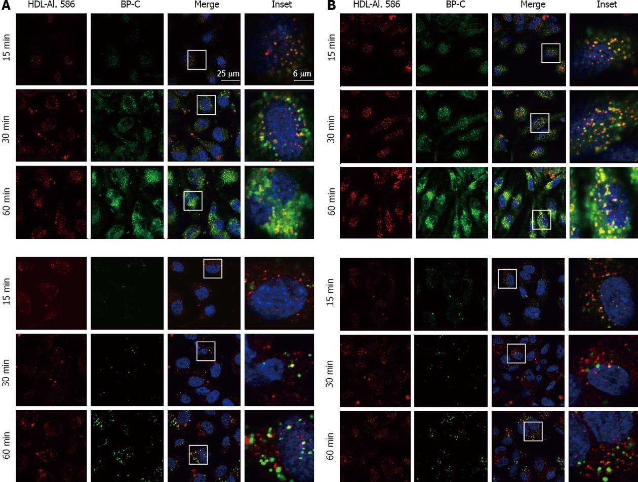
Figure 1 Detailed analysis of high density lipoprotein uptake and high density lipoprotein -derived lipid transfer in human umbilical vein endothelial cells and human coronary artery endothelial cells.
A: HUVECs; B: HCAECs. HUVECs and HCAECs were incubated for the indicated time with HDL-Alexa 586, which was additionally labeled with either Bodipy-cholesterol (BP-C; upper panel) or Bodipy-cholesteryl oleate (BP-CE; lower panel). Samples were fixed and imaged using confocal microscopy. Note that brightness and contrast were increased at the 15 min time point of the BP-CE panel for better visibility (lower panel). HUVECs: Human umbilical vein endothelial cells; HCAECs: Human coronary artery endothelial cells; HDL: High density lipoprotein.
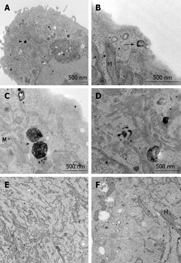
Figure 2 Ultrastructural analysis of high density lipoprotein uptake in human umbilical vein endothelial cells.
Cells incubated with HRP-labeled HDL for 30 min (A-D) or untreated controls (E, F) were processed for electron microscopy. HRP-positive clathrin-coated vesicles were seen near the cell surface (arrow in B), in tubular endosomes (asterisk in D), and densely packed MVBs were found in the perinuclear area (arrow heads in A and C). No specific staining was seen in control samples (E, F). HRP: Horseradish peroxidase; HDL: High density lipoprotein; M: Mitochondria.
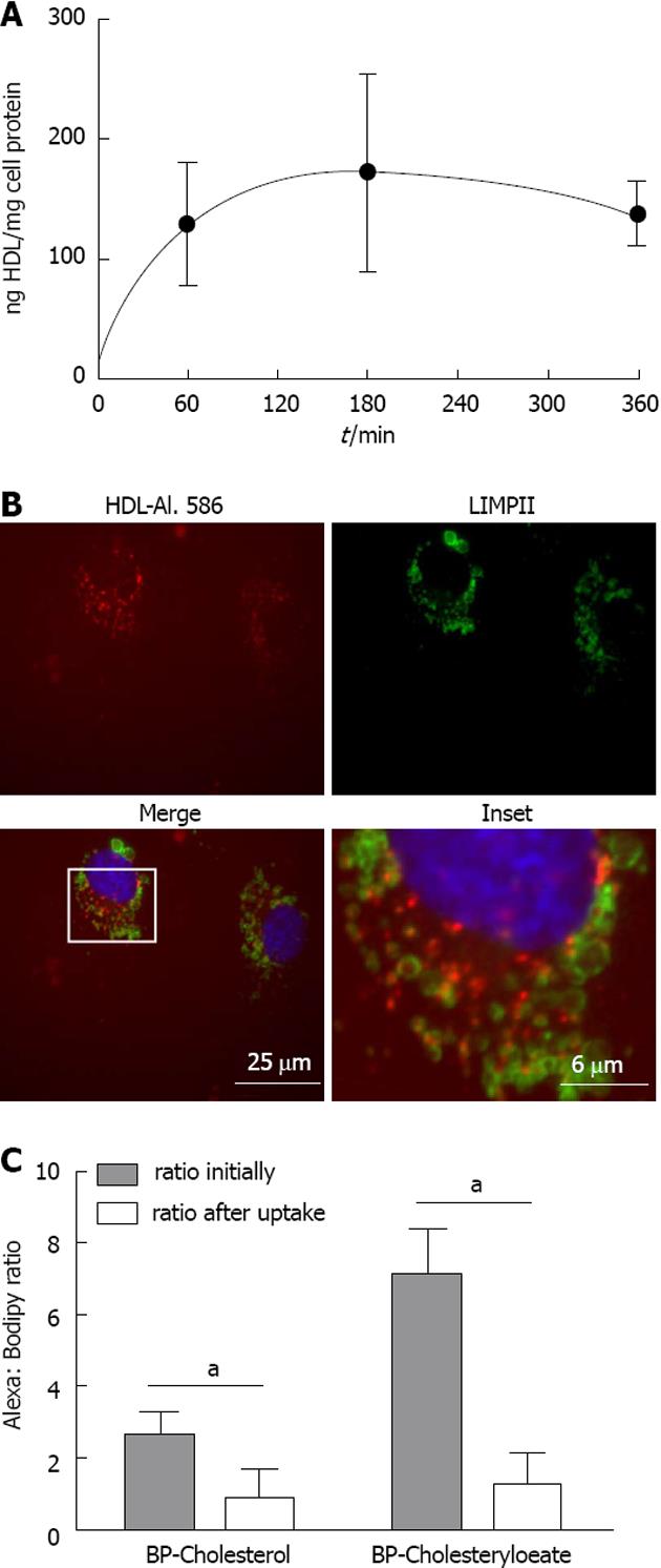
Figure 3 Quantification of high density lipoprotein uptake.
A: Time course of HDL uptake; HUVECs were incubated with iodinated HDL for up to 6 h and specific cell association was measured using a gamma counter. Measurements were normalized to protein content, determined by the Bradford protein assay; B: HUVECs were incubated with Alexa 568 labeled HDL for 1 h. Cells were fixed and immunofluorescene was performed using an antibody against LIMPII and visualized using fluorescence microscopy. No colocalization of LIMPII, a marker for the lysosomal compartment, and HDL-Alexa 568 was seen; C: HUVECs were incubated with HDL-Alexa-BP-C or -BP-CE for 60 min. Cells were then lyzed and the fluorescence intensity for each label was measured using a fluorometer. Results are expressed as the Alexa: Bodipy ratio initially and after the 60 min incubation period (n = 3). aP < 0.05 between groups. HDL: High density lipoprotein; BP-C: Bodipy-cholesterol; BP-CE: Bodipy-cholesteryl oleate; HUVECs: Human umbilical vein endothelial cells.
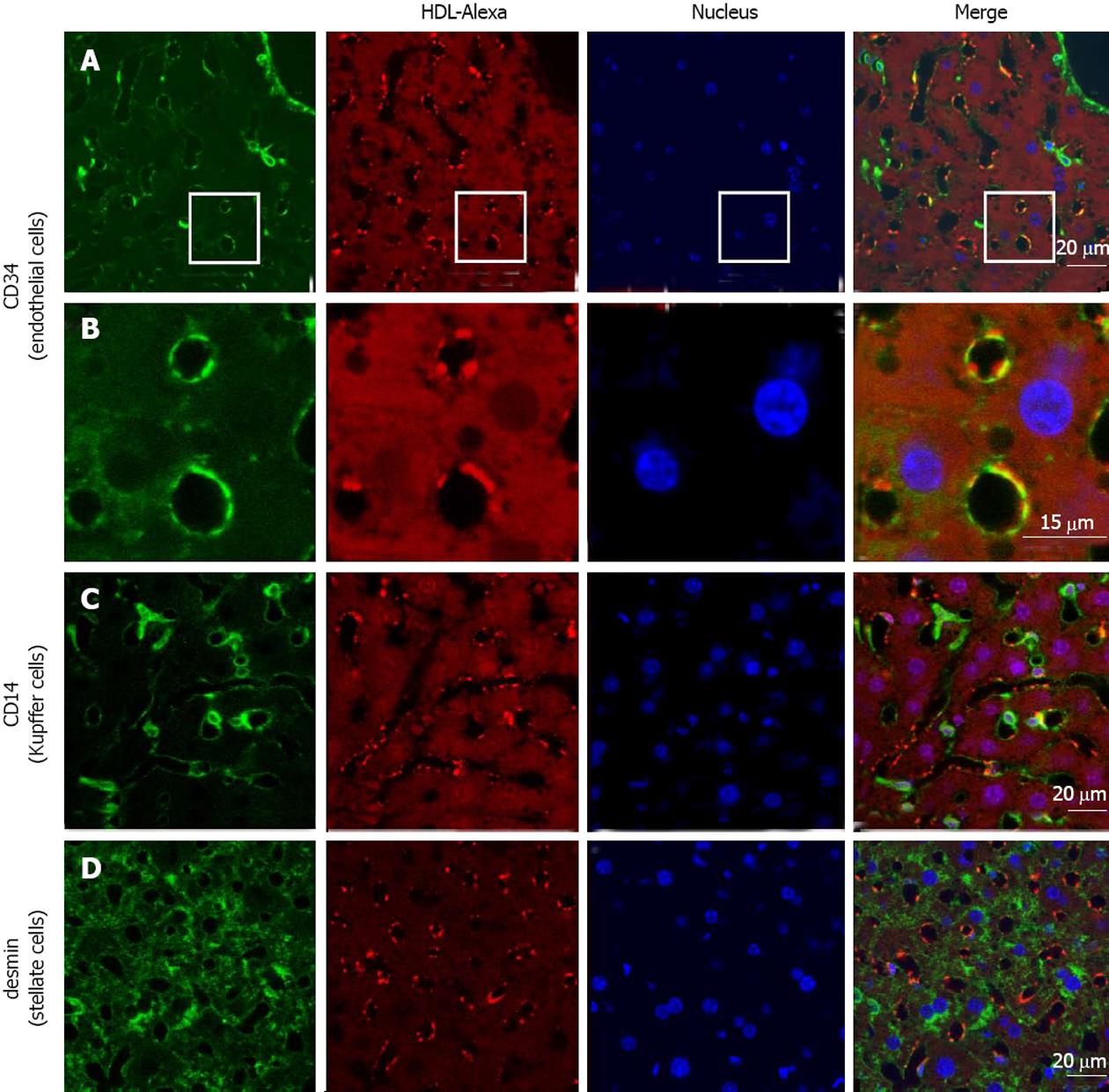
Figure 4 High density lipoprotein uptake in non-parenchymal liver cells.
Fluorescently labeled high density lipoprotein (HDL)-Alexa 568 was intravenously injected into C57BL/6 mice. After 60 min, tissues were fixed by transcardial perfusion. Liver sections were stained for cellular markers by immunofluorescence and analyzed by confocal microscopy. HDL is localized in endothelial cells [A and B (inset)] and to a limited amount in Kupffer cells (C), whereas stellate cells show no detectable HDL staining (D).
- Citation: Fruhwürth S, Pavelka M, Bittman R, Kovacs WJ, Walter KM, Röhrl C, Stangl H. High-density lipoprotein endocytosis in endothelial cells. World J Biol Chem 2013; 4(4): 131-140
- URL: https://www.wjgnet.com/1949-8454/full/v4/i4/131.htm
- DOI: https://dx.doi.org/10.4331/wjbc.v4.i4.131


