Minireviews Open Access
Copyright ©The Author(s) 2025. Published by Baishideng Publishing Group Inc. All rights reserved.
World J Gastrointest Surg. Jul 27, 2025; 17(7): 106471
Published online Jul 27, 2025. doi: 10.4240/wjgs.v17.i7.106471
Enhancing clinical practice: The role of digital rectal examination in diagnosing functional defecation disorders
Lian-Jun Zhu, School of Clinical Medicine, Chengdu University of Traditional Chinese Medicine, Chengdu 610075, Sichuan Province, China
Xing-Lin Zeng, Department of General Surgery, Jiangbei Campus of The First Affiliated Hospital of Army Medical University, Chongqing 400020, China
Xiang-Dong Yang, Department of Colorectal and Anal Surgery, Chengdu Anorectal Hospital, Chengdu 610015, Sichuan Province, China
ORCID number: Lian-Jun Zhu (0009-0004-7722-1650); Xing-Lin Zeng (0009-0006-8424-5428); Xiang-Dong Yang (0009-0007-5699-1949).
Author contributions: Zhu LJ and Yang XD contributed to the manuscript outline, composed the paper, and originated the concept for this manuscript; Zeng XL was responsible for sourcing and organizing relevant literature; Yang XD provided supervision, reviewed the paper, and finalized the manuscript; All authors have read and approved the final manuscript.
Conflict-of-interest statement: The authors declare that they have no conflict of interest.
Open Access: This article is an open-access article that was selected by an in-house editor and fully peer-reviewed by external reviewers. It is distributed in accordance with the Creative Commons Attribution NonCommercial (CC BY-NC 4.0) license, which permits others to distribute, remix, adapt, build upon this work non-commercially, and license their derivative works on different terms, provided the original work is properly cited and the use is non-commercial. See: https://creativecommons.org/Licenses/by-nc/4.0/
Corresponding author: Xiang-Dong Yang, PhD, Professor, Department of Colorectal and Anal Surgery, Chengdu Anorectal Hospital, No. 152 Daqiang East Street, Taisheng South Road, Chengdu 610015, Sichuan Province, China. y-xd@vip.163.com
Received: March 5, 2025
Revised: May 7, 2025
Accepted: June 4, 2025
Published online: July 27, 2025
Processing time: 140 Days and 4.3 Hours

Abstract

Digital rectal examination (DRE) is essential for diagnosing anorectal diseases, yet its widespread adoption remains suboptimal among practitioners. While modalities such as anorectal manometry, rectal balloon expulsion tests, pelvic floor electromyography, and dynamic imaging (defecography/pelvic floor magnetic resonance imaging) enable comprehensive assessment of anorectal function, many healthcare facilities lack advanced diagnostic tools and specialized personnel. DRE has notable diagnostic value in the primary evaluation of functional defecation disorders (FDDs), particularly for detecting dyssynergic defecation and structural pelvic floor defects. Its cost efficiency and universal accessibility render it indispensable in resource-limited environments where high-resolution diagnostic technologies (e.g., high-resolution manometry) are unavailable. This review delineates standardized DRE protocols for assessing FDDs and highlights characteristic imaging features of FDDs with the aim of improving the understanding of DRE. This review will hopefully encourage clinicians to perform DREs in diverse clinical settings.

Key Words: Digital rectal examination; Functional defecation disorders; Constipation; Dyssynergic defecation; Review

Core Tip: In this study, we delineate a standardized digital rectal examination protocol for diagnosing functional defecation disorders and explore clinical metrics validated by dynamic imaging. By incorporating high-resolution defecographic imaging from representative cases, we can establish anatomic-pathophysiological correlations to increase diagnostic precision. These visual didactic tools serve as pivotal clinical references for training and clinical decision-making. The protocol is based on pathognomonic physical signs and systematic characterization, enabling targeted differential diagnosis and evidence-based therapeutic stratification in routine clinical workflows.


INTRODUCTION

Constipation represents a prevalent clinical condition with significant epidemiological variation. According to epidemiologic studies employing the Rome IV criteria, the global prevalence of constipation is 10.1% [95% confidence interval (CI): 8.7-11.6] in the general population[1], indicating a significant sex disparity, with a greater prevalence among females. Notably, prevalence rates differ substantially across age groups: 14.4% (95%CI: 11.2-17.6) in pediatric populations[2] vs 18.9% (95%CI: 14.7-23.9) among older adults[3]. Of clinical relevance, obstructed defecation syndrome affects approximately 60% of individuals with constipation[4,5], whereas dyssynergic defecation constitutes 50% of chronic constipation cases referred to tertiary care centers[6,7]. Furthermore, according to current literature, up to 44% of patients with constipation have pelvic floor dysfunction[8]. Primary care settings and resource-limited regions lack specialized diagnostic modalities, including defecography and pelvic floor magnetic resonance imaging, and personnel with technical expertise, thus digital rectal examination (DRE) is not part of routine clinical practice. Notably, in even tertiary medical centers, specific imaging procedures are difficult to perform on pregnant patients or those with claustrophobia.

Functional defecation disorders (FDDs) encompass two predominant subtypes: Dyssynergic defecation and inadequate defecatory propulsion. Current evidence has revealed the remarkable diagnostic validity of DRE for detecting dyssynergic defecation, with a pooled sensitivity of 75.0%-93.2%, specificity of 58.7%-87.0%, and positive predictive value of 91%-97%[9-11]. Despite these robust metrics, DRE remains underutilized in both medical education and clinical workflows. Although major gastroenterological societies consistently recommend DRE in constipation evaluation, many clinicians omit this examination during patient assessments[9]. Alarmingly, 17% of medical school graduates report never having performed DRE during training[12], whereas only 31% of practicing clinicians express full confidence in their ability to perform DRE[13]. As with most clinical skills, proficiency is strongly correlated with operator experience, which enhances both clinician competence and patient compliance[13]. Critically, experienced practitioners achieve clinically meaningful diagnostic yield through DRE[14], reinforcing its status as an essential component of comprehensive physical examination[15-19]. These findings underscore the urgent need for systematic DRE training integration into medical curricula and primary care competency programs.

In this review, we interrogate the clinical validity of DRE in detecting pathognomonic features of pelvic floor dyssynergia and structural defects underlying FDDs. We also advocate for protocol standardization to enhance diagnostic reproducibility. Concurrently, we delineate tactile assessment limitations and explore pragmatic synergies between DRE and emerging point-of-care technologies to refine diagnostic workflows.

DIAGNOSTIC CRITERIA FOR FDDS

The diagnostic criteria for FDDs (encompassing dyssynergic defecation and inadequate defecatory propulsion) are as follows: (1) Functional constipation with symptoms persisting for ≥ 6 months and/or irritable bowel syndrome with constipation for the past 3 months according to the Rome IV criteria; and (2) Objective documentation of defecatory dysfunction manifesting as at least two of the following pathophysiological findings[20-22]: Abnormal balloon expulsion test indicating impaired rectal evacuation; Anorectal manometry or pelvic floor electromyography revealing paradoxical pelvic floor muscle contraction; Dynamic imaging evidence (e.g., defecography) of inadequate rectal propulsive forces or incomplete rectal emptying.

CLASSIFICATION OF FDDS

FDD classification is rooted in the Rome IV criteria for functional constipation, wherein dyssynergic defecation is subclassified into two pathophysiological subtypes: Structural pelvic floor dysfunction (e.g., rectocele, internal rectal prolapse, and descending perineum syndrome), characterized by impaired rectal motility, and nonrelaxing pelvic floor dysfunction (e.g., puborectalis muscle dyssynergia and spastic pelvic floor syndrome), resulting from paradoxical contraction of the pelvic floor muscles during defecation[23]. A third distinct entity, inadequate defecatory propulsion, refers to insufficient rectal contractile forces during evacuation, which constitutes the focus of this review regarding DRE applications. Notably, overlap between these subtypes is frequently observed in clinical practice[24,25].

DRE FOR FDDS
Preprocedural preparation

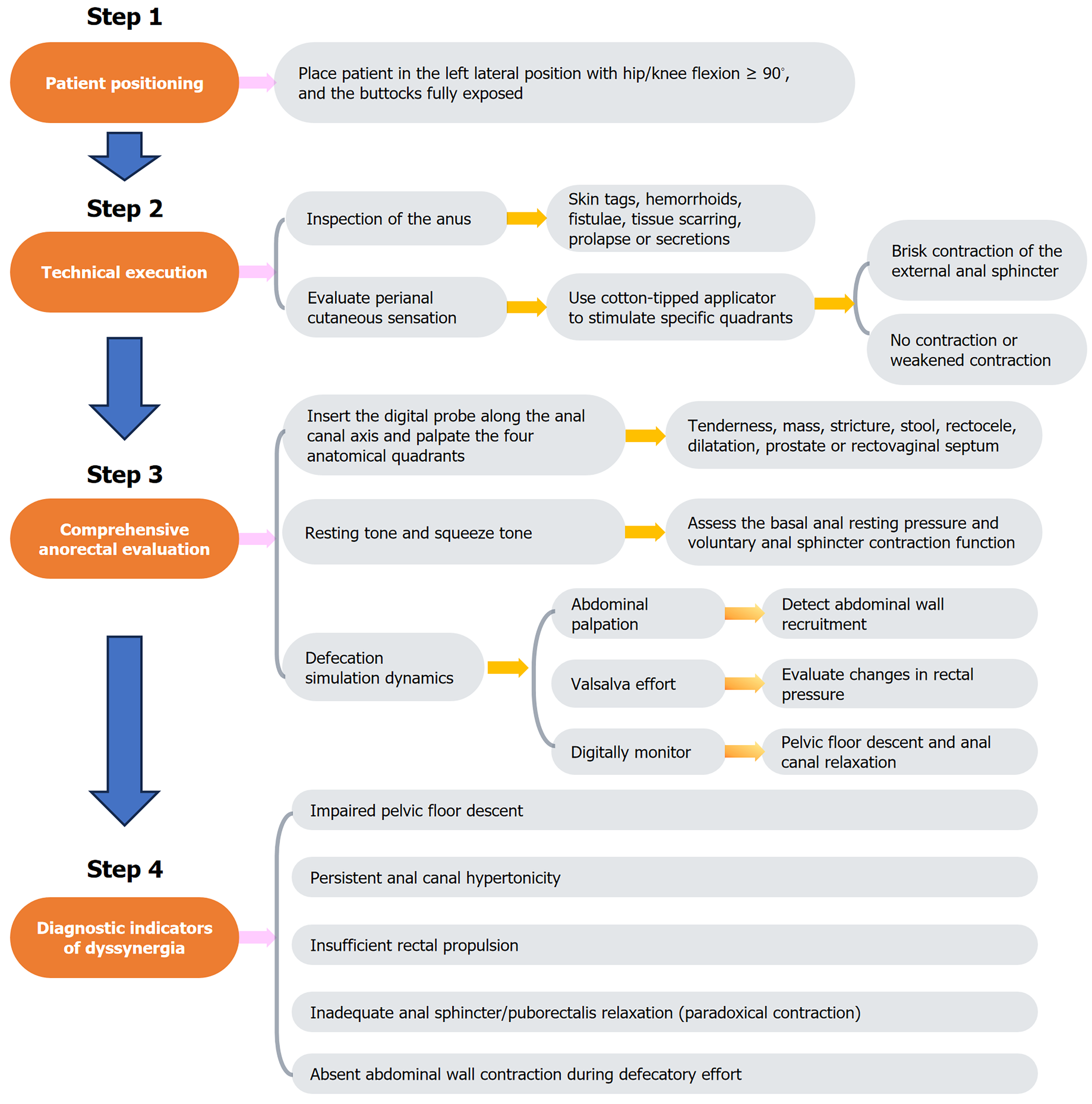
The key steps involved in DRE are illustrated in Figure 1. Prior to DRE, clinicians must establish clear communication with the patient, detailing the procedure’s clinical rationale and obtaining informed consent. A chaperone is strongly recommended to increase patient comfort and mitigate anxiety. The examination should be performed in private under adequate illumination and thermoregulation (20 °C-24 °C). Patients were placed in the left lateral decubitus position with hip/knee flexion ≥ 90° and the buttocks fully exposed via patient-assisted retraction (Figure 2).

Figure 1
Figure 1 Steps of digital rectal examination.
Figure 2
Figure 2 Patient positioning for digital rectal examination.
Technical execution

Using sterile gloves, the examiner systematically inspects the perianal region for pathological markers, including hemorrhoids, fistulae, tissue scarring, or neoplastic lesions. A standardized neurological assessment is performed using a cotton-tipped applicator to evaluate perianal cutaneous sensation via quadrant-specific stimulation (3:00, 6:00, 9:00, 12:00 positions) while observing transient external anal sphincter contractions.

Comprehensive anorectal evaluation

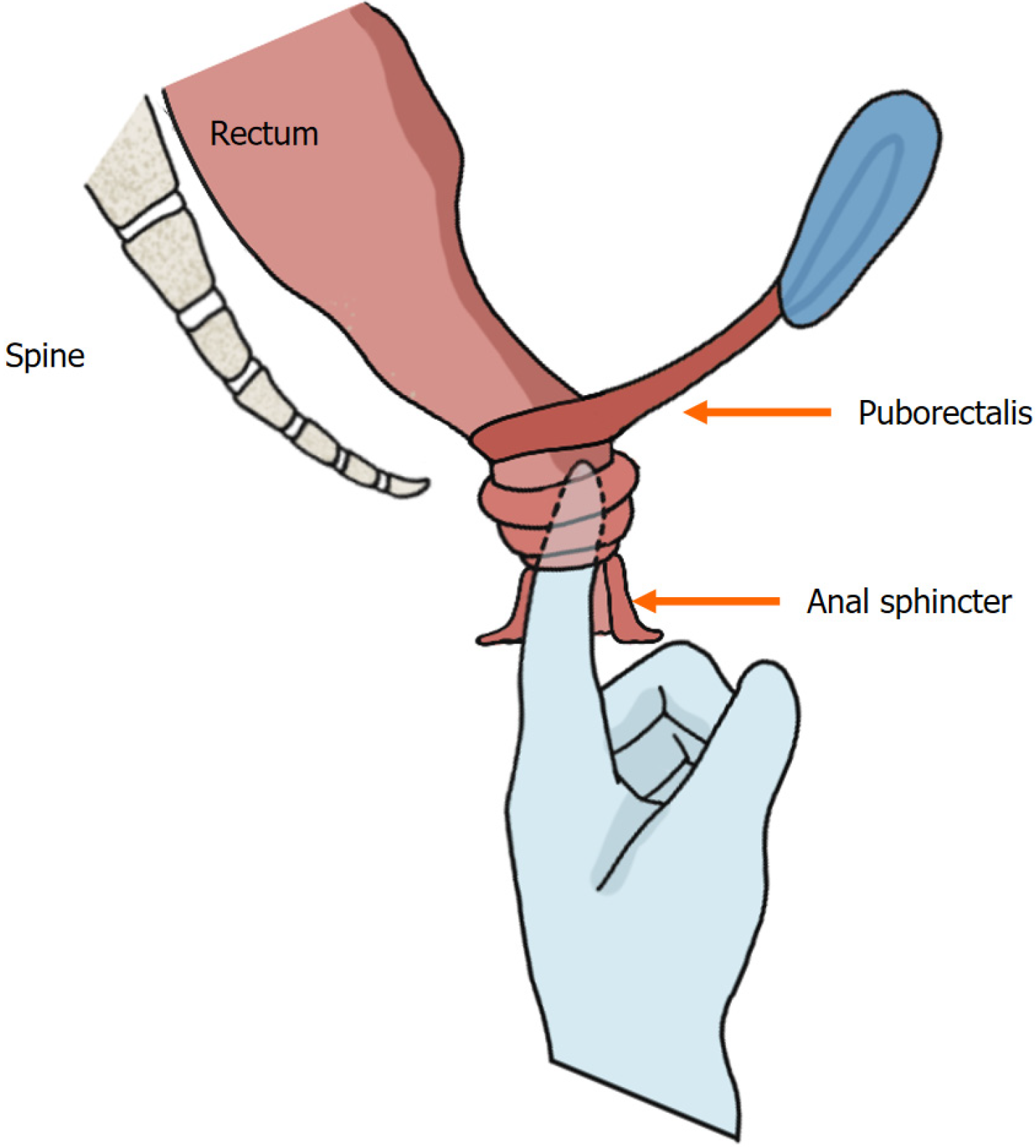
The perianal region is gently massaged with a lubricated gloved index finger to facilitate sphincter relaxation. The digital probe is inserted along the anal canal axis, and then the four anatomical quadrants are palpated (Figure 3). Basal anal resting pressure (measured during patient relaxation) and voluntary sphincter contraction (strength/duration) are then assessed. Defecation simulation dynamics: Simultaneous abdominal palpation (left hand supraumbilical) to detect abdominal wall recruitment; Rectal pressure elevation via Valsalva effort; Pelvic floor descent and anal canal relaxation monitored digitally.

Figure 3
Figure 3 Digital insertion and anorectal palpation.
Diagnostic indicators of dyssynergia

Pathognomonic findings include ≥ 1 of the following[26,27]: (1) Absent abdominal wall contraction during defecatory effort; (2) Inadequate anal sphincter/puborectalis relaxation (paradoxical contraction); (3) Failure to generate adequate rectal propulsive forces; (4) Impaired pelvic floor descent; and (5) Persistent anal canal hypertonicity.

CHARACTERISTICS OF DRE
Rectocele

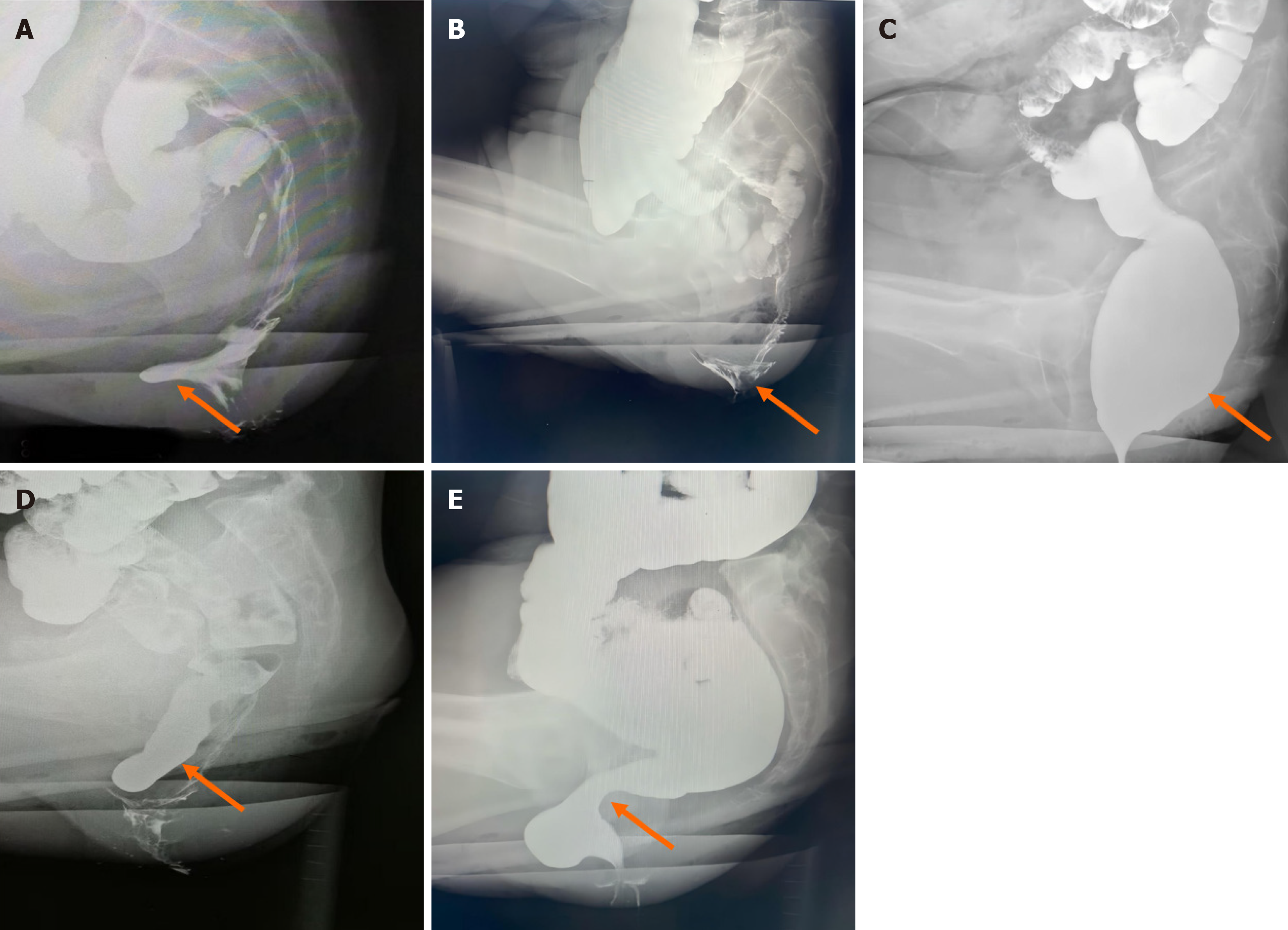
Patients manifest defecatory dysfunction characterized by digital vaginal pressure during evacuation to mechanically redirect fecal flow. Digital palpation at the proximal anal canal revealed an anterior rectal wall defect with reducible herniation into the vaginal lumen (Figure 4A), demonstrating diminished rectovaginal septum integrity and palpable posterior vaginal wall displacement. Provocative maneuvers (Valsalva) exacerbate the magnitude of the defect, with delayed/incomplete tissue recoil after examination. Anatomic severity is stratified by hernia depth: Mild (< 2 cm), moderate (2 cm-4 cm), or severe (> 4 cm)[28]. A clinically significant interpretation requires symptom correlation, given the high prevalence of asymptomatic rectoceles (detected in 80% of nulliparous women)[29].

Figure 4
Figure 4 Morphological changes demonstrated by defecography (indicated by the arrow). A: Sac-like protrusion of the anterior rectal wall; B: Internal rectal prolapse in a funnel-shaped configuration; C: Rectal luminal distension; D: The sigmoid colon descends below the pubococcygeal line; E: Evident pressure trace of the puborectalis muscle.
Internal rectal prolapse

Internal rectal prolapse represents intraluminal invagination of the rectal wall without external anal protrusion[30], which is distinct from external rectal prolapse characterized by complete rectal extrusion through the anal canal[31]. Digital examination revealed redundant rectal mucosa with a folded configuration and preserved mobility, smooth mucosal surface texture, and palpable fecal residue within the rectal lumen. During simulated defecation, the examiner perceives the circumferential mucosal encirclement of the digit, corresponding to “rectal intussusception” when thickened mucosa forms a complete annular obstruction. Positional changes (the squatting maneuver) elicit cervix-like palpation of the distal rectal component. Defecographic correlation demonstrated funnel-shaped mucosal descent (Figure 4B).

Rectal dilatation

Digital examination reveals a spacious rectal lumen with no mucosal wrapping of the examining digit, often requiring maximal digital flexion to achieve bowel wall contact. This finding correlates with radiographic evidence of colonic dilation on barium enema (Figure 4C). Affected patients exhibit diminished rectal contractility, resulting in ineffective fecal expulsion and frequent detection of high-consistency fecal impaction. Critical clinical observations include the following: Fecal consistency documentation; Patient awareness assessment of rectal content and impaired fecal perception suggesting compromised rectal sensitivity[27].

Insufficient rectal propulsion

Patients typically report symptoms of ineffective straining, incomplete evacuation, and rectal outlet obstruction. This condition stems from compromised rectal contractility, resulting in insufficient propulsive forces for effective fecal expulsion. Digital assessment during simulated defecation fails to detect appreciable expulsion forces against the examining digit. Current diagnostic paradigms lack standardized criteria, necessitating a multimodal approach that combines symptomatology, DRE findings, and objective functional testing[10].

Descending perineum syndrome

Descending perineum syndrome is defined as excessive descent of the pelvic organs below the pubococcygeal line[32] (Figure 4D), which constitutes approximately 10% of tertiary referrals for constipation[33]. This syndrome manifests a wide range of symptoms, including sensations of anal obstruction during defecation and incomplete evacuation, as well as urinary or fecal incontinence. On DRE, the pelvic floor muscles are palpated as abnormally flaccid, with reduced anal canal contraction tone. During simulated defecation maneuvers, perineal dilatation manifests as a hemispheric bulge. In female patients, pregnancy and vaginal delivery are considered the primary etiological factors for abnormal pelvic floor muscle relaxation[34,35].

Puborectalis muscle dyssynergia

During normal defecation, the puborectalis muscle relaxes, leading to an increased anorectal angle and dilatation of the anal canal, thereby facilitating fecal expulsion. At the termination of defecation, the puborectalis muscle recontracts, restoring anal canal closure[36]. In puborectalis syndrome, DRE reveals elevated anal canal pressure, progressive elongation of the anal canal, and a direct correlation between canal depth and pressure intensity. The hypertrophied puborectalis muscle exhibits palpable tenderness and firm consistency, with sharply defined margins observed in select cases. During simulated defecation maneuvers, paradoxical contraction of the puborectalis muscle is palpated (i.e., absence of relaxation), which exacerbates fecal obstruction despite increased straining efforts. Defecographic evaluation revealed characteristic puborectalis muscle compression patterns, as illustrated in Figure 4E.

Spastic pelvic floor syndrome

This syndrome is characterized by progressively worsening symptoms of strain during defecation, incomplete fecal evacuation, an anal blockage sensation, and persistent urges without successful evacuation, significantly impairing quality of life. It represents neuromuscular dysfunction of the pelvic floor muscles (external anal sphincter and puborectalis). DRE reveals sustained anal constriction with elevated resting sphincter tone, often preventing painless insertion of the examining finger. During simulated defecation, paradoxical contraction of the anal canal replaces the normal relaxation response.

OTHER ETIOLOGIES OF DEFECATION DISORDERS DETECTABLE THROUGH DRE
Anorectal stenosis

Patients in this group commonly present with symptoms such as straining during defecation. The condition is most frequently attributed to congenital malformations, chronic inflammation, trauma, or surgical interventions involving the anal or rectal region[37-39]. On clinical examination, the sites stenosis are typically located closer to the anal orifice, where DRE often reveals an inability to pass the index finger due to palpable scar tissue hyperplasia and compromised anal canal elasticity. In cases of postoperative anastomotic stenosis in rectal cancer patients, the stenotic segment is generally situated proximal to the surgical anal canal, presenting as a ring-like narrowing that is readily detectable through DRE.

Solitary rectal ulcer syndrome

Solitary rectal ulcer syndrome (SRUS) represents a rare inflammatory disorder characterized by chronic rectal ulceration with concomitant evacuation impairment. Compared with internal rectal prolapse and rectocele, SRUS is associated with a higher prevalence of constipation and elevated anal sphincter tone[40,41]. Digital examination detects irregular mucosal thickening with indurated mobility, often eliciting palpation-induced tenderness that necessitates differentiation from rectal malignancies. Although pathognomonic DRE features are lacking, SRUS findings serve as critical screening indicators that prompt definitive diagnostic investigations.

Rectal carcinoma

DRE serves as a valuable tool for the initial screening of rectal carcinoma. During DRE, irregular mucosal hyperplasia is typically detected, demonstrating poor mobility and varying degrees of luminal stenosis within the rectal cavity. Following withdrawal of the examining finger, residual pus, blood, mucus, and necrotic tissue are commonly identified on the glove. Particular attention should be given to thorough evaluation of the posterior rectal wall to prevent oversight of highly positioned or posteriorly located lesions. When a suspicious mass is palpated, its anatomical location, dimensions, morphological features, consistency, and involvement of the intestinal lumen must be documented in detail, as this information is critical for guiding subsequent diagnostic and therapeutic strategies[42].

LIMITATIONS AND FUTURE DIRECTIONS

While indispensable in routine practice, DRE is not without constraints that merit deliberate scrutiny. Chief among these is the need for operator expertise when performing this technique, where inter clinician discrepancies in discerning subtle anomalies-such as gradations in internal anal sphincter tone or the presence of non-relaxing puborectalis syndrome-remain a persistent challenge. These inconsistencies, rooted in the inherently subjective interpretation of tactile feedback and variable training exposure, highlight a critical need for standardized proficiency benchmarks. Moreover, the qualitative nature of DRE limits its capacity to provide serialized, objective metrics essential for monitoring disease trajectories or quantifying therapeutic responses in conditions like pelvic floor dyssynergia.

To transcend these limitations, a dual-pronged strategy integrating pedagogical innovation and technological augmentation is imperative. First, the development of accredited, simulation-based certification programs-incorporating validated tactile discrimination modules and competency assessments-would ensure uniform skill acquisition across training cohorts. Parallel to this, multicenter validation studies comparing structured DRE protocols against gold-standard functional imaging modalities (e.g., three dimensional high-resolution anorectal manometry, dynamic defecography) are crucial to establish population-adjusted diagnostic thresholds. On the technological frontier, the advent of haptic feedback gloves offers a promising avenue for real-time sphincter tone quantification, potentially reducing diagnostic subjectivity[43]. Concurrently, deep learning-assisted pattern recognition algorithms trained on multisite DRE datasets can standardize the interpretation of palpatory findings such as rectocele reducibility or mucosal prolapse severity. In resource-constrained environments, coupling DRE with portable transperineal ultrasound systems may deliver cost-effective, point-of-care triage without necessitating advanced infrastructure.

The aim of these synergistic approaches is not to supplant DRE’s clinical primacy but to refine its precision, ensuring its continued relevance in an era increasingly driven by quantitative biomarkers and personalized therapeutic algorithms.

CONCLUSION

DRE effectively identifies functional defecatory pathologies, including rectocele, internal rectal prolapse, and puborectalis muscle dyssynergia. While its diagnostic sensitivity exhibits operator-dependent variability inherent to manual palpation techniques, its clinical utility can be optimized through protocol-driven training and competency-based skill acquisition. As a cost-effective, noninvasive, and immediately deployable screening tool, DRE holds value in resource-constrained settings lacking advanced anorectal functional testing. Even in tertiary care centers, proficient DRE execution enables stratified diagnostic algorithms, guiding targeted therapeutic interventions to enhance constipation management efficacy. These attributes underscore DRE’s imperative for global clinical practice integration.

ACKNOWLEDGEMENTS

We extend our sincere appreciation to Dr. Zhen XB from the Department of Constipation at Chengdu Anorectal Hospital for providing select defecography images that greatly enriched this article. We are truly grateful for his valuable contribution.

Footnotes

Provenance and peer review: Unsolicited article; Externally peer reviewed.

Peer-review model: Single blind

Specialty type: Gastroenterology and hepatology

Country of origin: China

Peer-review report’s classification

Scientific Quality: Grade A

Novelty: Grade B

Creativity or Innovation: Grade B

Scientific Significance: Grade A

P-Reviewer: Yang WY S-Editor: Fan M L-Editor: A P-Editor: Xu ZH

References
1.  Barberio B, Judge C, Savarino EV, Ford AC. Global prevalence of functional constipation according to the Rome criteria: a systematic review and meta-analysis. Lancet Gastroenterol Hepatol. 2021;6:638-648.  [RCA]  [PubMed]  [DOI]  [Full Text]  [Cited by in Crossref: 203]  [Cited by in RCA: 180]  [Article Influence: 45.0]  [Reference Citation Analysis (2)]
2.  Tran DL, Sintusek P. Functional constipation in children: What physicians should know. World J Gastroenterol. 2023;29:1261-1288.  [RCA]  [PubMed]  [DOI]  [Full Text]  [Full Text (PDF)]  [Cited by in CrossRef: 30]  [Cited by in RCA: 20]  [Article Influence: 10.0]  [Reference Citation Analysis (33)]
3.  Salari N, Ghasemianrad M, Ammari-Allahyari M, Rasoulpoor S, Shohaimi S, Mohammadi M. Global prevalence of constipation in older adults: a systematic review and meta-analysis. Wien Klin Wochenschr. 2023;135:389-398.  [RCA]  [PubMed]  [DOI]  [Full Text]  [Cited by in Crossref: 9]  [Cited by in RCA: 24]  [Article Influence: 12.0]  [Reference Citation Analysis (1)]
4.  Hayat U, Dugum M, Garg S. Chronic constipation: Update on management. Cleve Clin J Med. 2017;84:397-408.  [RCA]  [PubMed]  [DOI]  [Full Text]  [Cited by in Crossref: 29]  [Cited by in RCA: 39]  [Article Influence: 5.6]  [Reference Citation Analysis (0)]
5.  Lucak S, Lunsford TN, Harris LA. Evaluation and Treatment of Constipation in the Geriatric Population. Clin Geriatr Med. 2021;37:85-102.  [RCA]  [PubMed]  [DOI]  [Full Text]  [Cited by in Crossref: 3]  [Cited by in RCA: 20]  [Article Influence: 4.0]  [Reference Citation Analysis (1)]
6.  Lee TH, Lee JS, Hong SJ, Jeon SR, Kwon SH, Kim WJ, Kim HG, Cho WY, Cho JY, Kim JO, Lee JS. Rectal hyposensitivity and functional anorectal outlet obstruction are common entities in patients with functional constipation but are not significantly associated. Korean J Intern Med. 2013;28:54-61.  [RCA]  [PubMed]  [DOI]  [Full Text]  [Full Text (PDF)]  [Cited by in Crossref: 13]  [Cited by in RCA: 11]  [Article Influence: 0.9]  [Reference Citation Analysis (0)]
7.  Choung RS, Locke GR 3rd, Schleck CD, Zinsmeister AR, Talley NJ. Cumulative incidence of chronic constipation: a population-based study 1988-2003. Aliment Pharmacol Ther. 2007;26:1521-1528.  [RCA]  [PubMed]  [DOI]  [Full Text]  [Cited by in Crossref: 91]  [Cited by in RCA: 108]  [Article Influence: 6.0]  [Reference Citation Analysis (0)]
8.  Brandler J, Camilleri M. Pretest and Post-test Probabilities of Diagnoses of Rectal Evacuation Disorders Based on Symptoms, Rectal Exam, and Basic Tests: a Systematic Review. Clin Gastroenterol Hepatol. 2020;18:2479-2490.  [RCA]  [PubMed]  [DOI]  [Full Text]  [Cited by in Crossref: 15]  [Cited by in RCA: 22]  [Article Influence: 4.4]  [Reference Citation Analysis (0)]
9.  Tantiphlachiva K, Rao P, Attaluri A, Rao SS. Digital rectal examination is a useful tool for identifying patients with dyssynergia. Clin Gastroenterol Hepatol. 2010;8:955-960.  [RCA]  [PubMed]  [DOI]  [Full Text]  [Cited by in Crossref: 173]  [Cited by in RCA: 153]  [Article Influence: 10.2]  [Reference Citation Analysis (0)]
10.  Chedid V, Vijayvargiya P, Halawi H, Park SY, Camilleri M. Audit of the diagnosis of rectal evacuation disorders in chronic constipation. Neurogastroenterol Motil. 2019;31:e13510.  [RCA]  [PubMed]  [DOI]  [Full Text]  [Cited by in Crossref: 27]  [Cited by in RCA: 28]  [Article Influence: 4.7]  [Reference Citation Analysis (0)]
11.  Soh JS, Lee HJ, Jung KW, Yoon IJ, Koo HS, Seo SY, Lee S, Bae JH, Lee HS, Park SH, Yang DH, Kim KJ, Ye BD, Byeon JS, Yang SK, Kim JH, Myung SJ. The diagnostic value of a digital rectal examination compared with high-resolution anorectal manometry in patients with chronic constipation and fecal incontinence. Am J Gastroenterol. 2015;110:1197-1204.  [RCA]  [PubMed]  [DOI]  [Full Text]  [Cited by in Crossref: 60]  [Cited by in RCA: 65]  [Article Influence: 6.5]  [Reference Citation Analysis (0)]
12.  Lawrentschuk N, Bolton DM. Experience and attitudes of final-year medical students to digital rectal examination. Med J Aust. 2004;181:323-325.  [RCA]  [PubMed]  [DOI]  [Full Text]  [Cited by in Crossref: 34]  [Cited by in RCA: 41]  [Article Influence: 2.0]  [Reference Citation Analysis (0)]
13.  Wong RK, Drossman DA, Bharucha AE, Rao SS, Wald A, Morris CB, Oxentenko AS, Ravi K, Van Handel DM, Edwards H, Hu Y, Bangdiwala S. The digital rectal examination: a multicenter survey of physicians' and students' perceptions and practice patterns. Am J Gastroenterol. 2012;107:1157-1163.  [RCA]  [PubMed]  [DOI]  [Full Text]  [Cited by in Crossref: 57]  [Cited by in RCA: 62]  [Article Influence: 4.8]  [Reference Citation Analysis (0)]
14.  Dekker L, van Reijn-Baggen DA, Han-Geurts IJM, Felt-Bersma RJF. To what extent are anorectal function tests comparable? A study comparing digital rectal examination, anal electromyography, 3-dimensional high-resolution anal manometry, and transperineal ultrasound. Int J Colorectal Dis. 2023;38:12.  [RCA]  [PubMed]  [DOI]  [Full Text]  [Cited by in Crossref: 1]  [Cited by in RCA: 9]  [Article Influence: 4.5]  [Reference Citation Analysis (0)]
15.  Liu J, Lv C, Huang Y, Wang Y, Wu D, Zhang C, Sun C, Wang W, Yu Y. Digital Rectal Examination Is a Valuable Bedside Tool for Detecting Dyssynergic Defecation: A Diagnostic Study and a Meta-Analysis. Can J Gastroenterol Hepatol. 2021;2021:5685610.  [RCA]  [PubMed]  [DOI]  [Full Text]  [Full Text (PDF)]  [Cited by in Crossref: 8]  [Cited by in RCA: 8]  [Article Influence: 2.0]  [Reference Citation Analysis (0)]
16.  Wang XJ. Diagnosing Dyssynergic Defecation with the Digital Rectal Exam: The New Digital Revolution? Dig Dis Sci. 2024;69:660-661.  [RCA]  [PubMed]  [DOI]  [Full Text]  [Cited by in RCA: 1]  [Reference Citation Analysis (0)]
17.  Bharucha AE, Coss-Adame E. Diagnostic Strategy and Tools for Identifying Defecatory Disorders. Gastroenterol Clin North Am. 2022;51:39-53.  [RCA]  [PubMed]  [DOI]  [Full Text]  [Cited by in Crossref: 3]  [Cited by in RCA: 8]  [Article Influence: 2.7]  [Reference Citation Analysis (0)]
18.  Pinto RA, Corrêa Neto IJF, Nahas SC, Froehner Junior I, Soares DFM, Cecconello I. Is the Physician Expertise in Digital Rectal Examination of Value in Detecting Anal Tone in Comparison to Anorectal Manometry? Arq Gastroenterol. 2019;56:79-83.  [RCA]  [PubMed]  [DOI]  [Full Text]  [Cited by in Crossref: 3]  [Cited by in RCA: 7]  [Article Influence: 1.2]  [Reference Citation Analysis (0)]
19.  Rajab TK, Bordeianou LG, von Keudell A, Rajab H, Zhou H. Digital Rectal Examination and Anoscopy. N Engl J Med. 2018;378:e30.  [RCA]  [PubMed]  [DOI]  [Full Text]  [Cited by in Crossref: 3]  [Cited by in RCA: 5]  [Article Influence: 0.7]  [Reference Citation Analysis (0)]
20.  Aziz I, Whitehead WE, Palsson OS, Törnblom H, Simrén M. An approach to the diagnosis and management of Rome IV functional disorders of chronic constipation. Expert Rev Gastroenterol Hepatol. 2020;14:39-46.  [RCA]  [PubMed]  [DOI]  [Full Text]  [Cited by in Crossref: 82]  [Cited by in RCA: 189]  [Article Influence: 37.8]  [Reference Citation Analysis (0)]
21.  O'Donnell MT, Haviland SM. Functional Constipation and Obstructed Defecation. Surg Clin North Am. 2024;104:565-578.  [RCA]  [PubMed]  [DOI]  [Full Text]  [Cited by in RCA: 2]  [Reference Citation Analysis (0)]
22.  Ciriza de Los Ríos C, Aparicio Cabezudo M, Zatarain Valles A, Rey Díaz-Rubio E. Obstructed defecation syndrome: a diagnostic and therapeutic challenge. Rev Esp Enferm Dig. 2020;112:477-482.  [RCA]  [PubMed]  [DOI]  [Full Text]  [Cited by in Crossref: 1]  [Cited by in RCA: 2]  [Article Influence: 0.4]  [Reference Citation Analysis (0)]
23.  Anorectal Branch of Chinese Medical Doctor Association; Clinical Guidelines Committee, Anorectal Branch of Chinese Medical Doctor Association;  Professional Committee on Anorectal Diseases, Chinese Society of Integrated Traditional Chinese and Western Medicine;  Anorectal Disease Committee of Chinese Medical Women's Association;  Chinese Constipation Medical Association. [Chinese expert consensus on the diagnosis and treatment of outlet obstructive constipation (2022 edition)]. Zhonghua Wei Chang Wai Ke Za Zhi. 2022;25:1045-1057.  [RCA]  [PubMed]  [DOI]  [Full Text]  [Cited by in RCA: 1]  [Reference Citation Analysis (0)]
24.  Pescatori M, Spyrou M, Pulvirenti d'Urso A. A prospective evaluation of occult disorders in obstructed defecation using the 'iceberg diagram'. Colorectal Dis. 2006;8:785-789.  [RCA]  [PubMed]  [DOI]  [Full Text]  [Cited by in Crossref: 64]  [Cited by in RCA: 64]  [Article Influence: 3.4]  [Reference Citation Analysis (0)]
25.  Bharucha AE, Lacy BE. Mechanisms, Evaluation, and Management of Chronic Constipation. Gastroenterology. 2020;158:1232-1249.e3.  [RCA]  [PubMed]  [DOI]  [Full Text]  [Cited by in Crossref: 218]  [Cited by in RCA: 335]  [Article Influence: 67.0]  [Reference Citation Analysis (1)]
26.  Rao SSC. Digital Rectal Examination: An Invaluable Clinical Tool. Gastro Hep Adv. 2024;3:592-593.  [RCA]  [PubMed]  [DOI]  [Full Text]  [Full Text (PDF)]  [Cited by in RCA: 1]  [Reference Citation Analysis (0)]
27.  Rao SS, Patcharatrakul T. Diagnosis and Treatment of Dyssynergic Defecation. J Neurogastroenterol Motil. 2016;22:423-435.  [RCA]  [PubMed]  [DOI]  [Full Text]  [Full Text (PDF)]  [Cited by in Crossref: 119]  [Cited by in RCA: 158]  [Article Influence: 17.6]  [Reference Citation Analysis (0)]
28.  Bunni J, Laugharne MJ. Pathophysiological basis, clinical assessment, investigation and management of patients with obstruction defecation syndrome. Langenbecks Arch Surg. 2023;408:75.  [RCA]  [PubMed]  [DOI]  [Full Text]  [Cited by in Crossref: 1]  [Cited by in RCA: 4]  [Article Influence: 2.0]  [Reference Citation Analysis (0)]
29.  Shorvon PJ, McHugh S, Diamant NE, Somers S, Stevenson GW. Defecography in normal volunteers: results and implications. Gut. 1989;30:1737-1749.  [RCA]  [PubMed]  [DOI]  [Full Text]  [Cited by in Crossref: 575]  [Cited by in RCA: 450]  [Article Influence: 12.5]  [Reference Citation Analysis (0)]
30.  Cariou de Vergie L, Venara A, Duchalais E, Frampas E, Lehur PA. Internal rectal prolapse: Definition, assessment and management in 2016. J Visc Surg. 2017;154:21-28.  [RCA]  [PubMed]  [DOI]  [Full Text]  [Cited by in Crossref: 16]  [Cited by in RCA: 18]  [Article Influence: 2.0]  [Reference Citation Analysis (0)]
31.  Oruc M, Erol T. Current diagnostic tools and treatment modalities for rectal prolapse. World J Clin Cases. 2023;11:3680-3693.  [RCA]  [PubMed]  [DOI]  [Full Text]  [Full Text (PDF)]  [Cited by in RCA: 11]  [Reference Citation Analysis (3)]
32.  Sharma A, Herekar A, Yan Y, Karunaratne T, Rao SSC. Dyssynergic Defecation and Other Evacuation Disorders. Gastroenterol Clin North Am. 2022;51:55-69.  [RCA]  [PubMed]  [DOI]  [Full Text]  [Cited by in Crossref: 1]  [Cited by in RCA: 18]  [Article Influence: 6.0]  [Reference Citation Analysis (0)]
33.  Wang XJ, Chedid V, Vijayvargiya P, Camilleri M. Clinical Features and Associations of Descending Perineum Syndrome in 300 Adults with Constipation in Gastroenterology Referral Practice. Dig Dis Sci. 2020;65:3688-3695.  [RCA]  [PubMed]  [DOI]  [Full Text]  [Cited by in Crossref: 10]  [Cited by in RCA: 12]  [Article Influence: 2.4]  [Reference Citation Analysis (0)]
34.  Chang J, Chung SS. An analysis of factors associated with increased perineal descent in women. J Korean Soc Coloproctol. 2012;28:195-200.  [RCA]  [PubMed]  [DOI]  [Full Text]  [Full Text (PDF)]  [Cited by in Crossref: 10]  [Cited by in RCA: 11]  [Article Influence: 0.8]  [Reference Citation Analysis (0)]
35.  Baek HN, Hwang YH, Jung YH. Clinical Significance of Perineal Descent in Pelvic Outlet Obstruction Diagnosed by using Defecography. J Korean Soc Coloproctol. 2010;26:395-401.  [RCA]  [PubMed]  [DOI]  [Full Text]  [Full Text (PDF)]  [Cited by in Crossref: 11]  [Cited by in RCA: 12]  [Article Influence: 0.8]  [Reference Citation Analysis (0)]
36.  Lalwani N, El Sayed RF, Kamath A, Lewis S, Arif H, Chernyak V. Imaging and clinical assessment of functional defecatory disorders with emphasis on defecography. Abdom Radiol (NY). 2021;46:1323-1333.  [RCA]  [PubMed]  [DOI]  [Full Text]  [Cited by in Crossref: 4]  [Cited by in RCA: 9]  [Article Influence: 2.3]  [Reference Citation Analysis (0)]
37.  He F, Yang F, Chen D, Tang C, Woraikat S, Xiong J, Qian K. Risk factors for anastomotic stenosis after radical resection of rectal cancer: A systematic review and meta-analysis. Asian J Surg. 2024;47:25-34.  [RCA]  [PubMed]  [DOI]  [Full Text]  [Cited by in Crossref: 9]  [Cited by in RCA: 9]  [Article Influence: 9.0]  [Reference Citation Analysis (0)]
38.  Kishida Y, Thorlacius H. Complete Stenosis After Endoscopic Submucosal Dissection of a Large Circumferential Rectal Lesion. Am J Gastroenterol. 2025;120:511.  [RCA]  [PubMed]  [DOI]  [Full Text]  [Cited by in RCA: 1]  [Reference Citation Analysis (0)]
39.  Zou R, Jiang X, Wang M, Wang Y, Zhao L, Fan Z. Successful endoscopic management of severe low rectal stenosis caused by chemical burns. Rev Esp Enferm Dig. 2024;.  [RCA]  [PubMed]  [DOI]  [Full Text]  [Cited by in RCA: 1]  [Reference Citation Analysis (0)]
40.  Felt-Bersma RJF. Solitary rectal ulcer syndrome. Curr Opin Gastroenterol. 2021;37:59-65.  [RCA]  [PubMed]  [DOI]  [Full Text]  [Cited by in Crossref: 2]  [Cited by in RCA: 6]  [Article Influence: 1.5]  [Reference Citation Analysis (0)]
41.  Morio O, Meurette G, Desfourneaux V, D'Halluin PN, Bretagne JF, Siproudhis L. Anorectal physiology in solitary ulcer syndrome: a case-matched series. Dis Colon Rectum. 2005;48:1917-1922.  [RCA]  [PubMed]  [DOI]  [Full Text]  [Cited by in Crossref: 39]  [Cited by in RCA: 33]  [Article Influence: 1.7]  [Reference Citation Analysis (0)]
42.  Lee J, Grubac V, Baxter N, Auer R, Porter G, Holm T, Moran B, Heald RJ, Simunovic M. Digital rectal examination in palpable rectal cancer: expert panel consensus on key elements and analysis of a case series. Br J Surg. 2021;108:e264-e265.  [RCA]  [PubMed]  [DOI]  [Full Text]  [Reference Citation Analysis (1)]
43.  Salvadores Fernandez C, Jaufuraully S, Bagchi B, Chen W, Datta P, Gupta P, David AL, Siassakos D, Desjardins A, Tiwari MK. A Triboelectric Nanocomposite for Sterile Sensing, Energy Harvesting, and Haptic Diagnostics in Interventional Procedures from Surgical Gloves. Adv Healthc Mater. 2023;12:e2202673.  [RCA]  [PubMed]  [DOI]  [Full Text]  [Cited by in Crossref: 2]  [Cited by in RCA: 1]  [Article Influence: 0.5]  [Reference Citation Analysis (1)]