Published online Jul 27, 2022. doi: 10.4240/wjgs.v14.i7.706
Peer-review started: March 2, 2022
First decision: April 25, 2022
Revised: April 30, 2022
Accepted: June 20, 2022
Article in press: June 20, 2022
Published online: July 27, 2022
Processing time: 146 Days and 17 Hours
Giant simple hepatic cysts causing intrahepatic duct dilatation and obstructive jaundice are uncommon. A variety of measures with different clinical efficacies and invasiveness have been developed. Nonsurgical management, such as percutaneous aspiration and sclerotherapy, is often applied.
The case is a 39-year-old female with a 5-mo history of cutaneous and scleral icterus, loss of appetite, and dark urine. Lab tests showed jaundice and liver function abnormalities. Imaging revealed a giant simple hepatic cyst obstructing the intrahepatic bile ducts. A combination of percutaneous catheter aspiration and lauromacrogol sclerotherapy was successfully performed and the effects were satisfactory with the size of cyst decreasing from 13.7 cm × 13.1 cm to 3.0 cm × 3.0 cm. Further literature review presented the challenges of managing giant simple hepatic cysts that cause obstructive jaundice and compared the safety and efficacy of a combination of percutaneous aspiration and lauromacrogol sclerotherapy with other management strategies.
Giant simple hepatic cysts can cause obstructive jaundice, and a combination of percutaneous catheter aspiration and sclerotherapy with lauromacrogol are suggested to treat such cases.
Core Tip: Giant simple hepatic cysts causing obstructive jaundice are uncommon. Here we presented the challenges of managing giant simple hepatic cysts causing obstructive jaundice and compared the safety and efficacy of percutaneous aspiration and lauromacrogol sclerotherapy with other management strategies. The case is a 39-year-old female with jaundice and liver function abnormalities. Images revealed a giant simple hepatic cyst with obstruction of intrahepatic bile ducts. A combination of percutaneous catheter aspiration and lauromacrogol sclerotherapy was conducted successively, achieving satisfactory efficacy. Therefore, a combination of percutaneous aspiration and lauromacrogol sclerotherapy may be suggested to solve such cases.
- Citation: He XX, Sun MX, Lv K, Cao J, Zhang SY, Li JN. Percutaneous aspiration and sclerotherapy of a giant simple hepatic cyst causing obstructive jaundice: A case report and review of literature. World J Gastrointest Surg 2022; 14(7): 706-713
- URL: https://www.wjgnet.com/1948-9366/full/v14/i7/706.htm
- DOI: https://dx.doi.org/10.4240/wjgs.v14.i7.706
Hepatic cysts occur in 2.5%-18% of the population[1-3]. They generally include a cluster of diseases with heterogeneous pathogenesis and etiology, including simple hepatic cysts, infectious cysts, cystic neoplasms, biliary duct-related cysts and some congenital polycystic liver diseases[4]. Most simple cysts are asymptomatic and are incidentally identified during imaging examinations, including ultrasonography (US), computed tomography (CT) or magnetic resonance imaging[5,6]. Only 5%-16% of simple hepatic cysts become symptomatic due to mass effects, rupture, hemorrhaging, or infection[5,7,8]. They mainly present as abdominal pain, nausea, vomiting and occasional jaundice[9,10].
The management of simple hepatic cysts widely differs according to clinical manifestations, imaging features, and, sometimes, patient preference. A watch-and-see strategy is acceptable for asymptomatic simple cysts, whereas interventions are required if cysts cause severe symptoms or complications. Various treatment methods with different clinical efficacies and levels of invasiveness have been developed. For nonsurgical management, percutaneous aspiration, sclerotherapy, and internal drainage are often used[8,9]. Surgical treatment mainly includes unroofing, cyst fenestration, hepatectomy, and open or laparoscopic liver transplantation[11]. Treatment selection depends on cyst location, size, surroundings and other factors[12,13].
Here, we report a case of a giant simple hepatic cyst in the hepatic hilum causing intrahepatic duct dilatation and obstructive jaundice. A combination of percutaneous aspiration and lauromacrogol sclerotherapy was performed and achieved satisfactory effects. The related literature was reviewed to better understand management in similar patients.
A 39-year-old female was admitted for cutaneous and scleral icterus, loss of appetite, and dark urine for 5 mo.
A 39-year-old female was admitted for cutaneous and scleral icterus, loss of appetite, and dark urine for 5 mo.
The patient used to be in good health and had no previous medical history.
The patient’s personal habits, customs, and family history were unremarkable.
Physical examination revealed moderate jaundice without abdominal tenderness, hepatomegaly, or Murphy’s sign.
Lab tests showed jaundice [total bilirubin (TBil) level was 149.8 μmol/L, and direct bilirubin (DBil) level was 118.7 μmol/L], liver function abnormalities (liver function test levels included the following: Alanine transaminase (ALT) was 175 U/L, aspartate aminotransferase (AST) was 130 U/L, gamma-glutamyl transpeptidase was 454 U/L, alkaline phosphatase was 314 U/L) and moderate anemia [the hemoglobin (HGB) level was 75 g/L]. Tumor markers were unremarkable except for a slightly elevated carcinoma embryonic antigen (CEA) level of 6.1 ng/mL (normal range: 0-5). Antibodies for hepatitis virus, primary biliary cholangitis and autoimmune hepatitis were all within the normal limits.
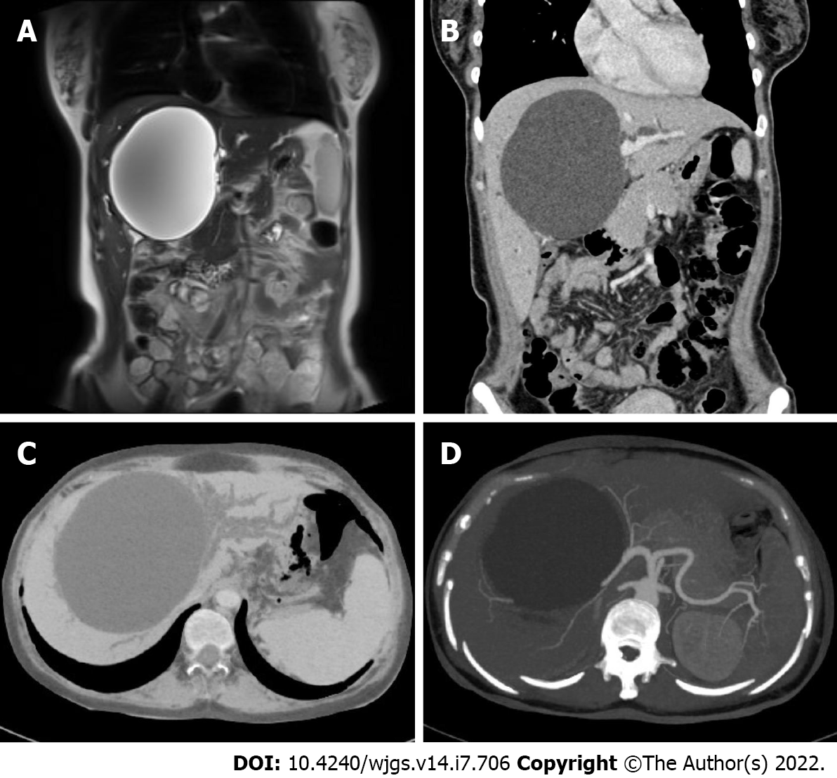
The abdominal US and the endoscopic US showed an enlarged liver (3.7 cm below the xiphoid process) and an anechoic area (increasing from 11.2 cm × 9.9 cm to 13.7 cm × 13.1 cm in three months) with a clear boundary and no peripheral blood flow, and the intrahepatic bile duct of the left lateral segment was approximately 0.6 cm wide. Magnetic resonance cholangiopancreatography showed several hepatic cysts. The largest cyst was approximately 9.5 cm × 11 cm in size, located in the hilum, and obstructed the intrahepatic bile ducts. Three-dimensional reconstruction of the biliary tract showed dilatated intrahepatic bile ducts and compressed hepatic vessels and branches of the portal vein (Figure 1).
Notably, esophagogastroduodenoscopy and colonoscopy were performed and excluded gastro
A giant simple hepatic cyst complicated with obstructive jaundice was the diagnosis.
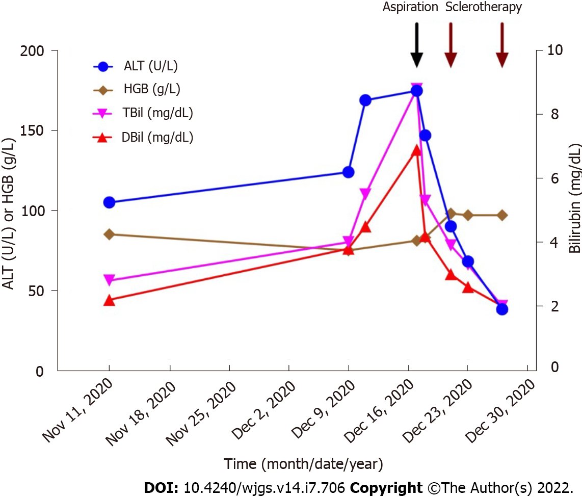
We successfully performed a combination of percutaneous catheter aspiration and sclerotherapy with lauromacrogol. During percutaneous catheter aspiration under the guidance of US, the giant cyst was punctured with an 18-gauge pig-tail catheter. Postoperative drainage was favorable, and a total of 800 milliliters of clear yellow fluid was drained; bilirubin levels, tumor markers (such as CEA level) and cytology tests were unremarkable. Jaundice (TBil was 66.4 μmol/L, DBil was 51.2 μmol/L) and liver function anomalies (ALT was 90 U/L, AST was 59 U/L) were significantly relieved soon after drainage.
Then, two sessions of sclerotherapy (lauromacrogol) of the hepatic cyst were performed (30 mL and 20 mL lauromacrogol mixed with triple amounts of air) at one week. Of note, before sclerotherapy, the communications of the cyst with the surrounding bile ducts were ruled out by injecting a diluted contrast medium into the cyst cavity. After sclerotherapy, a small amount of cyst fluid was drained, and the tube was removed. The patient was generally in good condition. He was discharged and experienced further improvement in his liver function (ALT level was 38 U/L, TBil level was 34.9 μmol/L, and DBil level was 33.5 μmol/L; Figure 2).
During follow-up, the patient reported continued resolution of his symptoms. Three months after treatment, the size of the liver cyst decreased to 6.5 cm × 5.6 cm, and liver function returned to normal limits. Fourteen months after treatment, the size of the cyst had decreased to 3.0 cm × 3.0 cm on US.
Most simple liver cysts are asymptomatic and stable in size and structure, which allows for observation. However, some of these tumors gradually grow and eventually cause symptoms due to large size, rupture, hemorrhaging, infection, or neoplasm in rare cases[8,14]. Symptoms, including abdominal discomfort or pain, nausea, vomiting, jaundice, early satiety, and even dyspnea[9,10], are largely related to cyst size and location and are more often attributed to larger cysts and right-sided cysts[9,15]. In a recent review, abdominal pain was reported to be the most common symptom of simple hepatic cysts and was reported by 60% (456 of 764) of the patients[16].
Obstructive jaundice caused by solitary simple liver cysts is quite rare. A total of 17 cases of simple or benign liver cysts accompanied by obstructive jaundice were reviewed (Table 1)[17-33]. The average age of the patients was 65.2 years old, with a 7:10 female to male ratio. These cysts tended to be large (greater than 10 cm) and centrally located when compression of the main intrahepatic duct or even the hepatic hilum was present. Treatment for these patients varied from aspiration to resection. In recent years, a combination of drainage, sclerosing agent injection, and deroofing seem to be the most common treatment methods. Choledochoscopy was also proven to effectively treat these patients[33]. In our patients, the giant liver cyst caused obstructive jaundice and dilatation of the intrahepatic bile duct of the left lateral segment of the liver, which largely accounted for the patient’s symptoms.
| No. | Ref. | Age/sex | Cyst (cm) | Location (segments) | Total bilirubin (mg/dL) | Treatment | Prognosis | Follow-up period |
| 1 | Caravati et al[17], 1950 | 33/M | NA | IV, V | NA | Aspiration + marsupialization | Improved | 7 mo |
| 2 | Hudson[18], 1963 | 55/F | 25 | III, IV, V | 14 | Cystenterostomy | Improved | 1 mo |
| 3 | Dardik et al[19], 1964 | 69/F | 15 | V | 9 | Cystectomy | Improved | 1 mo |
| 4 | Sacks et al[20], 1967 | 81/M | 20 | IV | 19 | Aspiration | Improved | 2 mo |
| 5 | Santman et al[21], 1977 | 61/M | 15 | IV | 29 | Partial resection | Improved | NA |
| 6 | Machell et al[22], 1978 | 67/F | NA | III, IV, V | NA | Drainage + transhepatic T-tube | Improved | 7 mo |
| 7 | Morin et al[23], 1980 | 80/M | 17 | IV, V | 15 | Aspiration only | Improved | 10 mo |
| 8 | Fernandez et al[24], 1984 | 61/F | 30 | III, IV, V | 22 | Partial resection | Improved | 24 mo |
| 9 | Clinkscales et al[25], 1985 | 80/M | 8 | IV | 8 | Aspiration only | Improved | 1 mo |
| 10 | Cappel et al[26], 1988 | 44/F | 12 | IV, V | 5 | Aspiration | Improved | 3 mo |
| 11 | Spivey et al[27], 1990 | 73/M | 11 | IV, V | 10 | Drainage + deroofing | Improved | NA |
| 12 | Terada et al[28], 1993 | 71/F | 12 | III, IV, V | 9 | Drainage + cystectomy | Improved | 1 mo |
| 13 | Yoshihara et al[29], 1996 | 88/M | 16 | IV, V | 8 | Drainage + minocycline injection | Improved | 9 mo |
| 14 | Kanai et al[30], 1999 | 71/M | 15 | IV, V, VIII | 5 | Drainage + deroofing | Improved | 15 mo |
| 15 | Ishikawa et al[31], 2002 | 70/M | 18 | IV, V, VIII | 9 | Drainage + minocycline injection | Improved | 20 mo |
| 16 | Ogawa et al[32], 2004 | 64/M | 9 | NA | NA | Drainage + minocycline injection | Improved | NA |
| 17 | Zhang et al[33], 2018 | 41/F | 5 | IV | 24 | Choledochoscopic high-frequency needle-knife electrotomy | Improved | 36 mo |
Aspiration is generally associated with high recurrence rates[34]. In recent years, percutaneous aspiration combined with sclerotherapy has been widely used as a minimally invasive procedure for simple hepatic cysts with satisfactory results[35-39]. During percutaneous aspiration and sclerotherapy, US- or CT-guided aspiration and drainage are combined with the injection of a sclerosing agent[7,40,41]. Sclerosing agents with good efficacy include ethanol, iophendylate, tetracycline chloride, doxycycline, minocycline chloride, and hypertonic saline solution[42].
While liquid sclerosing agents may mix with cyst contents and reduce sclerosing effects, foam sclerotherapy was initially used for vascular malformations and has evolved as an alternative for treating simple hepatic cysts[43]. The agents in a foam vehicle can completely destroy the intimal barrier after 2 min of exposure, causing endothelial edema, exfoliation from the tunica media, and thrombogenesis in the tunica media in 30 min[44]. Sclerotherapy using lauromacrogol foam is rarely reported for treating hepatic cysts. In one case report, laparoscopic lauromacrogol sclerotherapy surgery was reported to be safe and effective in patients with IVa, VII and VIII segment simple hepatic cysts, but more studies are needed to confirm their conclusion[45]. Our case report is the first to combine percutaneous aspiration with sclerotherapy using lauromacrogol in treating a giant simple hepatic cyst, thus proving the safety and efficacy of the therapy. Single or multiple sessions of percutaneous aspiration and sclerotherapy for persistent or recurrent symptoms are adaptable based on cyst features, efficacy and doctor or patient preference[7]. In our patients, sclerotherapy with lauromacrogol was planned and administered twice to achieve a better sclerosing effect.
Surgical treatment of simple hepatic cysts, such as open or laparoscopic cyst deroofing or hepatectomy, can be effective but may contribute to recurrence and complications[46,47]. Generally, percutaneous aspiration combined with sclerotherapy and laparoscopic deroofing is reasonable for most symptomatic simple hepatic cysts. A systematic review showed that the outcome of percutaneous aspiration and sclerotherapy was excellent, with symptoms that persisted in less than 4% of patients, and both complication and recurrence rates were < 1%[16]. Major complications were reported in 2/265 (0.8%), 6/348 (1.7%) and 3/123 (2.4%), and cyst recurrence rates were 0.0%, 5.6% and 7.7% in patients treated with percutaneous aspiration and sclerotherapy and laparoscopic and open surgery, respectively[16]. Other studies on the advantage of percutaneous aspiration and sclerotherapy compared to surgical techniques reported similar results[13]. These results supported the safety and efficacy of percutaneous aspiration and sclerotherapy in treating symptomatic simple hepatic cysts prior to surgical procedures. Our patient’s outcome suggested that percutaneous aspiration and sclerotherapy could effectively treat simple giant hepatic cysts. Studies concerning cost, hospitalization time, and quality of life are needed to further compare these measures.
Giant simple hepatic cysts can obstruct the intrahepatic bile ducts and cause obstructive jaundice. A combination of percutaneous catheter aspiration and sclerotherapy using lauromacrogol can achieve satisfactory results without evident complications compared to surgical interventions.
| 1. | Caremani M, Vincenti A, Benci A, Sassoli S, Tacconi D. Ecographic epidemiology of non-parasitic hepatic cysts. J Clin Ultrasound. 1993;21:115-118. [RCA] [PubMed] [DOI] [Full Text] [Cited by in Crossref: 100] [Cited by in RCA: 82] [Article Influence: 2.5] [Reference Citation Analysis (0)] |
| 2. | Carrim ZI, Murchison JT. The prevalence of simple renal and hepatic cysts detected by spiral computed tomography. Clin Radiol. 2003;58:626-629. [RCA] [PubMed] [DOI] [Full Text] [Cited by in Crossref: 217] [Cited by in RCA: 210] [Article Influence: 9.1] [Reference Citation Analysis (0)] |
| 3. | European Association for the Study of the Liver (EASL). EASL Clinical Practice Guidelines on the management of benign liver tumours. J Hepatol. 2016;65:386-398. [RCA] [PubMed] [DOI] [Full Text] [Cited by in Crossref: 286] [Cited by in RCA: 355] [Article Influence: 35.5] [Reference Citation Analysis (2)] |
| 4. | Taylor BR, Langer B. Current surgical management of hepatic cyst disease. Adv Surg. 1997;31:127-148. [PubMed] |
| 5. | Cowles RA, Mulholland MW. Solitary hepatic cysts. J Am Coll Surg. 2000;191:311-321. [RCA] [PubMed] [DOI] [Full Text] [Cited by in Crossref: 88] [Cited by in RCA: 74] [Article Influence: 2.8] [Reference Citation Analysis (1)] |
| 6. | Lantinga MA, Gevers TJ, Drenth JP. Evaluation of hepatic cystic lesions. World J Gastroenterol. 2013;19:3543-3554. [RCA] [PubMed] [DOI] [Full Text] [Full Text (PDF)] [Cited by in CrossRef: 119] [Cited by in RCA: 94] [Article Influence: 7.2] [Reference Citation Analysis (3)] |
| 7. | Karam AR, Connolly C, Fulwadhva U, Hussain S. Alcohol sclerosis of a giant liver cyst following failed deroofings. J Radiol Case Rep. 2011;5:19-22. [RCA] [PubMed] [DOI] [Full Text] [Cited by in Crossref: 2] [Cited by in RCA: 10] [Article Influence: 0.7] [Reference Citation Analysis (0)] |
| 8. | Nisenbaum HL, Rowling SE. Ultrasound of focal hepatic lesions. Semin Roentgenol. 1995;30:324-346. [RCA] [PubMed] [DOI] [Full Text] [Cited by in Crossref: 41] [Cited by in RCA: 34] [Article Influence: 1.1] [Reference Citation Analysis (0)] |
| 9. | Lai EC, Wong J. Symptomatic nonparasitic cysts of the liver. World J Surg. 1990;14:452-456. [RCA] [PubMed] [DOI] [Full Text] [Cited by in Crossref: 42] [Cited by in RCA: 43] [Article Influence: 1.2] [Reference Citation Analysis (1)] |
| 10. | Karavias DD, Tsamandas AC, Payatakes AH, Solomou E, Salakou S, Felekouras ES, Tepetes KN. Simple (non-parasitic) liver cysts: clinical presentation and outcome. Hepatogastroenterology. 2000;47:1439-1443. [PubMed] |
| 11. | Gomez A, Wisneski AD, Luu HY, Hirose K, Roberts JP, Hirose R, Freise CE, Nakakura EK, Corvera CU. Contemporary Management of Hepatic Cyst Disease: Techniques and Outcomes at a Tertiary Hepatobiliary Center. J Gastrointest Surg. 2021;25:77-84. [RCA] [PubMed] [DOI] [Full Text] [Full Text (PDF)] [Cited by in Crossref: 5] [Cited by in RCA: 12] [Article Influence: 2.4] [Reference Citation Analysis (0)] |
| 12. | Macutkiewicz C, Plastow R, Chrispijn M, Filobbos R, Ammori BA, Sherlock DJ, Drenth JP, O'Reilly DA. Complications arising in simple and polycystic liver cysts. World J Hepatol. 2012;4:406-411. [RCA] [PubMed] [DOI] [Full Text] [Full Text (PDF)] [Cited by in Crossref: 30] [Cited by in RCA: 35] [Article Influence: 2.5] [Reference Citation Analysis (0)] |
| 13. | Erdogan D, van Delden OM, Rauws EA, Busch OR, Lameris JS, Gouma DJ, van Gulik TM. Results of percutaneous sclerotherapy and surgical treatment in patients with symptomatic simple liver cysts and polycystic liver disease. World J Gastroenterol. 2007;13:3095-3100. [RCA] [PubMed] [DOI] [Full Text] [Full Text (PDF)] [Cited by in CrossRef: 46] [Cited by in RCA: 45] [Article Influence: 2.4] [Reference Citation Analysis (0)] |
| 14. | Monteagudo M, Vidal G, Moreno M, Bella R, Díaz MJ, Colomer O, Santesmasses A. Squamous cell carcinoma and infection in a solitary hepatic cyst. Eur J Gastroenterol Hepatol. 1998;10:1051-1053. [RCA] [PubMed] [DOI] [Full Text] [Cited by in Crossref: 18] [Cited by in RCA: 14] [Article Influence: 0.5] [Reference Citation Analysis (0)] |
| 15. | Sanchez H, Gagner M, Rossi RL, Jenkins RL, Lewis WD, Munson JL, Braasch JW. Surgical management of nonparasitic cystic liver disease. Am J Surg. 1991;161:113-8; discussion 118. [RCA] [PubMed] [DOI] [Full Text] [Cited by in Crossref: 80] [Cited by in RCA: 73] [Article Influence: 2.1] [Reference Citation Analysis (1)] |
| 16. | Furumaya A, van Rosmalen BV, de Graeff JJ, Haring MPD, de Meijer VE, van Gulik TM, Verheij J, Besselink MG, van Delden OM, Erdmann JI; Dutch Benign Liver Tumor Group. Systematic review on percutaneous aspiration and sclerotherapy versus surgery in symptomatic simple hepatic cysts. HPB (Oxford). 2021;23:11-24. [RCA] [PubMed] [DOI] [Full Text] [Cited by in Crossref: 23] [Cited by in RCA: 26] [Article Influence: 5.2] [Reference Citation Analysis (0)] |
| 17. | Caravati CM, Watts TD. Benign solitary non-parasitic cyst of the liver. Gastroenterology. 1950;14:317-320. [PubMed] |
| 18. | Hudson EK. Obstructive jaundice from solitary hepatic cyst. Am J Gastroenterol. 1963;39:161-164. [PubMed] |
| 19. | Dardik H, Glotzer P, Silver C. Congenital Hepatic Cyst Causing Jaundice: Report Of A Case And Analogies With Respiratory Malformations. Ann Surg. 1964;159:585-592. [RCA] [PubMed] [DOI] [Full Text] [Cited by in Crossref: 38] [Cited by in RCA: 39] [Article Influence: 1.3] [Reference Citation Analysis (0)] |
| 20. | Sacks HJ, Robbins LS. Fistulization of a solitary hepatic cyst. JAMA. 1967;200:415-417. [PubMed] |
| 21. | Santman FW, Thijs LG, Van Der Veen EA, Den Otter G, Blok P. Intermittent jaundice: a rare complication of a solitary nonparasitic liver cyst. Gastroenterology. 1977;72:325-328. [PubMed] |
| 22. | Machell RJ, Calne RY. Solitary non-parasitic hepatic cyst presenting with jaundice. Br J Radiol. 1978;51:631-632. [RCA] [PubMed] [DOI] [Full Text] [Cited by in Crossref: 8] [Cited by in RCA: 8] [Article Influence: 0.2] [Reference Citation Analysis (0)] |
| 23. | Morin ME, Baker DA, Vanagunas A, Tan A, Sue HK. Solitary nonparasitic hepatic cyst causing obstructive jaundice. Am J Gastroenterol. 1980;73:434-436. [PubMed] |
| 24. | Fernandez M, Cacioppo JC, Davis RP, Nora PF. Management of solitary nonparasitic liver cyst. Am Surg. 1984;50:205-208. [PubMed] |
| 25. | Clinkscales NB, Trigg LP, Poklepovic J. Obstructive jaundice secondary to benign hepatic cyst. Radiology. 1985;154:643-644. [RCA] [PubMed] [DOI] [Full Text] [Cited by in Crossref: 16] [Cited by in RCA: 16] [Article Influence: 0.4] [Reference Citation Analysis (4)] |
| 26. | Cappell MS. Obstructive jaundice from benign, nonparasitic hepatic cysts: identification of risk factors and percutaneous aspiration for diagnosis and treatment. Am J Gastroenterol. 1988;83:93-96. [PubMed] |
| 27. | Spivey JR, Garrido JA, Reddy KR, Jeffers LJ, Schiff ER. ERCP documentation of obstructive jaundice caused by a solitary, centrally located, benign hepatic cyst. Gastrointest Endosc. 1990;36:521-523. [RCA] [PubMed] [DOI] [Full Text] [Cited by in Crossref: 6] [Cited by in RCA: 6] [Article Influence: 0.2] [Reference Citation Analysis (0)] |
| 28. | Terada N, Shimizu T, Imai Y, Kobayashi T, Terashima M, Furukawa K, Kumazawa S, Kiyosawa K. Benign, non-parasitic hepatic cyst causing obstructive jaundice. Intern Med. 1993;32:857-860. [RCA] [PubMed] [DOI] [Full Text] [Cited by in Crossref: 17] [Cited by in RCA: 15] [Article Influence: 0.5] [Reference Citation Analysis (0)] |
| 29. | Yoshihara K, Yamashiro S, Koizumi S, Matsuo Y, Shigeru J, Kanegae S, Oda Y. Obstructive jaundice caused by non-parasitic hepatic cyst treated with percutaneous drainage and instillation of minocycline hydrochloride as a sclerosing agent. Intern Med. 1996;35:373-375. [RCA] [PubMed] [DOI] [Full Text] [Cited by in Crossref: 8] [Cited by in RCA: 9] [Article Influence: 0.3] [Reference Citation Analysis (0)] |
| 30. | Kanai T, Kenmochi T, Takabayashi T, Hangai N, Kawano Y, Suwa T, Yonekawa H, Miyazawa N. Obstructive jaundice caused by a huge liver cyst riding on the hilum: report of a case. Surg Today. 1999;29:791-794. [RCA] [PubMed] [DOI] [Full Text] [Cited by in Crossref: 9] [Cited by in RCA: 10] [Article Influence: 0.4] [Reference Citation Analysis (0)] |
| 31. | Ishikawa H, Uchida S, Yokokura Y, Iwasaki Y, Horiuchi H, Hiraki M, Kinoshita H, Shirouzu K. Nonparasitic solitary huge liver cysts causing intracystic hemorrhage or obstructive jaundice. J Hepatobiliary Pancreat Surg. 2002;9:764-768. [RCA] [PubMed] [DOI] [Full Text] [Cited by in Crossref: 46] [Cited by in RCA: 36] [Article Influence: 1.6] [Reference Citation Analysis (0)] |
| 32. | Ogawa M, Kubo S, Uenishi T, Hirohashi K, Tanaka H, Shuto T, Yamamoto T, Takemura S. Nonoprerative management of obstructive jaundice caused by a benign hepatic cyst. Osaka City Med J. 2004;50:95-99. [PubMed] |
| 33. | Zhang C, Ma YF, Yang YL. Jaundice caused by protrusion of a hepatic cyst into common bile duct that was resolved by choledochoscopic needle-knife electrotomy: a case report. BMC Gastroenterol. 2018;18:90. [RCA] [PubMed] [DOI] [Full Text] [Full Text (PDF)] [Cited by in Crossref: 2] [Cited by in RCA: 2] [Article Influence: 0.3] [Reference Citation Analysis (1)] |
| 34. | Koperna T, Vogl S, Satzinger U, Schulz F. Nonparasitic cysts of the liver: results and options of surgical treatment. World J Surg. 1997;21:850-4; discussion 854. [RCA] [PubMed] [DOI] [Full Text] [Cited by in Crossref: 54] [Cited by in RCA: 51] [Article Influence: 1.8] [Reference Citation Analysis (0)] |
| 35. | Vardakostas D, Damaskos C, Garmpis N, Antoniou EA, Kontzoglou K, Kouraklis G, Dimitroulis D. Minimally invasive management of hepatic cysts: indications and complications. Eur Rev Med Pharmacol Sci. 2018;22:1387-1396. [RCA] [PubMed] [DOI] [Full Text] [Cited by in RCA: 8] [Reference Citation Analysis (0)] |
| 36. | Wijnands TF, Görtjes AP, Gevers TJ, Jenniskens SF, Kool LJ, Potthoff A, Ronot M, Drenth JP. Efficacy and Safety of Aspiration Sclerotherapy of Simple Hepatic Cysts: A Systematic Review. AJR Am J Roentgenol. 2017;208:201-207. [RCA] [PubMed] [DOI] [Full Text] [Cited by in Crossref: 70] [Cited by in RCA: 67] [Article Influence: 7.4] [Reference Citation Analysis (0)] |
| 37. | Wijnands TF, Ronot M, Gevers TJ, Benzimra J, Kool LJ, Vilgrain V, Drenth JP. Predictors of treatment response following aspiration sclerotherapy of hepatic cysts: an international pooled analysis of individual patient data. Eur Radiol. 2017;27:741-748. [RCA] [PubMed] [DOI] [Full Text] [Full Text (PDF)] [Cited by in Crossref: 12] [Cited by in RCA: 17] [Article Influence: 1.9] [Reference Citation Analysis (0)] |
| 38. | Yang CF, Liang HL, Pan HB, Lin YH, Mok KT, Lo GH, Lai KH. Single-session prolonged alcohol-retention sclerotherapy for large hepatic cysts. AJR Am J Roentgenol. 2006;187:940-943. [RCA] [PubMed] [DOI] [Full Text] [Cited by in Crossref: 48] [Cited by in RCA: 47] [Article Influence: 2.4] [Reference Citation Analysis (0)] |
| 39. | Yu JH, Du Y, Li Y, Yang HF, Xu XX, Zheng HJ, Li B. Effectiveness of CT-guided sclerotherapy with estimated ethanol concentration for treatment of symptomatic simple hepatic cysts. Clin Res Hepatol Gastroenterol. 2014;38:190-194. [RCA] [PubMed] [DOI] [Full Text] [Cited by in Crossref: 8] [Cited by in RCA: 8] [Article Influence: 0.7] [Reference Citation Analysis (0)] |
| 40. | Debs T, Kassir R, Reccia I, Elias B, Ben Amor I, Iannelli A, Gugenheim J, Johann M. Technical challenges in treating recurrent non-parasitic hepatic cysts. Int J Surg. 2016;25:44-48. [RCA] [PubMed] [DOI] [Full Text] [Cited by in Crossref: 11] [Cited by in RCA: 15] [Article Influence: 1.4] [Reference Citation Analysis (0)] |
| 41. | Wijnands TF, Lantinga MA, Drenth JP. Hepatic cyst infection following aspiration sclerotherapy: a case series. J Gastrointestin Liver Dis. 2014;23:441-444. [RCA] [PubMed] [DOI] [Full Text] [Cited by in Crossref: 8] [Cited by in RCA: 13] [Article Influence: 1.1] [Reference Citation Analysis (0)] |
| 42. | Blonski WC, Campbell MS, Faust T, Metz DC. Successful aspiration and ethanol sclerosis of a large, symptomatic, simple liver cyst: case presentation and review of the literature. World J Gastroenterol. 2006;12:2949-2954. [RCA] [PubMed] [DOI] [Full Text] [Full Text (PDF)] [Cited by in CrossRef: 46] [Cited by in RCA: 44] [Article Influence: 2.2] [Reference Citation Analysis (2)] |
| 43. | Itou C, Koizumi J, Hashimoto T, Myojin K, Kagawa T, Mine T, Imai Y. Foam sclerotherapy for a symptomatic hepatic cyst: a preliminary report. Cardiovasc Intervent Radiol. 2014;37:800-804. [RCA] [PubMed] [DOI] [Full Text] [Full Text (PDF)] [Cited by in Crossref: 15] [Cited by in RCA: 13] [Article Influence: 1.1] [Reference Citation Analysis (0)] |
| 44. | Orsini C, Brotto M. Immediate pathologic effects on the vein wall of foam sclerotherapy. Dermatol Surg. 2007;33:1250-1254. [RCA] [PubMed] [DOI] [Full Text] [Cited by in Crossref: 12] [Cited by in RCA: 18] [Article Influence: 0.9] [Reference Citation Analysis (0)] |
| 45. | Xu S, Rao M, Pu Y, Zhou J, Zhang Y. The efficacy of laparoscopic lauromacrogol sclerotherapy in the treatment of simple hepatic cysts located in posterior segments: a refined surgical approach. Ann Palliat Med. 2020;9:3462-3471. [RCA] [PubMed] [DOI] [Full Text] [Cited by in Crossref: 4] [Cited by in RCA: 10] [Article Influence: 2.0] [Reference Citation Analysis (0)] |
| 46. | Moorthy K, Mihssin N, Houghton PW. The management of simple hepatic cysts: sclerotherapy or laparoscopic fenestration. Ann R Coll Surg Engl. 2001;83:409-414. [PubMed] |
| 47. | Katkhouda N, Mavor E. Laparoscopic management of benign liver disease. Surg Clin North Am. 2000;80:1203-1211. [RCA] [PubMed] [DOI] [Full Text] [Cited by in Crossref: 25] [Cited by in RCA: 25] [Article Influence: 1.0] [Reference Citation Analysis (0)] |
Open-Access: This article is an open-access article that was selected by an in-house editor and fully peer-reviewed by external reviewers. It is distributed in accordance with the Creative Commons Attribution NonCommercial (CC BY-NC 4.0) license, which permits others to distribute, remix, adapt, build upon this work non-commercially, and license their derivative works on different terms, provided the original work is properly cited and the use is non-commercial. See: https://creativecommons.org/Licenses/by-nc/4.0/
Provenance and peer review: Unsolicited article; Externally peer reviewed.
Peer-review model: Single blind
Specialty type: Gastroenterology and hepatology
Country/Territory of origin: China
Peer-review report’s scientific quality classification
Grade A (Excellent): 0
Grade B (Very good): 0
Grade C (Good): C, C
Grade D (Fair): 0
Grade E (Poor): 0
P-Reviewer: Ajiki T, Japan; Elshimi E, Egypt S-Editor: Yan JP L-Editor: A P-Editor: Yan JP


